Cop
yright
© ABE&M t
odos os dir
eit
os r
eser
vados
.
1 Departamento de Fisiologia, Faculdade de Medicina de Ribeirão Preto, Universidade de São Paulo (FMRP-USP), Ribeirão Preto, SP, Brasil 2 Departamento de Clínica Médica, FMRP-USP, Ribeirão Preto, SP, Brasil
Correspondence to:
Paula C. L. Elias
Divisão de Endocrinologia, Departamento de Clínica Médica, Faculdade de Medicina de Ribeirão Preto, USP
Av. Bandeirantes, 3.900
14049-900 − Ribeirão Preto, SP, Brasil [email protected]
Received on Oct/15/2009 Accepted on Nov/5/2009
Clinical features and molecular
analysis of arginine-vasopressin
neurophysin II gene in long-term
follow-up patients with idiopathic
central
diabetes insipidus
Apresentação clínica e análise molecular do gene da arginina-vasopressina neuroisina II de pacientes com diabetes insípido central idiopático com longo seguimento
Sergio L. Batista1, Ayrton C. Moreira2, Jose Antunes-Rodrigues1,
Margaret de Castro2, Lucila L. K. Elias1, Paula C. L. Elias2
ABSTRACT
Introduction: Central diabetes insipidus (DI) characterized by polyuria, polydipsia and inability to concentrate urine, has different etiologies including genetic, autoimmune, post-traumatic, among other causes. Autosomal dominant central DI presents the clinical feature of a progres-sive decline of arginine-vasopressin (AVP) secretion. Objective: In this study, we characterized the clinical features and sequenced the AVP-NPII gene of seven long-term follow-up patients with idiopathic central DI in an attempt to determine whether a genetic cause would be invol-ved. Methods: The diagnosis of central DI was established by luid deprivation test and hyper-tonic saline infusion. For molecular analysis, genomic DNA was extracted and the AVP-NPII gene was ampliied by polymerase chain reaction and sequenced. Results: Sequencing analy-sis revealed a homozygous guanine insertion in the intron 2 (IVS2 +28 InsG) of the AVP-NPII gene in four patients, which represents an alternative gene assembly. No mutation in the code region of the AVP-NPII gene was found. Conclusions: The homozygous guanine insertion in intron 2 (IVS2 +28 InsG) is unlikely to contribute to the AVP-NPII gene modulation in DI. In addi-tion, the etiology of idiopathic central DI in children may not be apparent even after long-term follow-up, and requires continuous etiological surveillance. Arq Bras Endocrinol Metab. 2010;54(3):269-73
Keywords
Central diabetes insipidus; AVP-NPII gene; PCR; sequencing; mutation
RESUMO
Introdução: O diabetes insípido (DI) central, caracterizado por poliúria, polidipsia e inabilida-de em concentrar a urina, apresenta diferentes etiologias, incluindo causas genética, autoi-mune, pós-traumática, entre outras. O DI central autossômico dominante apresenta a carac-terística clínica de falência progressiva da secreção da arginina-vasopressina (AVP). Objetivo: No presente estudo, caracterizou-se a apresentação clínica e sequenciou-se o gene AVP-NPII de sete pacientes com DI central idiopático seguidos de longa data na tentativa de determinar se uma causa genética estava envolvida na etiologia. Métodos: O diagnóstico do DI central foi estabelecido por meio do teste de jejum hídrico e infusão de salina hipertônica. Para a rea-lização da análise molecular, o DNA genômico foi extraído e o gene AVP-NPII foi ampliicado pela reação em cadeia da polimerase e, posteriormente, sequenciado. Resultados: A análise do sequenciamento do gene AVP-NPII revelou uma inserção em homozigose de uma guanina no íntron 2 (IVS2 +28 InsG) em quatro pacientes, correspondendo a um arranjo alternativo do gene. Nenhuma mutação da região codiicadora do gene AVP-NPII foi encontrada. Conclu-sões: A inserção em homozigose de uma guanina no íntron 2 (IVS2 +28 InsG) provavelmente não contribui na modulação do gene AVP-NPII no DI. Adicionalmente, a etiologia do DI central idiopático em crianças pode não se tornar evidente mesmo após um longo período de segui-mento, necessitando de contínua vigilância da etiologia. Arq Bras Endocrinol Metab. 2010;54(3):269-73
Descritores
Cop
yright
© ABE&M t
odos os dir
eit
os r
eser
vados
.
INTRODUCTION
C
entral diabetes insipidus (DI) is a heterogeneous disease characterized by polyuria and polydipsia and failure to concentrate urine leading to a conse-quent excretion of large volumes of dilute urine. It is caused by a deicient secretion of arginine-vasopressin (AVP) by the neurohypophysis (1).Although rare, familial inherited forms account for some cases of central DI (2-4). Genetic causes of cen-tral DI are usually due to gene mutations that encode AVP and its intracellular binding protein, neurophysin-II (AVP-NPII gene) and include autosomal dominant (2) and recessive forms (5). The AVP-NPII gene is located on chromosome 20 and contains three exons: exon 1 encodes the signal peptide, AVP peptide and the amino-terminal region of neurophysin-II; exon 2 encodes the central region of neurophysin-II; and exon 3 encodes the carboxy-terminal region of neurophysin-II and co-peptin, a glycoprotein (6). Most of the mu-tations causing autosomal dominant central DI have been described in the gene portion encoding neuro-physin-II (2,7,8), leading to an improper folding and dimerization of neurophysin-II which accumulates in the endoplasmic reticulum (3,9,10). The onset of vaso-pressin deiciency in autosomal dominant familial cen-tral neurohypophyseal DI (adFNDI) usually becomes gradually apparent during the irst decade of life, and is associated with a progressive degeneration of vaso-pressin-producing magnocellular neurons in the supra-optic and paraventricular nuclei of the hypothalamus (2,8,11). The neuron degeneration is probably due to a cytotoxic effect of the retained vasopressin-neurophy-sin-II in the magnocellular neurons (3,9,12). In addi-tion, the dimerizationbetween the wild type and the mutant precursors may occur, leading to a dominant negative mechanism that may contribute to the patho-genesisof adFNDI (13).
The destruction or degeneration of vasopressiner-gic neurons can be due to different acquired etiolo-gies such as intrasellar tumors, granulomatous disease, trauma, inlammatory and vascular disease (1). Never-theless, central DI is still considered idiopathic in 15% to 50% of patients (14-16) and its etiology should be pursued, especially in those patients with other ante-rior pituitary deiciencies and pituitary stalk thicken-ing (17). Therefore, periodic clinical, laboratorial and imaging evaluations by magnetic resonance imaging (MRI) are required during the follow-up of patients with idiopathic central DI in order to rule out
associ-ated anterior pituitary deiciencies or cerebral diseases (17,18). However, even after long-term follow-up, the etiology of the so-called idiopathic central DI remains not established in many patients (19). In the present study we aimed to evaluate the clinical presentation and the molecular analysis of the AVP-NPII gene in patients with idiopathic central DI with onset of symp-toms during childhood.
PATIENTS AND METHODS
We studied seven patients (5 male, 2 female) with idio-pathic central DI after obtaining informed consent and the approval of the University Hospital Ethics Commit-tee. The diagnosis of central DI was based on the luid deprivation test (20) or on hypertonic saline infusion (21). Anterior pituitary function was evaluated at base-line or stimulated pituitary hormone measurements us-ing standard previously published immunoassays (22). Central nervous system MRI was performed in all but one patient who was lost to follow-up in 1995, be-fore the technique was available at the hospital.
Molecular analysis
Genomic DNA was extracted from peripheral blood samples using a QIAmp kit (Qiagen Inc., Valencia, CA, USA), and the AVP-NPII gene was ampliied by polymerase chain reaction (PCR) using speciic prim-ers. For exon 1, the sense TGG CGG CCG CGT CTC GCC TCC ACG GGA ACA and antisense GCT ATG GCT GCC CTG AGA TGG CCC ACA GTG primers, were used; for exons 2 and 3 and their intronic region, the sense TCG CTG CGT TCC CCT CCA ACC CCT CGA CTC and antisense CCT CTC TCC CCT TCC CTC TTC CCG CCA GAG primers, were used. The PCR reaction was carried out with 10% DMSO, using the hot start method followed by forty cycles of amplii-cation (exon 1: 1 minute at 95°C, 1 minute at 66°C and 1 minute at 72°C; exons 2 and 3: 1 minute at 95°C, 1 minute at 60°C and 1 minute at 72°C). PCR products were visualized in 1% agarose gel followed by automat-ed sequencing (ABI 377; Appliautomat-ed Biosystems). DNA sequencing was compared to data described by Bahnsen and cols. (23) (GenBank access number X62890).
RESULTS
Cop
yright
© ABE&M t
odos os dir
eit
os r
eser
vados
.
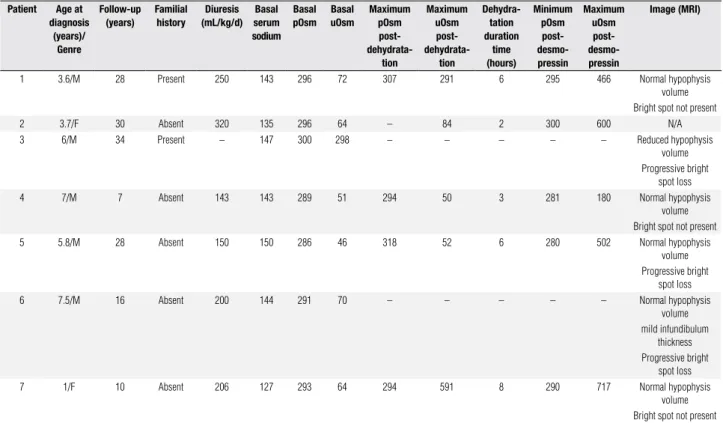
Table 1. Clinical, radiological and laboratory data for patients with central diabetes insipidus Patient Age at
diagnosis (years)/
Genre
Follow-up (years)
Familial history
Diuresis (mL/kg/d)
Basal serum sodium
Basal pOsm
Basal uOsm
Maximum pOsm post-
dehydrata-tion
Maximum uOsm post-
dehydrata-tion
Dehydra-tation duration
time (hours)
Minimum pOsm post- desmo-pressin
Maximum uOsm post- desmo-pressin
Image (MRI)
1 3.6/M 28 Present 250 143 296 72 307 291 6 295 466 Normal hypophysis
volume Bright spot not present
2 3.7/F 30 Absent 320 135 296 64 – 84 2 300 600 N/A
3 6/M 34 Present – 147 300 298 – – – – – Reduced hypophysis
volume Progressive bright
spot loss
4 7/M 7 Absent 143 143 289 51 294 50 3 281 180 Normal hypophysis
volume Bright spot not present
5 5.8/M 28 Absent 150 150 286 46 318 52 6 280 502 Normal hypophysis
volume Progressive bright
spot loss
6 7.5/M 16 Absent 200 144 291 70 – – – – – Normal hypophysis
volume mild infundibulum
thickness Progressive bright
spot loss
7 1/F 10 Absent 206 127 293 64 294 591 8 290 717 Normal hypophysis
volume Bright spot not present
M: male; F: female; pOsm: plasma osmolality; uOsm: urinary osmolality; MRI: magnetic resonance image.
history of diabetes insipidus except patients 1 and 3 who were third-degree cousins but had no other relatives af-fected. Also, no autoimmune disease was identiied in any of the nine patients. Age at onset of polyuria and polydipsia varied from 1 to 7 years of age, and dura-tion of symptoms at diagnosis varied between 2 months and 5 years. Polyuria and polydipsia were the presenting symptoms in all patients, whose urinary volume at diag-nosis ranged from 3 to 13.3 L/24h (143 to 320 mL/ kg/day). The luid deprivation test and vasopressin ana-log responses were consistent with the diagnosis of total central DI in all but one patient that had a response compatible with partial central DI (Patient 6). Growth retardation and bone age delay were observed in pa-tients 1 and 7. Adrenal and thyroid functions and pu-bertal development were normal in all patients. Patients were followed for 22 years at our out-patient clinic.
No expansive sella turcica lesions were visualized by imaging study in any patient, except patient 6 who showed mild infundibulum thickness and a pars inter-media cyst, which disappeared during follow-up. Initial sella turcica MRI studies in six patients resulted in the absence of the posterior pituitary hyperintense signal in three patients and its presence in three patients also; which lost the bright spot of the neurohypophysis in
subsequent MRI during follow-up. Only one patient showed a reduced anterior pituitary.
Sequencing of AVP-NPII gene revealed no muta-tions in exons 1, 2 and 3 in any patients. However, a homozygous insertion of an additional guanine in in-tron 2 (IVS2 +28 InsG) was found in 4 patients. Due to the latter inding, we also sequenced AVP-NPII
gene from controls and the same insertion in intron 2 was found in three out of nine controls.
DISCUSSION
In this study we presented seven patients with central DI diagnosed during the infancy with no apparent eti-ology after long-term follow-up. Molecular analysis of
AVP-NPII gene showed no mutations in the coding region of this gene.
Cop
yright
© ABE&M t
odos os dir
eit
os r
eser
vados
.
the onset of symptoms and the diagnosis, with previ-ous reports showing an interval of 4 years (24). The delay of the diagnosis of diabetes insipidus in pediatric patients suggests that clinicians may not be aware of the need to investigate the symptoms of polyuria and polydipsia in children.
Growth retardation and bone age delay were ob-served in patients 1 and 7, which are in accordance with the 20% to 35% of patients with idiopathic central DI and short stature previously described (16,24,25).
All seven patients in this series had no clear etiology for the vasopressin deiciency, despite the follow-up of 3 to 34 years. In an attempt to uncover the etiology of the disease in these patients, we carried out molecular analysis of the AVP-NPII gene. Although we found no mutation in the AVP-NPII gene in this series of DI patients, Rutishauser and cols. (10) previously reported a de novo AVP-NPII gene mutation in a patient with early onset of central DI and no family history, suggest-ing that genetic testsuggest-ing may be useful in patients who develop idiopathic DI during childhood.
While no mutation in the coding region was found, we identiied a homozygous guanine insertion in intron 2 (IVS2 +28 InsG) in 4 patients. This insertion has also been described by Bahnsen and cols. (23) (GenBank access X62891) with a concomitant mutation in the coding region (Gly57Ser). As the authors performed direct gene sequencing in only one member of the kin-dred of 6 affected members, we do not know whether the G insertion was also present in their other patients or non-affected individuals, and the authors do not discuss this particular issue. Moreover, an error due to PCR/sequencing technique problems is less likely to account for our indings since the result was found in both, sense and antisense sequences as well as in repeat-ed PCR experiments. Inderepeat-ed, inding the G insertion in 3 out of 9 controls indicated that this variation might represent an alternative gene assembly that is unlikely to contribute to the AVP-NPII gene modulation (26).
The presence of vasopressin-secreting cell autoan-tibodies in association with pituitary stalk thickening suggests autoimmunity in almost 100% of patients with apparent idiopathic central DI (27). However, circu-lating human hypothalamus vasopressin-secreting cell autoantibodies have also been reported in patients with different causes of central DI (19,27-29), including Langerhans cell histiocytosis and germinoma; therefore, the presence of these antibodies may not be a reliable marker of autoimmune central DI (29). In the present
study, determination of autoantibodies to vasopressin secreting cells was not available, therefore, we could not rule out an autoimmune cause of central DI, especially in patient 6, who showed pituitary stalk thickness.
Although, pituitary stalk thickening in patients with central DI can be reversible and transient, as reported by De Buyst and cols. (24), the inding of pituitary stalk thickening in patients with central DI strongly indicates the need for long-term follow-up, since organic cause of central DI, such as germinoma and Langerhans cell histiocytosis, has been associated with isolated central DI with pituitary stalk thickening (18,30).
In the present study, brightness signal of the poste-rior lobe on magnetic resonance T1-weighted images was absent in ive patients. The absence of posterior pi-tuitary bright signal has been associated with central DI (31-33). On the other hand, presence of the signal may not indicate normal vasopressin secretion (31). Maho-ney and cols. (34) studied the kindred with adFNDI by magnetic resonance imaging and showed the presence of the posterior pituitary hypertensive signal in all af-fected children, but an absent or barely visible signal in all adult patients but one, suggesting a progressive loss of the posterior pituitary signal in this inherited form of DI. In fact, three out of six patients in the current study presented a normal posterior pituitary signal, which dis-appeared later during follow-up, indicating progressive development of MRI features, similar to the decrease in AVP secretion (8).
It is important to point out that vascular abnormal-ity should also be considered as a plausible cause of vasopressin deiciency in the present series of patients, since abnormal arterial blood low affecting posterior pituitaryblood supply has been previously described in patients with idiopathic central DI and normalanterior pituitary and pituitary stalk size with absence of poste-riorpituitary bright signal in the MRI (35).
In conclusion, we found a homozygous guanine in-sertion in intron 2 (IVS2 +28 InsG) in the AVP-NPII
gene in DI patients as well as in controls, suggesting an alternative gene assembly that is unlikely to contribute to the AVP-NPII gene modulation. In addition, we con-irm that the etiology of idiopathic central DI in children may not be apparent even after long-term follow-up, and requires continuous etiological surveillance.
Acknowledgements: This work was supported by Fapesp (grant number 07/58365-3).
Cop
yright
© ABE&M t
odos os dir
eit
os r
eser
vados
.
REFERENCES
1. Verbalis JG. Diabetes insipidus. Rev Endocr Metab Disord. 2003;4(2):177-85.
2. Christensen JH, Rittig S. Familial neurohypophyseal diabetes in-sipidus − an update. Semin Nephrol. 2006;26(3):209-23.
3. Ito M, Jameson JL, Ito M. Molecular basis of autosomal dominant neurohypophyseal diabetes insipidus. Cellular toxicity caused by the accumulation of mutant vasopressin precursors within the endoplasmic reticulum. J Clin Invest. 1997;99(8):1897-905. 4. Rittig S, Robertson GL, Siggaard C, Kovacs L, Gregersen N,
Ny-borg J, et al. Identiication of 13 new mutations in the vasopres-sin-neurophysin II gene in 17 kindreds with familial autosomal dominant neurohypophyseal diabetes insipidus. Am J Hum Ge-net. 1996;58(1):107-17.
5. Willcutts MD, Felner E, White PC. Autosomal recessive fami-lial neurohypophyseal diabetes insipidus with continued se-cretion of mutant weakly active vasopressin. Hum Mol Genet. 1999;8(7):1303-7.
6. Sausville E, Carney D, Battey J. The human vasopressin gene is linked to the oxytocin gene and is selectively expressed in a cul-tured lung cancer cell line. J Biol Chem. 1985;260(18):10236-41. 7. Siggaard C, Christensen JH, Corydon TJ, Rittig S, Robertson GL,
Gregersen N, et al. Expression of three different mutations in the arginine vasopressin gene suggests genotype-phenotype corre-lation in familial neurohypophyseal diabetes insipidus kindreds. Clin Endocrinol (Oxf). 2005; 63(2):207-16.
8. Elias PC, Elias LL, Torres N, Moreira AC, Antunes-Rodrigues J, Castro M. Progressive decline of vasopressin secretion in familial autosomal dominant neurohypophyseal diabetes insipidus pre-senting a novel mutation in the vasopressin-neurophysin II gene. Clin Endocrinol (Oxf). 2003;59(4):511-8.
9. Siggaard C, Rittig S, Corydon TJ, Andreasen PH, Jensen TG, An-dresen BS, et al. Clinical and molecular evidence of abnormal processing and traficking of the vasopressin preprohormone in a large kindred with familial neurohypophyseal diabetes insipi-dus due to a signal peptide mutation. J Clin Endocrinol Metab. 1999;84(8):2933-41.
10. Rutishauser J, Kopp P, Gaskill MB, Kotlar TJ, Robertson GL. Clini-cal and molecular analysis of three families with autosomal do-minant neurohypophyseal diabetes insipidus associated with a novel and recurrent mutations in the vasopressin-neurophysin II gene. Eur J Endocrinol. 2002;146(5):649-56.
11. Nijenhuis M, Zalm R, Burbach JP. Mutations in the vasopressin pro-hormone involved in diabetes insipidus impair endoplasmic reticu-lum export but not sorting. J Biol Chem. 1999;274(30):21200-8. 12. Beuret N, Rutishauser J, Bider MD, Spiess M. Mechanism of
en-doplasmic reticulum retention of mutant vasopressin precursor caused by a signal peptide truncation associated with diabetes insipidus. J Biol Chem. 1999;274(27):18965-72.
13. Ito M, Yu RN, Jameson JL. Mutant vasopressin precursors that cause autosomal dominant neurohypophyseal diabetes insipi-dus retain dimerization and impair the secretion of wild-type pro-teins. J Biol Chem. 1999;274(13):9029-37.
14. Greger NG, Kirkland RT, Clayton GW, Kirkland JL. Central diabetes insipidus. 22 years’ experience. Am J Dis Child. 1986;140(6):551-4. 15. Wang LC, Cohen ME, Duffner PK. Etiologies of central diabetes
insipidus in children. Pediatr Neurol. 1994;11(4):273-7.
16. Maghnie M, Cosi G, Genovese E, Manca-Bitti ML, Cohen A, Zecca S, et al. Central diabetes insipidus in children and young adults. N Engl J Med. 2000;343(14):998-1007.
17. Maghnie M. Diabetes insipidus. Horm Res. 2003;59(Suppl 1):42-54. 18. Mootha SL, Barkovich AJ, Grumbach MM, Edwards MS, Gitel-man SE, Kaplan SL, et al. Idiopathic hypothalamic diabetes insi-pidus, pituitary stalk thickening, and the occult intracranial ger-minoma in children and adolescents. J Clin Endocrinol Metab. 1997;82(5):1362-7.
19. De Bellis A, Colao A, Bizzarro A, Di Salle F, Coronella C, Solimeno S, et al. Longitudinal study of vasopressin-cell antibodies and of hypothalamic-pituitary region on magnetic resonance imaging in patients with autoimmune and idiopathic complete central diabe-tes insipidus. J Clin Endocrinol Metab. 2002;87(8):3825-9. 20. Miller M, Dalakos T, Moses AM, Fellerman H, Streeten DH.
Recog-nition of partial defects in antidiuretic hormone secretion. Ann Intern Med. 1970;73(5):721-9.
21. Zerbe RL, Robertson GL. A comparison of plasma vasopressin measurements with a standard indirect test in the differential diagnosis of polyuria. N Engl J Med. 1981;305(26):1539-46. 22. Amato MC, Elias LL, Elias J, Santos AC, Bellucci AD, Moreira AC,
et al. Endocrine disorders in pediatric − onset Langerhans Cell Histiocytosis. Horm Metab Res. 2006;38(11):746-51.
23. Bahnsen U, Oosting P, Swaab DF, Nahke P, Richter D, Schmale H. A missense mutation in the vasopressin-neurophysin precursor gene cosegregates with human autosomal dominant neurohypo-physeal diabetes insipidus. EMBO J. 1992;11(1):19-23.
24. De Buyst J, Massa G, Christophe C, Tenoutasse S, Heinrichs C. Cli-nical, hormonal and imaging indings in 27 children with central diabetes insipidus. Eur J Pediatr. 2007;166(1):43-9.
25. Czernichow P, Pomarede R, Basmaciogullari A, Brauner R, Rappa-port R. Diabetes insipidus in children. III. Anterior pituitary dys-function in idiopathic types. J Pediatr. 1985;106(1):41-4.
26. Ye L, Li X, Chen Y, Sun H, Wang W, Su T, et al. Autosomal dominant neurohypophyseal diabetes insipidus with linkage to chromoso-me 20p13 but without mutations in the AVP-NPII gene. J Clin En-docrinol Metab. 2005;90(7):4388-93.
27. Pivonello R, De Bellis A, Faggiano A, Di Salle F, Petretta M, Di Somma C, et al. Central diabetes insipidus and autoimmunity: relationship between the occurrence of antibodies to arginine vasopressin-secreting cells and clinical, immunological, and ra-diological features in a large cohort of patients with central diabe-tes insipidus of known and unknown etiology. J Clin Endocrinol Metab. 2003;88(4):1629-36.
28. Scherbaum WA, Hauner H, Pfeiffer EF. Vasopressin cell surface an-tibodies in central diabetes insipidus detected on cultured human foetal hypothalamus. Horm Metab Res. 1985;17(11):622. 29. Maghnie M, Ghirardello S, De Bellis A, Di Iorgi N, Ambrosini L,
Secco A, et al. Idiopathic central diabetes insipidus in children and young adults is commonly associated with vasopressin-cell antibodies and markers of autoimmunity. Clin Endocrinol (Oxf). 2006;65(4):470-8.
30. Leger J, Velasquez A, Garel C, Hassan M, Czernichow P. Thi-ckened pituitary stalk on magnetic resonance imaging in chil-dren with central diabetes insipidus. J Clin Endocrinol Metab. 1999;84(6):1954-60.
31. Maghnie M, Villa A, Arico M, Larizza D, Pezzotta S, Belufi G, et al. Correlation between magnetic resonance imaging of posterior pi-tuitary and neurohypophyseal function in children with diabetes insipidus. J Clin Endocrinol Metab. 1992;74(4):795-800.
32. Gudinchet F, Brunelle F, Barth MO, Taviere V, Brauner R, Rappaport R, et al. MR imaging of the posterior hypophysis in children. Am J Roentgenol. 1989;153(2):351-4.
33. Fujisawa I. Magnetic resonance imaging of the hypothalamic-neu-rohypophyseal system. J Neuroendocrinol. 2004;16(4):297-302. 34. Mahoney CP, Weinberger E, Bryant C, Ito M, Jameson JL, Ito M.
Effects of aging on vasopressin production in a kindred with au-tosomal dominant neurohypophyseal diabetes insipidus due to the DeltaE47 neurophysin mutation. J Clin Endocrinol Metab. 2002;87(2):870-6.