Authors Luis F. Arias1 Carlos A. Jiménez2 Mariam J Arroyave1
1 Department of Pathology, University of Antioquia. 2 Department of Pathology, Universidad de Antioquia (Department of Pathology, Valle del Lili Foundation).
Submitted on: 01/16/2013. Approved on: 04/20/2013.
Correspondence to:
Luis F. Arias.
PRYT Group, Department of Pathology, Faculty of Medicine, University of Antioquia, Medellín, Colombia.
Department of Pathology, Faculty of Medicine, University of Antioquia, Carrera 51D, nº 62-29, Medellín, Colombia.
E-mail: [email protected] Tel: (+57) 4219-2412.
Fax: (+57) 4263-0253. University of Antioquia.
Histologic variants of primary focal segmental
glomerulos-clerosis: presentation and outcome
Introduction: The clinical significance of histologic variants of primary focal segmental glomerulosclerosis (FSGS) remains unclear. With the aim to determine presentation and outcome of the variants of FSGS in a hispanic population, we studied our cases of this glomerulopathy. Methods: In this retrospective study, all renal biopsies with FSGS (1998-2009), were classified according to the Columbia's classification. We analyzed histological, clinical and follow-up data and compared among variants. Results: Among 291 cases, 224 (77.0%) corresponded to NOS variant, 40 cases (13.7%) to tip variant (TIP), 14 cases (4.8%) to perihilar (PH), 10 cases (3.4%) to collapsing (COLL) and three cases (1.0%) to cellular variant (CELL). Median age: 26 years (range 1 to 79); 74 patients (25.4%) were < 15 years of age. Hypertension and renal dysfunction were more frequent in PH and COLL cases. PH presented frequently as non-nephrotic proteinuria. There were fewer histologic chronic lesions in TIP cases. There was remission in 23.5% of patients with NOS, 57.7% of patients with TIP, 22.2% of patients with COLL and 0 patients with PH (p < 0.01). Chronic kidney disease (CKD) was less frequent in TIP than in the other variants (p = 0.03). There were not statistical differences for end-stage renal disease among variants.
Conclusion: Glomerular histological appearance is not a good indicator of outcome. COLL is a disease with many differences to the other variants and bad prognosis; PH is a variant mainly of adults, with frequent evolution to CKD. TIP appears as a less aggressive, although not benign, variant.
A
BSTRACTKeywords: glomerulosclerosis, focal segmental; kidney glomerulus; nephrotic syndrome; podocytes.
I
NTRODUCTIONAlthough we refer to “focal and segmental glomerulosclerosis” (FSGS) as a glomerular disease, at present, this glomerular “mor-phological change” is considered a “pattern of injury”1 associated to diverse factors
(se-condary) or without any “known” asso-ciated factor or cause (primary); in fact, it is a morphologic presentation common to diverse mechanisms of disease. Its diagnosis is based on morphological changes: focal and segmental sclerosis and/or hyalinosis, and absence of diffuse immune deposits on immunopathology. In the future, when we will know more about the etiology and pa-thogenesis, the term “FSGS” will be obsole-te, and each case will be renamed according to its cause or physiopathology.2 The term
“FSGS” is a misnomer, as it is not always fo-cal or segmental, or even sclerotic (in some cases the lesions are hyaline or collapsing);3
nevertheless the denomination “FSGS” is extensively used. FSGS is considered the main cause of nephrotic syndrome in some ethnic/geographical groups, and it is one of the three main causes of the syndrome worldwide.4-8 In our country and in Latin
America FSGS is the more frequent primary glomerulopathy diagnosed by biopsy.4,5
Define, diagnose, and treat a disease that is heterogeneous in morphology and clinical presentation is a difficult task, as it is to compare its prognosis and treat-ment among different centers. Our igno-rance has led us to try dividing the disease according to their morphological features. Several histological variants has been described, and now the more used mor-phologic classification is known as “the Columbia classification” of FSGS,9 with
five pathologic variants: collapsing (COLL), cellular (CELL), tip (TIP), perihilar (PH) and not otherwise specified (NOS). However the prognostic and ther-apeutic utility of this classification remains unclear, largely because studies that have assessed the clinical relevance of the histologic variants of primary FSGS are few and conflicting, and several with short follow-up time.
The aim of this retrospective work was determine the clinical and histological features, and outcome of the morphologic variants of FSGS in a different geographical population.
M
ETHODSThis is a descriptive, retrospective, clinicopathologic study. All native renal biopsies diagnosed as FSGS in our department between August 1998 and December 2009 were revised and classified according to the Columbia classification of FSGS.9 Light microscopic
examination of slides (21 to 42 sections) stained with haematoxylin and eosin, Masson’s trichrome, PAS, and methenamine-silver provided the diagnosis of FSGS and categorization into one of the five variants. Immunofluorescence (for IgA, IgG, IgM, C3, C1q,
κ, and λ) and clinical information were used to ex-clude nonprimary causes of FSGS or other glomeru-lopathies. Cases with low serum complement levels, no proteinuria, systemic disease, chronic viral in-fection, any suspect of immune-mediated disease, congenital nephrotic syndrome, or familial history of renal disease suggesting a hereditary glomeru-lopathy were excluded. All the cases diagnosed as minimal change disease were also revised, and those with tip lesions (n = 3) were included in the study as TIP cases. All the specimens were cylinders obtained by core biopsy. Criteria to assign each case in a category of the Columbia classification was based exclusively in the paper by D’Agati et al.9 (the original publication of the classification)
(Figure 1); to diagnose TIP or PH it was required to identify the proximal tubular pole or the glo-merular vascular pole, respectively, in the defining glomerulus. Glomeruli with global sclerosis (GS) and glomeruli with segmental lesions were quanti-fied as percentage of total glomeruli or percentage of viable glomeruli, respectively. The percentage of interstitial fibrosis was semiquantitatively calculated as no fibrosis or mild, moderate or severe, according to Banff schema for renal allograft classification.10
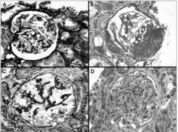
Figure 1. A: Glomerulus with a synechia of the tuft just at the tubular origin, a lesion defining the tip variant; B: Glomerular sclerosis and hyalinosis in the vascular pole; in some cases serial sections are required to identify the vascular pole; C: Collapsing lesion with notorious hypertrophy and hyperplasia of podocytes; D: Segmental and focal endocapillary proliferation. In an adequate immunopathologic and clinical context, this alteration could correspond to the cellular variant of FSGS; A-C: Methenamine-silver stain; B: PAS stain, and D: Hematoxylin-eosin stain, all x400.
Arteriolar hyalinosis was registered as present or absent. The histological evaluation was blinded to clinical and follow-up data.
All biopsies came from patients considered as hispanic, according to geographical origin, physical ap-pearance/skin colour, and self-identification, although we know that these features do not exactly indicate racial/ethnic origin because genetic heterogeneity exists and patients could have either Caucasian or African genetic background;11,12 Hispanic race/ethnicity is a
particular mix of ancestors from different races. Demographic, clinical, and laboratory information at the time of renal biopsy and at follow-up (when possible) was obtained, from medical records, on each patient. Patient’s data included gender, age, blood pressure, level of protein excretion, serum creatinine (SCr), and measured creatinine clearance (CrCl) at pre-sentation. Presentation was defined as the time when proteinuria was first detected. Hypertension was de-fined as systolic blood pressure equal to or greater than 140 mmHg and/or diastolic blood pressure equal to or greater than 90 mmHg, according to the Seventh Report of the Joint National Committee on Prevention, Detection, Evaluation, and Treatment of High Blood Pressure.13 At the end of follow-up SCr,
The number of glomeruli for evaluation by light microscopy was 16.7 ± 12.9 (range 6-104; median: 14) and there was no significant difference among groups (Table 2). There were significantly less global glomer-ulosclerosis, glomerular segmental lesions, interstitial fibrosis and arteriolar hyalinosis in TIP cases than in the other variants (Table 2). In COLL cases there were significantly more glomerular segmental lesions, and in PH significantly more arteriolar hyalinosis than in the other variants (Table 2). In TIP, glomerular lesions included glomerular tip lesion alone in 25 cases (62.5%) and tip lesion with peripheral and/or indeterminate lesions in 15 (37.5%).
CLINICAL OUTCOME
Follow-up data were available on 151 patients (51.9%), 8 of them developed ESRD before 2 years of follow-up (at 6-18 months). The number of cases wi-th follow-up data for variant was: NOS: 105 (46.9% within this variant); tip: 26 (65.0%); perihilar: 8 (57.1%); COLL: 9 (90%). In the 3 cases of cellular variant there was follow-up (25, 28 and 36 months). The median of follow-up in patients who did not develop ESRD before 2 years was 40.0 months (24.3-160.0); 65.2% of the cases with > 36 months.
Treatment was very variable among patients; many received several immunosuppressants, including pred-nisone, cyclosporine, mycophenolate, azathioprine and/ or cyclophosphamide, for a variable time. Although an adequate comparison for treatment among variants was not possible, there were not statistical differences for percentage of cases receiving immunosuppressants.
Remission (complete or partial) was attained in 23.5% of patients with NOS, 57.7% of patients with TIP, 22.2% of patients with COLL and in 0 pa-tients with PH (p < 0.01) (Table 3). CKD was signifi-cantly less frequent in TIP than in the other variants (p = 0.03). With our follow-up time, there was not statistical difference for ESRD among variants. The statistical differences for remission and CKD were lost in the group of patients < 15 years old (Table 3).
The median of follow-up in patients who developed CKD was 50.0 months (24.3-160.0) and in patients no developing CKD was 49.2 months (24.4-159) (p = 0.47). There were not statistical differences for follow-up time between patients with and without CKD in the groups with NOS, TIP, PH, and COLL. In patients with CELL, two developed CKD, one with ESRD; the other one had complete remission.
min. Complete remission was defined as proteins in urine < 0.3 g/24h in adults, and < 4 mg/m2/h in < 15
years old, and partial remission was defined as protein-uria between 0.31 and 2.5 g/24h in adults and between 4 and 40 mg/m2/h in < 15 years old. We compare
clini-cal and morphologiclini-cal features among the histologiclini-cal variants of FSGS. ESRD was registered in all the cases with this event, although for the analyses of outcome, CKD was taken as the endpoint. For follow-up analyses we included only patients with at least 2 years of follow-up, or patients developing the end-point before two years.
STATISTICAL ANALYSES
Data are expressed as mean ± SD or, where indicated, as median and ranges, according to the variables and Kolmogorov-Smirnov test for normality. χ2 test, or
Fisher’s exact test were used to compare percentages. Unpaired t test or Mann-Whitney test were used to compare means, according to normality test. P values < 0.05 were considered statistically significant in two tailed tests. All analysis was done using SPSS®
soft-ware, version 16.0 (SPSS Inc, Chicago, IL).
R
ESULTSTABLE 1 CLINICALCHARACTERISTICSATPRESENTATION
Characteristics NOS TIP Perihilar Collapsing p
n 224 40 14 10
-Male 54% 60% 78.6% 60% 0.23
Agea 28 (1-79) 17 (1-65) 44 (18-65) 11 (3-66) < 0.01
Hypertensionb 67.1% 50% 83.3% 100% 0.03
Creatininec 1.2 ± 1.0 1.1 ± 1.0 1.4 ± 0.6 2.8 ± 3.8 0.03
Creatinine clearanced 80.5 ± 33.4 92.1 ± 33.0 74.3 ± 32.2 50.1 ± 41.2 0.07
Proteinuria adults (g/24h) (n = 217) 6.9 ± 5.9 6.3 ± 5.0 3.9 ± 2.4 9.7 ± 6.4 0.45
Proteinuria < 15y (mg/m2/h) (n = 74) 153 ± 110 127 ± 98 No cases 198 ± 104 0.02
Nephrotic proteinuriab 81.7% 95% 57.1% 100% < 0.01
a Age: median (minimal - maximal values); b Percentages of cases with hypertension and nephrotic proteinuria, respectively; c mg/dL; d mL/minute.
TABLE 2 HISTOLOGICCOMPARISONAMONGGROUPS
Characteristics NOS TIP Perihilar Collapsing p
n 224 40 14 10
-Total glomeruli 15.8 ± 12.9 19.4 ± 9.4 16.7 ± 13.5 25.4 ± 19.7 0.06
Global GS (%) 8.0 (0-86) 0.0 (0-30) 9.4 (0-65) 5.0 (0-50) < 0.01
Segmental lesions (%) 24.2 (2-100) 16.3 (3-57) 27.9 (14-67) 50.0 (22-100) < 0.01
Total glomeruli with lesions (%) 35.8 (3-100) 16.3 (4-70) 42.2 (14-87) 57.8 (22-100) < 0.01
Interstitial fibrosisa 18.8 2.5 21.4 20.0 0.03
Arteriolar hyalinosisb 42.4 12.5 78.6 20 < 0.01
a Percentage of cases with interstitial fibrosis > 25% (moderate or severe); b % o cases with any degree of arteriolar hyalinosis
TABLE 3 OUTCOMECOMPARISONAMONGHISTOLOGICVARIANTS
NOS TIP Perihilar Collapsing p
n 105 (45.5%) 26 (65.0%) 8 (57.1%) 9 (90%)
-Follow-up (months) 38 (24-159) 52 (24-87) 44 (27-160) 40 (26-61) 0.34
Remission 24 (23.5%) 15 (57.7%) 0 (0.0%) 2 (22.2%) < 0.01
Complete 12 (11.8%) 8 (30.8%) 0 (0.0%) 1 (11.1%) 0.07
Partial 12 (11.8%) 7 (26.9%) 0 (0.0%) 1 (11.1%) 0.16
CKD 55 (53.9%) 8 (30.8%) 7 (87.5%) 5 (55.6%) 0.03
ESRD 20 (19.6%) 5 (19.2) 1 (12.5%) 3 (33.3%) 0.74
Adults (≥ 15 years) n = 69 n = 17 n = 8 n = 4
-Complete remission 6 (8.7%) 7 (41.2%) 0 (0.0%) 0 (0.0%) < 0.01
CKD 44 (63.8%) 3 (17.6%) 7 (87.5%) 3 (75%) < 0.01
ESRD 13 (18.8%) 3 (17.6%) 1 (12.5%) 2 (50%) 0.46
Children (< 15 years) n = 33 n = 9 n = 0 n = 5
-Complete remission 6 (18.2%) 1 (11.1%) - 1 (20%) 0.95
CKD 11 (33.3%) 5 (55.6%) - 2 (40%) 0.51
ESRD 7 (21.2%) 2 (22.2%) - 1 (20%) 1.00
NOS: Not otherwise specified variant; TIP: Tip variant; CKD: Chronic kidney disease; ESRD: End-stage renal disease.
D
ISCUSSIONIn this work we reviewed the clinical presentation, histopathologic findings and outcome of FSGS, according to the Columbia’s histologic variant of
FSGS in Latin America. In a previous report from Brazil14 analyzing immunohistochemical podocyte
markers, the authors reported percentage frequencies almost similar to our study for TIP, PH and CELL variants (14.5%, 6.9% and 3.8% respectively), however, the percentage frequency for COLL (36.6%) was notoriously higher in the Brazilian series. We do not know the reason for this difference; it is possible that population characteristics or environmental fac-tors influence the results of these works. Our series increases the worldwide information about the cli-nical or prognosis relevance of the FSGS variants. FSGS is the more frequent glomerular disease in our population, but we do not know the cause of this in-creased frequency in our region. Proportions of the Columbia’s variants appear not very different that in other series,1,15-17 and several results on presentation,
chronic histologic lesions and outcome are not diffe-rent than in other previous published works, however, some of our results show interesting aspects of these variants, as we will discuss in the next paragraphs.
According to definition of CELL, the diagnosis requires the presence of endocapillary hypercellular-ity with occlusion of capillary lumina, with foam cells and/or macrophages and/or endothelial cells and/or neutrophils and/or lymphocytes; there may be pyknotic or karyorrhectic debris and endocapillary fibrin.9 As we can deduce from this microscopic
features, in our 3 cases, lesions were similar to en-docapillary proliferative glomerulonephritis (in im-mune mediated glomerular diseases). As “neither hyalinosis nor segmental sclerosis are required fea-tures”,9 it was essential in these cases a rigorous
ex-amination and analysis of immunofluorescence (IF), other histological features, clinical manifestations, and, in two cases, electron microscopy. The ultra-structural alterations can be essential to distinguish CELL variant from other glomerulopathies. We do not know if the low frequency of this diagnosis is ac-tually due to rarity of this variant or if the difficulty on its diagnosis contributes to this low frequency. In our series cellular variant correspond to 1.0%; in all three, the diagnosis required search for a second opinion from an experienced nephropathologist. In the series by Chun et al.,1 with 87 patients with
FSGS diagnosis, there were not cases of cellular vari-ant, as described by the Columbia classification. Endocapillary hypercellularity, a diagnostic criterion of the cellular variant, can be also found in collapsing
variant cases.16 Overlapping histological findings in
the collapsing and cellular variants sometimes cause difficulty in identifying the variant; further stud-ies are needed to identify clear differences between these variants to apply to cases with histologically overlapping findings.17,18 On the other hand,
endo-capillary hypercellularity involving the tip domain rules out the cellular variant, as endocapillary hy-percellularity may characterize lesions in tip variant. Cellular variant may include cases of unsampled tip or collapsing lesions.16 Implications for cellular
vari-ant diagnosis are unknown as so few patients are registered in most series reported. To the best of our knowledge, the work with the largest number of cases was that by Stokes et al. (n = 22),16 and
the authors reported rates of remission and ESRD no statistically different to NOS. In our 3 cases, one developed ESRD, other one CKD, not yet terminal (28 months of follow-up) and the remaining patient presented remission.
It has been postulated that tip lesion simply rep-resents a response to heavy proteinuria,19 and cases
of nephrotic syndrome with tip lesions, but no other histological abnormalities, may represent a form of minimal change nephropathy.20-22 However, others
have reported that such lesions have a clinical course similar to that of primary FSGS.1,23 The definition of
the genuine glomerular tip lesion in the papers by the original authors has been the presence of changes at the tubular origin and nowhere else, in glomeruli that are otherwise normal on light microscopy.20,24 In
the Columbia classification are included cases with peripheral and/or indeterminate lesions, which mean that some at least are not the glomerular tip lesion as originally defined. We found that there was a lower percentage of CKD in patients with tip lesions alone (neither peripheral nor indeterminate lesions) than in patients with tip lesions and other glomerular seg-mental lesions (included in the TIP variant as defined in the Columbia classification), but this difference was not statistically significant.25 It appears that GTL,
whether defined by the original definition20,24 or the
less restrictive in the Columbia Classification,9
repre-sents a heterogeneous group of lesions that can behave like minimal change disease or like FSGS.26 Routine
change disease, or vice versa, may prove helpful in this regard, for now, only the response to treatment can predict the outcome of the GTL.27
Several reports in nephrotic patients with TIP suggest an excellent response to steroids and/or more favourable course;20-22,24,28-30 but other authors
have reported that the response and course are sim-ilar to that of patients with NOS and question the clinical significance of this feature.24,31-34 We found
that although TIP presents with less chronic histo-logic alterations, the prognostic implications for this diagnosis were not “favourable”: CKD developed in 30.8% and ESRD in 19.2% of our patients. Although the outcome was worse in the other variants, our data suggest that the tip variant should not be considered a prognostically favourable disease. In fact, it could be a more early stage of a severe glomerular disease. Also in the work by Howie et al.,26 they conclude that
“many patients have a good outcome but that some will progress pathologically and clinically” and that “progression of GTL to FSGS (NOS) is not uncom-mon”, then, it is not a “benign” disease.
It was interesting in our work that in TIP cases, remission was lower and CKD was higher in < 15 years old compared to ≥ 15 years old, but the case number in each of these two groups was low (9 and 17 patients, respectively) and these differences can be spurious.
The term “collapsing glomerulopathy” was used for the first time in 1986,35 and it was assumed to be
a variant of FSGS.30,36,37 However, COLL is
frequent-ly an aggressive disease, with massive proteinuria and rapidly progressive renal disease. In our series, 55.5% of cases progressed to CKD in the 5 years after initial presentation. The notoriously more ag-gressive outcome in COLL that in the other variants of FSGS, and the phenotypic features of podocytes suggesting a different pathogenesis,38,39 have led
some authors to propose that collapsing glomeru-lopathy is not a variant of FSGS; we think that it is a different disease, and it should not be considered a variant of FSGS.
It is believed that PH variant is commonly associated with secondary forms to hyperfiltration or nephron loss or glomerular hypertension.9 In our
series PH presented more frequently than the other variants with non-nephrotic proteinuria: 42.9%. This lower level of proteinuria has been also re-ported by other authors.17 All our patients were > 15
years old (18-65) and 85.0% of cases with follow-up developed CKD. Histological chronic lesions were more severe in PH than in NOS and TIP, and near to COLL. These facts suggest that morphologi-cal lesions develop gradually and not in an abrupt manner as in the other variants, resulting clinically in disorders of insidious onset and slow but steady progression.
NOS variant is a heterogeneous glomerulopa-thy; in fact, it is a common morphological lesion in many glomerular and non-glomerular diseases. It is the most common histological form of FSGS: 77% of our cases. It is very interesting that all the other 4 variants may evolve into this pattern in the course of disease progression and increasing chronicity: i.e. it is an unspecific glomerulopathy with many known and unknown etiologies. It is a “collage” of glomerular al-terations that converge on a common morphological pattern. The NOS variant is a “junk drawer” of mul-tiple glomerular alterations with this common pattern of lesion, with a mixture of pathophysiological mechanisms that do not allow for now, based only on morphological findings, determine etiology or effective treatment.
This work was based on a retrospective clinical information collection, leading to limitations because this methodology: to determine with precision the moment of the clinical presentation it is very difficult; time between presentation and biopsy can be variable among patients; treatment is different according to treating nephrologists; adherence to treatment is very difficult to determine in clinical charts, and possibly other limitations inherent to a retrospective methodology.
C
ONCLUSIONA
CKNOWLEDGMENTSThanks to Dr. Luiz A. Moura for his help in prepa-ring the manuscript.
R
EFERENCES1. Chun MJ, Korbet SM, Schwartz MM, Lewis EJ. Focal segmental glomerulosclerosis in nephrotic adults: presentation, prognosis, and response to therapy of the histologic variants. J Am Soc Nephrol 2004;15:2169-77. http://dx.doi.org/10.1097/01. ASN.0000135051.62500.97 PMid:15284302
2. Cameron JS. Focal segmental glomerulosclerosis in adults. Nephrol Dial Transplant 2003;18:vi45-51. http://dx.doi. org/10.1093/ndt/gfg1058 PMid:12953042
3. Meyrier A. Focal and segmental glomerulosclerosis: multiple pathways are involved. Semin Nephrol 2011;31:326-32. http://dx.doi.org/10.1016/j.semnephrol.2011.06.003 PMid:21839365
4. Arias LF, Henao J, Giraldo RD, Carvajal N, Rodelo J, Arbeláez M. Glomerular diseases in a Hispanic population: review of a regional renal biopsy database. Sao Paulo Med J 2009;127:140-4. http://dx.doi.org/10.1590/S1516-31802009000300006 PMid:19820874
5. Polito MG, de Moura LA, Kirsztajn GM. An overview on fre-quency of renal biopsy diagnosis in Brazil: clinical and patholo-gical patterns based on 9,617 native kidney biopsies. Nephrol Dial Transplant 2010;25:490-6. http://dx.doi.org/10.1093/ndt/ gfp355 PMid:19633091
6. Korbet SM, Genchi RM, Borok RZ, Schwartz MM. The ra-cial prevalence of glomerular lesions in nephrotic adults. Am J Kidney Dis 1996;27:647-51. http://dx.doi.org/10.1016/S0272-6386(96)90098-0
7. Rivera F, López-Gómez JM, Pérez-García R; Spsnish Registry of Glomerulonephritis. Frequency of renal pathology in Spain 1994-1999. Nephrol Dial Transplant 2002;17:1594-2. http:// dx.doi.org/10.1093/ndt/17.9.1594 PMid:12198210
8. Gesualdo L, Di Palma AM, Morrone LF, Strippoli GF, Sche-na FP; Italian Immunopathology Group, Italian Society of Nephrology. The Italian experience of the national registry of renal biopsies. Kidney Int 2004;66:890-4. http://dx.doi. org/10.1111/j.1523-1755.2004.00831.x PMid:15327376 9. D'Agati VD, Fogo AB, Bruijn JA, Jennette JC. Pathologic
clas-sification of focal segmental glomerulosclerosis: a working proposal. Am J Kidney Dis 2004;43:368-82. http://dx.doi. org/10.1053/j.ajkd.2003.10.024 PMid:14750104
10. Racusen LC, Solez K, Colvin RB, Bonsib SM, Castro MC, Cavallo T, et al. The Banff 97 working classification of renal allograft pathology. Kidney Int 1999;55:713-23. http://dx.doi. org/10.1046/j.1523-1755.1999.00299.x PMid:9987096 11. Chernin G, Heeringa SF, Vega-Warner V, Schoeb DS,
Nürn-berg P, Hildebrandt F. Adequate use of allele frequencies in Hispanics--a problem elucidated in nephrotic syndrome. Pedia-tr Nephrol 2010;25:261-6. http://dx.doi.org/10.1007/s00467-009-1315-6 PMid:19876656 PMCid:2899680
12. Parra FC, Amado RC, Lambertucci JR, Rocha J, Antunes CM, Pena SD. Color and genomic ancestry in Brazilians. Proc Natl Acad Sci U S A 2003;100:177-82. http://dx.doi.org/10.1073/ pnas.0126614100 PMid:12509516 PMCid:140919
13. Chobanian AV, Bakris GL, Black HR, Cushman WC, Green LA, Izzo JL Jr, et al.; Joint National Committee on Prevention, Detection, Evaluation, and Treatment of High Blood Pressu-re. National Heart, Lung, and Blood Institute; National High Blood Pressure Education Program Coordinating Committee. Seventh report of the Joint National Committee on Prevention, Detection, Evaluation, and Treatment of High Blood Pressure. Hypertension 2003;42:1206-52. http://dx.doi.org/10.1161/01. HYP.0000107251.49515.c2 PMid:14656957
14. Testagrossa L, Azevedo Neto R, Resende A, Woronik V, Malheiros D. Immunohistochemical expression of podocyte markers in the variants of focal segmental glomerulosclero-sis. Nephrol Dial Transplant 2013;28:91-8. http://dx.doi. org/10.1093/ndt/gfs325 PMid:22859792
15. Thomas DB, Franceschini N, Hogan SL, Ten Holder S, Jennette CE, Falk RJ, et al. Clinical and pathologic characteristics of focal segmental glomerulosclerosis pathologic variants. Kidney Int 2006;69:920-6. http://dx.doi.org/10.1038/sj.ki.5000160 PMid:16518352
16. Stokes MB, Valeri AM, Markowitz GS, D'Agati VD. Cellular focal segmental glomerulosclerosis: Clinical and pathologic fea-tures. Kidney Int. 2006;70:1783-92. http://dx.doi.org/10.1038/ sj.ki.5001903 PMid:17021605
17. Taneda S, Honda K, Uchida K, Nitta K, Yumura W, Oda H, et al. Histological heterogeneity of glomerular segmental lesions in focal segmental glomerulosclerosis. Int Urol Nephrol 2012;44:183-96. http://dx.doi.org/10.1007/s11255-011-9932-y PMid:21424374 PMCid:3253997
18. Schwartz MM, Evans J, Bain R, Korbet SM. Focal segmental glomerulosclerosis: prognostic implications of the cellular le-sion. J Am Soc Nephrol 1999;10:1900-7. PMid:10477141 19. Haas M, Yousefzadeh N. Glomerular tip lesion in minimal
change nephropathy: a study of autopsies before 1950. Am J Kidney Dis 2002;39:1168-75. http://dx.doi.org/10.1053/ ajkd.2002.33386 PMid:12046027
20. Howie AJ, Brewer DB. The glomerular tip lesion: a previously undescribed type of segmental glomerular abnormality. J Pathol 1984;142:205-20. http://dx.doi.org/10.1002/path.1711420308 PMid:6707787
21. Howie AJ, Lee SJ, Green NJ, Newbold KM, Kizaki T, Koram A, et al. Different clinicopathological types of segmental scle-rosing glomerular lesions in adults. Nephrol Dial Transplant 1993;8:590-9. PMid:8396741
22. Ito H, Yoshikawa N, Aozai F, Hazikano H, Sakaguchi H, Aka-matsu R, et al. Twenty-seven children with focal segmental glo-merulosclerosis: correlation between the segmental location of the glomerular lesions and prognosis. Clin Nephrol 1984;22:9-14. PMid:6478668
23. Schwartz MM, Korbet SM, Rydell J, Borok R, Genchi R. Pri-mary focal segmental glomerular sclerosis in adults: prognostic value of histologic variants. Am J Kidney Dis 1995;25:845-52. http://dx.doi.org/10.1016/0272-6386(95)90566-9
24. Howie AJ, Brewer DB. Further studies on the glomerular tip lesion: early and late stages and life table analysis. J Pathol 1985;147:245-55. http://dx.doi.org/10.1002/path.1711470403 PMid:4087076
25. Arias LF, Franco-Alzate C, Rojas SL. Tip variant of focal segmental glomerulosclerosis: outcome and comparison to 'not otherwise specified' variant. Nephrol Dial Transplant 2011;26:2215-21. http://dx.doi.org/10.1093/ndt/gfq668 PMid:21068139 26. Howie AJ, Pankhurst T, Sarioglu S, Turhan N, Adu D.
Evolu-tion of nephrotic-associated focal segmental glomerulosclerosis and relation to the glomerular tip lesion. Kidney Int 2005;67:987-1001. http://dx.doi.org/10.1111/j.1523-1755.2005.00162.x PMid:15698437
27. Haas M. The glomerular tip lesion: what does it really mean? Kidney Int 2005;67:1188-9. http://dx.doi.org/10.1111/j.1523-1755.2005.00188.x PMid:15698461
28. Howie AJ, Agarwal A, Sebire NJ, Trompeter RS. Glomerular tip changes in childhood minimal change nephropathy. Pediatr Nephrol 2008;23:1281-6 http://dx.doi.org/10.1007/s00467-008-0823-0 PMid:18446377
30. D'Agati VD, Alster JM, Jennette JC, Thomas DB, Pullman J, Savino DA, et al. Association of histologic variants in FSGS clinical trial with presenting features and outcomes. Clin J Am Soc Nephrol 2013;8:399-6. http://dx.doi.org/10.2215/ CJN.06100612 PMid:23220425
31. Arias LF, Franco-Alzate C, Rojas SL. Tip variant of focal segmen-tal glomerulosclerosis: outcome and comparison to 'not otherwi-se specified' variant. Nephrol Dial Transplant 2011;26:2215-21. http://dx.doi.org/10.1093/ndt/gfq668 PMid:21068139 32. Paik KH, Lee BH, Cho HY, Kang HG, Ha IS, Cheong HI, et al.
Primary focal segmental glomerular sclerosis in children: clinical course and prognosis. Pediatr Nephrol 2007;22:389-95. http:// dx.doi.org/10.1007/s00467-006-0301-5 PMid:17058050 33. Morita M, White RH, Coad NA, Raafat F. The clinical
signi-ficance of the glomerular location of segmental lesions in focal segmental glomerulosclerosis. Clin Nephrol 1990;33:211-9. PMid:2354557
34. Huppes W, Hené RJ, Kooiker CJ. The glomerular tip lesion: a distinct entity or not? J Pathol 1988;154:187-90. http://dx.doi. org/10.1002/path.1711540212 PMid:3280767
35. Weiss MA, Daquioag E, Margolin EG, Pollak VE. Nephrotic syndrome, progressive irreversible renal failure, and glomerular "collapse": a new clinicopathologic entity? Am J Kidney Dis 1986;7:20-8. PMid:3510532
36. Detwiler RK, Falk RJ, Hogan SL, Jennette JC. Collapsing glo-merulopathy: a clinically and pathologically distinct variant of focal segmental glomerulosclerosis. Kidney Int 1994;45:1416-24. http://dx.doi.org/10.1038/ki.1994.185 PMid:8072254 37. Valeri A, Barisoni L, Appel GB, Seigle R, D'Agati V.
Idiopa-thic collapsing focal segmental glomerulosclerosis: a clinico-pathologic study. Kidney Int 1996;50:1734-46. http://dx.doi. org/10.1038/ki.1996.493 PMid:8914044
38. Hodgin JB, Borczuk AC, Nasr SH, Markowitz GS, Nair V, Martini S, et al. A molecular profile of focal segmental glo-merulosclerosis from formalin-fixed, paraffin-embedded tissue. Am J Pathol 2010;177:1674-86. http://dx.doi.org/10.2353/ ajpath.2010.090746 PMid:20847290 PMCid:2947265 39. Shkreli M, Sarin KY, Pech MF, Papeta N, Chang W, Brockman