1 Physical Therapist, Ribeirão Preto, SP, Brazil
2 Department of Biomechanics, Medicine and Rehabilitation of the Locomotor Apparatus, School of Medicine of Ribeirão Preto,
Universidade de São Paulo (USP), Ribeirão Preto, SP, Brazil Received: 07/11/2012 Revised: 01/08/2013 Accepted: 03/15/2013
a r t i c l e
Effects of dorsal and lateral decubitus on
peak expiratory flow in healthy subjects
Heloisa H. Gianinis1, Bianca O. Antunes1, Rita C. V. Passarelli1, Hugo C. D. Souza2, Ada C. Gastaldi2
ABSTRACT | Background: One of the measures of the pulmonary function is the peak expiratory low (PEF) that can be deined as the major low obtained in an expiratory pressure after a complete inspiration to the level of the total lung capacity. This measure depends on the effort and strength of expiratory muscles, the airway diameter and the lung volume. Objective: To compare the results of the peak expiratory low in healthy male and female obtained in a seated position and dorsal decubitus (DD), right lateral decubitus (RLD) and left lateral decubitus (LLD). Method: Thirty
young subjects with mean age 22.7 years, healthy and non-smokers were included at the study, 15 of male sex. They did spirometry and IPAQ questionnaire to check the normal pulmonary function and physical activity level. The measures of PEF were performed in four positions, being performed 3 measures in which position, in a random order. Statistical analysis was performed according to Student’s t test, with signiicance level set at 5%. Results: There was a difference between the values obtained in sitting position(481±117.1 L/min) with DD(453.2±116.3 L/min) and RLD (454±112.9 L/min) (p<0.05), however, did not ind a signiicant difference between the sitting position and LLD (469±83 L/min). Conclusions: Body position affects the values of PEF, with decreasing values in DD and RLD. The LLD can be an alternative to optimize the expiratory low in situations of constraint to the sitting position.
Keywords: peak expiratory low rate; meter; physical therapy.
HOW TO CITE THIS ARTICLE
Gianinis HH, Antunes BO, Passarelli RCV, Souza HCD, Gastaldi AC. Effects of dorsal and lateral decubitus on peak expiratory low in healthy subjects. Braz J Phys Ther. 2013 Sept-Oct; 17(5):435-441. http://dx.doi.org/10.1590/S1413-35552012005000116
Introduction
Several tests are available to assess pulmonary function. Lung volume and flow are the most commonly used measures. Lung volume does not directly assess pulmonary function; however, changes in lung volumes and lows are associated with pathological respiratory conditions1. Pulmonary function tests play a key role in the diagnosis and
quantiication of the intensity of respiratory disorders and management of patients with pulmonary diseases,
both in adult and pediatric populations2.The peak
expiratory low (PEF) rate is a measure of pulmonary function that may be deined as the greatest low
assessed in a forced expiration from a full inspiration
at total lung capacity level1,2.
PEF is dependent on volunteer effort, expiratory muscle strength, airway caliber, lung volume and
strength of lung elastic recoil3. The muscles activated during inspiration cause the increase in thoracic
volume and consequent pleural and alveolar pressure
drop to sub-atmospheric values, enabling ambient air, at atmospheric pressure, to move into the lungs. Upon expiration, the thorax and lungs passively return to the rest position moving the air out of the lungs
at the expense of their elastic forces. Upon forced
expiration, however, several thoracic and abdominal wall muscle groups should be contracted to decrease the thoracic volume4-6.
PEF values may be related to the ability of coughing. Effective coughing requires generating high PEF rates at a level suficient to move secretions from the airways surface. Coughing protects the airways
from inhalation of foreign substances in healthy people and is responsible for eliminating the excess
mucus in hypersecretory diseases. High lows may be
generated by forced contraction of expiratory muscles,
which generate high thoracic and abdominal pressure
of expiratory muscles provides high lows in the cough explosive phase, and that high low transfers
kinetic energy from the air to the secretion or foreign
body, removing them from the bronchial wall and transporting them into the pharynx or mouth, where they may be removed4.
The simplicity of the PEF measuring method is its main advantage and it may be performed using different instruments, including portable devices7,8.
The portable peak low meter is a simple, reliable, inexpensive device, which is easy to transport, handle
and understand. The gauges of adults typically
range from 100 to 850 L/min (no less than 100 L/ min)9. The satisfactory completion of PEF is volume
effort-dependent. The forced expiratory effort should
be launched from a neutral position because neck
extension increases and flexion decreases PEF
through changes in tracheal complacency9. At least three measurements should be performed at each
session. The maneuver should be repeated until three readings fall within a range of less than 20 L/min from
each other10. The highest value of the three readings should be recorded9.
Although PEF measurements are affected by the different devices and measuring methods11,
some studies show there are no differences between
measurements assessed in the sitting or standing up positions12-14, while others suggest differences15.
Regardless of those controversies, the guidelines of
international associations16 recommend performing
the maneuver in the standing up position16.However,
hospitalized patients, in particular, may have
limitations and difficulties in performing the measurement standing up15.
It is known that the regional distribution of ventilation in the supine position may be affected by body posture, with a predominance of ventilation in areas dependent on gravity16-19. However, the effect
of different postures on PEF, particularly relevant for physical therapy, has yielded controversial results. Respiratory therapy uses several techniques that may associate forced expirations, cough and various body positions aiming to remove secretions, with improved pulmonary ventilation and gas exchange20.
This study aimed to compare the results of PEF in young and healthy adult men and women assessed in the sitting position with measures assessed in dorsal decubitus (DD), right lateral decubitus (RLD) and left lateral decubitus (LLD).
Method
This research consisted in a cross-sectional observational study. Young adults were recruited according to the following inclusion criteria: age between 18 and 30 years, nonsmokers, without neurological and/or respiratory diseases, able to adequately perform the maneuvers and not using
stimulants.
The study was approved by the Research Ethics Committee of the University Hospital of the School of Medicine of Ribeirão Preto, University of São Paulo (Universidade de São Paulo – USP), Ribeirão Preto, São Paulo (SP), Brazil, HCRP (University Hospital of Ribeirão Preto) process No. 8731/2010. All volunteers read and freely signed the informed
consent form.
The physical evaluation of the volunteers was subsequently performed, and data were recorded on an evaluation form, which contained the name, age, vital signs, peripheral oxygen saturation and PEF data. All subjects performed spirometry towards assessing the normality of lung function, and illed out the International Physical Activity Questionnaire (IPAQ)21 to measure the level of physical activity.
The evaluations were performed by the same rater, always in the afternoon period.
Spirometry
Each volunteer performed a rest period of ive to ten minutes before the test. The procedure was
carefully described and conducted according to the
guidelines for pulmonary function tests of 20029.
A previously calibrated spirometer (Koko PFT system version 4.11, 2007 nSpire Health, Inc; Pulmonary Data Services, United States) was used for the measurements. The volunteers sat upright, feet lat and head in a neutral position, avoiding lexion and extension, using a nose clip to prevent air from
escaping through the nose.
The volunteers were instructed to put the mouthpiece in their mouths to avoid air leakage and breathe at tidal volume. Shortly after, they were instructed to perform a laminar low inspiration to total lung capacity (TLC), a forced and maximum expiration and again inspiration until TLC.
Evaluation of peak expiratory flow
A Mini Wright® (Clement Clarke International
Ltd, England) portable device, with disposable
mouthpiece, easily handled and belonging to
the institution was used to perform the PEF
measurements.
The measurements were performed at four positions: sitting and lying on DD, LLD and RLD. The position at which the volunteer should start the measurements was randomly deined by draw towards avoiding the effect of the order of measures and consequent fatigue in the last measurements.
The volunteers performed three measurements at each position, with one minute of rest between measurements, and the readings should not have more than 20 L/min difference between them. The highest value found among the measurements performed was considered the inal value.
Statistical analysis
The variables analyzed followed a normal distribution according to the Shapiro-Wilk test. The sitting position was compared to the DD, LLD and RLD positions according to the paired Student t test, with signiicance level set at 5% probability.
Results
A total of 30 volunteers, with 15 females and 15 males, with a mean age of 22.2±2.4 years were studied. The male subgroup mean age was 22.13±1.9 and the female subgroup mean age was 22.27±2.4
years.
The mean body mass index (BMI) was 26±4.95 Kg/cm2 in the male subgroup and 22.5±2.28 Kg/cm2 in the female subgroup.
According to the IPAQ, most subjects were classified as sedentary (n=23) or with irregular physical activity (n=7).
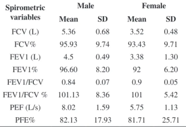
The results of spirometry are outlined in Table 1, showing that all subjects were healthy, with normal
lung function test.
The PEF values found in the sitting, DD, LLD and RLD positions for healthy male and female volunteers are shown in Table 2.
Comparing the results found in the whole group or
separately for males and females, the sitting position
with lying on DD, LLD and RLD positions, the values found in the sitting position were higher than those
assessed in DD and RLD (p<0.05), albeit without signiicant differences between the sitting and LLD positions (Figure 1).
Discussion
The purpose of this study was to evaluate the changes that occur in the values of PEF in healthy
adults at different positions. The results suggest
a decrease in PEF values at DD and RLD when compared with the sitting position, albeit without signiicant differences regarding the LLD.
The effect of DD in healthy people on different variables of pulmonary function is already well established in the literature, with a decrease in forced vital capacity (FVC) and forced expiratory volume in one second (FEV1)22, in addition to increased
airway resistance18 and decreased maximal expiratory
pressure (MEP)18,23.
Regarding PEF in healthy subjects, some authors found decreased values in the supine position24, while
others found similar values14,15. Furthermore, others
found a decrease only when the volunteers were in the head down position8.
The decrease in at lung volumes and low rates at DD has already been described in obese subjects with asthma also. Haffejee25 studied the effect of
supine position on PEF values in asthmatic children, aged between four and 11 years, using the Mini Wright portable meter. A irst measurement was performed with the child in orthostatic position, subsequently lying down the child and performing the measurements every 30 minutes for four hours. The results showed a signiicant decrease in PEF in
asthmatic children in the supine position.
A similar effect was also found in obese subjects. Domingos-Benício et al.26 compared the FVC, FEV
1
and FEV1/FVC values in the standing, sitting and lying down positions in volunteers in the age group from 20 to 40 years, eutrophic and obese, sedentary, by spirometry. They found a 20% reduction in the FVC of both eutrophic and obese subjects when subjects shift into the lying down position.
Table 1. Means and standard deviations of spirometric variables in male volunteers and female.
Spirometric variables
Male Female
Mean SD Mean SD
FCV (L) 5.36 0.68 3.52 0.48
FCV% 95.93 9.74 93.43 9.71
FEV1 (L) 4.5 0.49 3.38 1.30
FEV1% 96.60 8.20 92 6.20
FEV1/FCV 0.84 0.07 0.9 0.05
FEV1/FCV % 101.13 8.36 101 5.42
PEF (L/s) 8.02 1.59 5.75 1.13
PFE% 82.13 17.93 81.71 25.71
FVC: forced vital capacity; FEV1: forced expiratory volume in one second; FEV1/FVC: index Tiffenau; PEF: Peak expiratory low; SD: Standard deviation.
Table 2. Means and standard deviations of peak expiratory low
in L/min for males and females in the sitting, supine, left lateral
and right lateral position.
Position Males Female
Mean SD Mean SD
Sitting 558 116.7 404.7 45.1
Supine position 530 118.2 375.7 37.4
Left lateral
decubitus
545.3 132.2 393 38.7
Right lateral
decubitus
526.7 117 382.3 39.7
Figure 1. Peak expiratory low in healthy men and women and for the whole group in the sitting position, supine position (SP), left
lateral decubitus (LLD) and right (DLD). * Signiicant difference compared to sitting position (p < 0,05)
Meysman and Vincken22 evaluated the effect
of the sitting, DD, RLD and LLD positions on the low-volume curve, showing that PEF at the sitting position was higher than at decubitus positions and the FEV1/PEF ratio was higher in the lateral decubitus
positions. The authors explain that diaphragm strength
is affected by the supine position, with an increase in intrathoracic blood volume and consequent decrease in lung volume. Furthermore, the size of the pharynx is smaller at DD, given the gravitational force acting on the tongue and soft palate, and LD may decrease the resistance of the upper airways.
Badr et al.23 studied the PEF and MEP in 25
healthy adults between 18 and 65 years of age and 11 with chronic airlow limitation. The PEF measurements were performed using a spirometer, and the results showed that the standing position had the highest values, followed by the sitting position. There was no signiicant difference between the decubitus positions (dorsal and lateral), although the LD values were slightly lower than at supine. The explanation provided by the authors indicates
that the dependent hemithorax expandability may
have decreased.
Elkins et al.24 evaluated PEF and MEP in patients
spirometry and manovacuometry in eight different positions and showed differences in the values of PEF between the positions, with sitting, at 45°, supine, LD and head down tilt showing results signiicantly lower than both standing and sitting with legs stretched, and the largest difference occurred between the standing (6.35 L/s) and supine (5.79 L/s) positions. However, they found no differences between the lateral decubitus positions. Conversely, they found that MEP values at LD were signiicantly lower than
standing and sitting on a chair.
The results of Shinde and Shinde8, who evaluated eight different positions in healthy adults and patients
with chronic obstructive pulmonary disease (COPD) and found no signiicant trend to higher values of PEF at RLD in relation to LLD in both groups, are also controversial. The authors attributed that result to the higher volume of the right lung and a reduction in the compression of the heart on the lungs at RLD8.
This mechanism does not explain the results of the
present study. The LD position enables the forward
displacement of the abdominal content. Thus, the
authors speculate that the hemidiaphragm, which is in the position dependent on gravity, is elongated, thereby improving its ability to generate pressure, while the non-dependent hemidiaphragm remains less
elongated8. Conversely, the larger volume of the right
lung may have inferably contributed to an increased
elongation of the left hemidiaphragm on our results
because the diaphragm position was not assessed in
any of the studies.
The therapist should deine the method of PEF
assessment in clinical practice, including patient
positioning, enabling to compare PEF values between different periods or between different patients15.
Subjects should be positioned with the torso at a higher position to aid the removal of secretions from the airways8,15, especially those with reduced
ability to cough, considering that PEF is important for performing an effective cough, requiring a peak low >180 L/min for an effective cough27 and the
body position may affect the PEF. The low and
rate of air at the time of cough are the main factors
responsible for the clearance of airways4,5. Thus, it is
important to consider that LLD may be an alternative,
especially for bedridden patients, although it is crucial
to know and understand the physiological effects of
different decubitus positions, especially unilateral,
and different pathologies on lung function, in the selection process.
Roquejani et al.28 analyzed the position effects on
the values of muscle strength, which may also affect the PEF. The authors studied the maximal inspiratory and expiratory pressures (MIP and MEP) in healthy adults with ages ranging from 18 to 55 years, using a manovacuometer. The measurements were performed in seven positions and the results of that study showed that signiicant interactions occurred between the body position and the subject’s gender. The highest values of MIP were found at 45° in women and at RLD in men, whereas the lowest values were found in the Trendelenburg position. However, only the MEP values assessed in the male group were higher, albeit with no effect from the decubitus position.
We chose to show the results separately for men and women, given the predicted differences according to gender in several pulmonary function variables, established by different predictive equations for each gender, although we emphasize that the differences found were confirmed in the whole group and
in the male and female groups separately. In our
study, we established an age group for inclusion of subjects from 18 to 30 years because aging triggers
a reduction of lung elasticity and decreased elastic recoil pressure, contributing to a reduction in
respiratory muscle strength, and PEF has its highest peak between 18 and 20 years of age and maintains this level until 30 years of age7,29. Only sedentary
subjects or those without regular physical activity were evaluated towards reducing the confounding factors because physical activity may contribute to the increase in PEF5,30. Additionally, they were randomly
positioned, with no fatigue effect on the results. Regarding limitations, PEF measurements were
performed at different positions using only the
portable device; thus, we are not sure whether the results would change when measured by spirometry.
Other pulmonary function tests assessing other
measures, including residual volume or regional ventilation, were not performed either, or imaging tests towards conirming the position of the dependent
hemidiaphragm.
Conclusion
the sitting and LLD positions. Thus, the LLD may be an alternative to optimize the expiratory low in
situations restricting the sitting position.
References
1. Boaventura CM, Amuy FF, Franco JH, Sgarbi ME, Matos LB, Matos LB. Valores de referência de medida de pico de luxo expiratório máximo em escolares. Arq Med ABC. 2007;32(Supl.2):S30-4.
2. Ruchys VC, Dias RM, Sakurai E, Camargos PAM. Acurácia de medidores do pico de luxo expiratório (peak-low) da marca MiniWright. J Pediatr. 2000;76:447-52. 3. Quanjer PH, Tammeling GJ, Cotes JE, Pedersen OF,
Peslin R, Yernault JC. Lung volumes and forced ventilatory flows: official statement of the European Respiratory Society. Eur Respir J. 1993;6(16):5-40. 4. Freitas FS, Parreira VF, Ibiapina CC. Aplicação clínica do
pico de luxo da tosse: uma revisão de literatura. Fisioter Mov. 2010;23(3):495-502. http://dx.doi.org/10.1590/ S0103-51502010000300016
5. Freitas FS, Ibiapina CC, Alvim CG, Britto RR, Parreira VF. Relação entre força de tosse e nível funcional em um grupo de idosos. Rev Bras Fisioter. 2010;14(6):470-6. http://dx.doi.org/10.1590/S1413-35552010000600004 6. Smith JA, Aliverti A, Quaranta M, McGuinness K,
Ketsall A, Earis J, et al. Chest wall dynamics during voluntary and induced cough in healthy volunteers. J Physiol. 2012;590(3):563-574. PMid:22144580 PMCid:PMC3379701.
7. Dikshit MB, Raje S, Agrawal MJ. Lung functions with spirometry: an Indian perspective-I. Peak expiratory low rates. Indian J Physiol Pharmacol. 2005;49(1):8-18. PMid:15881854.
8. Shinde N, Shinde KJ. Peak expiratory low rate: Effect of body positions in patients with chronic obstructive pulmonary disease. Indian Journal of Basic & Applied Medical Research. 2012;1(4):357-362.
9. Pereira CAC, Jasen JM, Barreto SSM, Marinho J, Sulmonett N, Dias RM. Espirometria. In: Diretrizes para testes de função pulmonar. J Pneumol. 2002;28(3):S1-S82. 10. Ayres JG, Turpin PJ. Measurement, recording and
analysis of peak low records. In: Peak low measurement. Chapman & Hall Medical; 1997. p. 13-32.
11. Bongers T, O’Driscoll BR. Effects of equipment and technique on peak low measurements. BMC Pulm Med. 2006; 6(14) [cited 2013 Feb 20]. Available from: http:// www.biomedcentral.com/1471-2466/6/14. http://dx.doi. org/10.1186/1471-2466-6-14
12. Vaswani R, Moy R, Vaswani SK. Evaluation of Factors Affecting Peak Expiratory Flow in Healthy Adults:Is It Necessary to Stand Up? J Asthma. 2005;42:793-794. PMid:16316876. http://dx.doi. org/10.1080/02770900500308528
13. McCoy EK, Thomas LJ, Sowell RS, George C, Finch KC, Tolley EA. An Evaluation of Peak Expiratory Flow Monitoring: A Comparison of Sitting Versus Standing Measurements. J Am Board Fam Med.
2010;23(2):166-170. PMid:20207926. http://dx.doi. org/10.3122/jabfm.2010.02.090120
14. Badaruddin M, Uddin MB, Khatun MF, Ahmad K. Study on Peak Expiratory Flow Rate in Different Positions. Dinajpur Med Col J. 2010;3(1):17-18.
15. Wallace JL, George CM, Tolley EA, Winton JC, Fasanella D, Finch CK, et al. Peak expiratory flow in bed? A comparison of 3 positions. Respir Care. 2013;58(3):494-497. PMid:22906434.
16. Global Initiative for Asthma. Global strategy for asthma management and prevention. 2012 [cited 2013 Jan 8]. Available from: http://www.ginasthma.org/guidelines-gina-report-global-strategy-for-asthma.html.
17. Kaneko K, Milic-Emili J, Dolovich MB, Dawson A, Bates DV. Regional distribution of ventilation and perfusion as a function of body position. J Appl Physiol. 1966;21(3):767-777. PMid:5912746.
18. Behrakis PK, Baydur A, Jaeger MJ, Milic-Emili J. Lung mechanics in sitting and horizontal body positions. Chest. 1983;83(4):643-6. PMid:6831953. http://dx.doi. org/10.1378/chest.83.4.643
19. Alcoforado L, Pessôa LC Fº, Brandão DC, Galvão AM, Reinaux CM, Andrade AD. Inluence of change in lateral decubitus on pulmonary aerosol deposition. Rev Bras Fisioter. 2011;15(4):278-83. PMid:21971723. http:// dx.doi.org/10.1590/S1413-35552011000400004 20. Gomes EL, Postiaux G, Medeiros DR, Monteiro KK,
Sampaio LM, Costa D. Chest physical therapy is effective in reducing the clinical score in bronchiolitis: randomized controlled trial. Rev Bras Fisioter. 2012;16(3):241-7. PMid:22499404. http://dx.doi.org/10.1590/ S1413-35552012005000018
21. Craig CI, Marshall AL, Sjostrom M, Baumann AE, Booth ML, Ainsworth BE. International Physical Activity Questionnaire: 12-Country Reliability and Validity. Med Sci Sports Exerc. 2003;35(8):1381-1395. PMid:12900694. http://dx.doi.org/10.1249/01.MSS.0000078924.61453.FB 22. Meysman M, Vincken W. Effect of body posture on
spirometric values and upper airway obstruction indices derived from the low-volume loop in young nonobese subjects. Chest. 1998;114(4):1042-7. http://dx.doi. org/10.1378/chest.114.4.1042
23. Badr C, Elkins MR, Ellis ER. The effect of body position on maximal expiratory pressure and flow. Aust J Physiother. 2002;48(2):95-102. PMid:12047207. 24. Elkins MR, Alison JA, Bye PTP. Effect of Body Position
on Maximal Expiratory Pressure and Flow in Adults With Cystic Fibrosis. Pediatr Pulmonol. 2005;40:385-391. PMid:16130087. http://dx.doi.org/10.1002/ppul.20287 25. Haffejee IE. Effect of supine posture on peak expiratory
low rates in asthma. Arch Dis Child. 1988;63:127-129. PMid:3348658 PMCid:PMC1778739. http://dx.doi. org/10.1136/adc.63.2.127
27. Toussaint M, Boitano LJ, Gathot V, Steen M, Soundon P. Limits of Effective Cough-Augmentation Techniques in Patients With Neuromuscular Disease. Respir Care. 2009;54(3):359-366. PMid:19245730.
28. Roquejani AC, Araújo S, Oliveira RARA, Dragosavac D, Falcão ALE, Terzi RGG, et al. Inluência da Posição Corporal na Medida de Pressão Inspiratória Máxima e Pressão Expiratória Máxima em Voluntários Adultos Sadios. Rev Bras Ter Intensiva. 2004;16(4):215-218. 29. Wun YT, Chan MS, Wong NM, Kong AY, Lam TP. A
Curvilinear Nomogram of Peak Expiratory Flow Rate for the Young. J Asthma. 2013;50(1):39-44. PMid:23174006. http://dx.doi.org/10.3109/02770903.2012.743152
30. Bemanian MH, Shirkhoda S, Nakhjavani M, Mozafari H. Effect of Swimming on Peak Expiratory Flow Rate of Atopic Children Iran. J Allergy Asthma Immunol. 2009;8(2):121-123.
Correspondence
Ada Clarice Gastaldi Av. Bandeirantes, 3900