Evaluation of the relation between the horizontal condylar angle
and the internal derangement of the TMJ – a magnetic resonance
imaging study
Avaliação da relação entre o ângulo horizontal do côndilo e o
desarranjo interno da ATM, por meio de ressonância
magnética
Iêda Margarida Rocha Crusoé-Rebello* Paulo Sérgio Flores Campos**
Izabel Regina Fischer Rubira** Jurandyr Panella***
Carlos Maurício Cardeal Mendes****
ABSTRACT:This research aimed at assessing the relation between the horizontal condylar angle (HCA) and the inter-nal derangement (ID) of the temporomandibular joint (TMJ), as a result of interference by the TMJ disk, in individuals undergoing magnetic resonance (MR) scans. The sample included a total of 144 TMJs (sagittal and coronal views) of 72 subjects, 15 of whom were male and 57 female, with ages ranging from 15 to 70. The scans were made in a Signa system (GE) model at a magnetic field magnitude of 1.5 T. Sixty-eight TMJs were found to be normal, while 46 showed anterior displacement with reduction. Of these, 41 had some kind of adaptive change in the condyle, while 5 showed degenerative changes. Anterior displacement without reduction was found in 29 joints, 12 of which showed adaptative changes in the condyle, while 17 showed degenerative changes. Only one posterior displacement of the articular disk was recorded. For the TMJs in which disk displacement was found, such values achieved 24.69º on the right side, and 22.94º on the left side. Hence, it was possible for us to conclude that the HCA tends to increase in those TMJs where ID is present. For contralateral TMJs, a strong association was observed between HCA values (57.8%), state of normality (69.7%), and ID (66.7%). To corroborate such findings, a correlation between contralateral HCA values (63.31%) and the diagnosis for contralateral TMJs (68.05%) was determined. Thus, we could infer that there is a tendency between contralateral TMJs to share characteristics and conditions.
DESCRIPTORS:Temporomandibular joint; Nuclear magnetic resonance; Temporomandibular joint disk.
RESUMO:Esta pesquisa teve por objetivo avaliar a relação entre o valor do ângulo horizontal do côndilo (AHC) e o de-sarranjo interno (DI), por interferência do disco da articulação temporomandibular (ATM), de indivíduos indicados para exame por ressonância magnética (RM). A amostra perfez um total de 144 ATMs (cortes sagital e coronal) de 72 in-divíduos, na faixa etária de 15 a 70 anos, 15 do gênero masculino e 57 do gênero feminino. Os exames foram procedi-dos em um sistema modelo Signa (GE), magnitude de 1,5 T para o campo magnético. Sessenta e oito ATMs apresenta-ram uma condição de normalidade e 46 apresentaapresenta-ram deslocamento anterior do disco articular com redução. Dessas, 41 expressaram algum tipo de alteração adaptativa no côndilo e cinco, alterações degenerativas. Vinte e nove articula-ções mostraram deslocamento anterior do disco articular sem redução, sendo que 12 dessas apresentaram alteraarticula-ções adaptativas do côndilo e 17, alterações degenerativas. Foi registrado apenas um caso de deslocamento posterior do disco articular. O valor médio encontrado para o AHC do lado direito foi de 22,09º e para o lado esquerdo foi de 21,47º, para aquelas ATMs que se apresentaram em condições de normalidade. Para as ATMs com deslocamento de disco, es-ses valores foram de 24,69º para o lado direito e 22,94º para o lado esquerdo. Desse modo, foi-nos possível concluir que existe uma tendência ao aumento do AHC nas ATMs com DI. Observamos forte associação, para as ATMs contra-laterais, entre os valores do AHC (57,8%), condição de normalidade (69,7%) e DI (66,7%). E, corroborando esses resul-tados, registramos correlação para os valores dos AHC contralaterais (63,31%) e o diagnóstico para as ATMs contrala-terais (68,05%). Assim, pudemos inferir que existe tendência das ATMs contralacontrala-terais apresentarem as mesmas características e condições.
DESCRITORES:Articulação temporomandibular; Ressonância magnética nuclear; Disco da articulação temporoman-dibular.
* PhD, Professor, FAPESB Researcher (Fundação de Amparo à Pesquisa do Estado da Bahia – Research Support Foundation of the State of Bahia).
** PhD, Professor, Department of Propaedeutics and Integrated Clinic, School of Dentistry; ****PhD, Professor, Collective Health Institute, School of Medicine – Federal University of Bahia.
INTRODUCTION
The expression Temporomandibular Dysfuncti-on (TMD) has been used in a generic way to diagno-se facial pain and dysfunction of the jaws (Sano15
, 2000). Although referred to as a syndrome, nowa-days researches support the hypothesis that TMD is a group of disorders related to the masticatory system, including intra-articular morphologic ab-normalities, myalgias, different types of disk dis-placement, articular degenerative diseases, in-flammatory arthritis, synovitis, and congenital and neoplastic conditions that affect the Temporo-mandibular Joint (TMJ). Since disk displacement is the most frequent among these disorders, the use of a more specific term could undoubtedly enhance clinicians’ and researchers’ understan-ding of the problem. To this end, the term Internal Derangement (ID) has been applied to refer to a disk’s abnormal relationship with the articular bone components. This disorder is often characte-rized by an anterior articular disk displacement, with or without reduction (Paesani et al.12
, 1992; Dolwicket al.3
, 1983; Tallentset al.16
, 1996). Concerning the relationship between the disk and the articular bone components, Magnetic Res-onance (MR) and arthrography are the imaging scans that have the greatest ability of providing the most necessary and optimized information for the establishment of accurate diagnoses (Heffezet al.5
, 1995). As a method, however, MR has the advan-tages of being non-invasive, not using ionizing ra-diation and causing less discomfort for patients (Katzberg8
, 1989).
In medical orthopedics, the spatial relationship between the articular bone components has been a discovery of great clinical importance. Alterations in the alignment of the knee joint components, for instance, are associated with local alterations in flexibility and articular compression, and osteoarthritis is a probable consequence (Pullinger et al.13, 1986).
Studies in dentistry have suggested that the Horizontal Condylar Angle (HCA), established be-tween its long axis (medial-lateral) and the coronal plan, is higher in joints affected with ID than in normal joints, with approximate values ranging from 0º to 30º (average values ranging from 15º to 20º) in joints without disk displacement, in post-mortem specimens (Westesson, Liedberg18
, 1987; Westesson et al.19
, 1991; Katzberg, Westesson7, 1994).
In a correlation with the knee joint, whose in-vestigation has reached a more mature state than that of the TMJ, Fuet al.4
(1994) affirmed that an-gular deformities may promote articular diseases, such as arthritis, and that the opposite is also true: deformities may happen as a consequence of the evolution of an articular degenerative disease. Thus, the aim of the present research was to evaluate the relationship between HCA magnitude and ID as a result of TMJ disk interference in indi-viduals who had been referred to MR scans.
MATERIAL AND METHODS
One hundred and forty-four MR file images of the TMJ area were appraised from November 2000 to July 2001, according to the parameters de-scribed in Table 1. The subjects, 57 females and 15 males, had ages from 15 to 70 years.
As a rule, the clinical exam included directing the anamnesis of all the subjects to the main com-plaint and palpating TMJs when maximum open-ing of the mouth was achieved.
The MR scans were made in a Signa system model (General Electric, Milwaukee, WIS, USA), at a magnetic field magnitude of 1.5 T, using a bilat-eral radiofrequency surface coil of 6.5 x 6.5 cm in size.
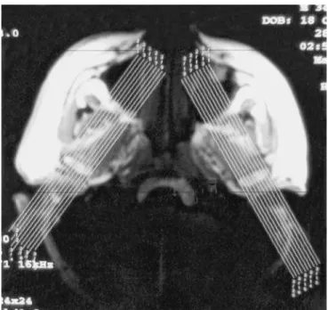
For the acquisition of the final scan in T1, an axial scout was performed. Based on that, the condyle was located and a record of the horizontal angle (supplied by the system) was made. Hence,
TABLE 1 -Parameters for obtaining TMJ T1 images.
Image TR (msec) TE (msec) NEX FOV (cm) Slice thickness (mm)
Matrix Imaging time
Axial localizer (closed jaw) 400 13 1 24 4 256 x 256 58’’
Parassagital (closed jaw) 400 13 4 14 2 256 x 256 5’ 12’’
Parassagital (open jaw) 400 13 4 14 2 256 x 256 5’ 12’’
the sequence of perpendicular cuts along the condyle axis (Figure 1) was determined.
The scans were interpreted by an experienced examiner who used the same diagnosis criteria as proposed by Katzberg, Westesson7 (1994), and Milanoet al.9
(2000). For the purpose of data anal-ysis, only two groups were identified: normal TMJs and TMJs affected with articular disk displace-ment. Therefore, the aspect of reduction or non-re-duction was not separately considered. The same applies to the different types of articular disk dis-placement, although the discussion that follows has considered all of the aspects involved.
Additionally, bone changes were interpreted as either adaptive or degenerative. To be considered adaptive, a change could be faceted, but show no cortical erosion or affection of the subchondral portion. The presence of the latter aspects was in-terpreted as an evidence of a degenerative change, especially if accompanied by subchondral sclero-sis, osteophytes or subchondral cysts.
In order to perform a statistical analysis, de-scriptive parameters of the distribution of the vari-ables of interest, measured to the minimum in an ordinal scale, were estimated. In addition, the Shapiro-Wilk test of adherence to normal distribu-tion and the Spearman test (r) of correlation of
po-sitions (to check the degree of compliance among
techniques), as well as the t-Student and the Pearson chi-square tests, were made.
RESULTS
Of the 144 appraised joints, 68 fell within nor-mality and 46 showed an anterior disk displace-ment with reduction. Of these, 41 featured some type of condylar adaptive change, whereas 5 pre-sented degenerative changes. Twenty-nine joints showed an anterior disk displacement without re-duction, of which 12 featured condylar adaptive changes and 17, degenerative changes. In one case, a posterior disk displacement was observed. The type of displacement showed no predomi-nance as regards gender and age groups (Table 2).
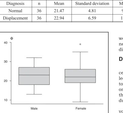
The distribution of the HCA values followed a similar behavior both for the male and female groups (Graph 1).
On the other hand, it was not possible to estab-lish a correlation between left and right HCA val-ues and the subjects’ age (Graph 2).
The average HCA values for joints whose disks were in a normal position were 22.09º (RAV) and 21.47º (LAV). For joints presenting disk displace-ment, such values were 24.69º (RAV) and 22.94º (LAV) (Table 3 and 4).
The group with a disk displacement diagnosis presented larger average values than the group with a normal diagnosis. In addition, their HCA values fell within a higher range (Graph 3).
A statistically significant association was ob-served between HCA values (57.8%), normal posi-tion diagnoses (69.7%) and articular disk displace-ment diagnoses (66.7%), for contralateral TMJs.
FIGURE 1 -Axial localizer exhibiting the orientation tool for the parassagittal cuts, which is also the one that al-lows automatic measurement of the horizonal condylar angle.
TABLE 2 - Descriptive statistics of all right horizonal condylar angle values obtained for the respective diag-nosis groups in the sample.
Age Female Male
BD UD WD BD UD WD
10-19 1 4 2 2 1
-20-29 3 7 3 1 2 1
30-39 7 5 7 - 1 2
40-49 5 1 2 - - 2
50-59 3 1 3 1 2
-60-69 1 - - - -
-70-79 2 - - - -
When contralateral horizonal condylar angles were compared, a correlation of 63.31% was obtai-ned. Concerning the diagnosis of the articular con-dition, a correlation of 68.05% was verified.
DISCUSSION
Since MR is an imaging diagnostic method of re-cent and still limited use in stomatology, all know-ledge resulting from its application will contribute to more precise indications. In the field of research on MR, the work of Campos1
(2001) stands out for the vast number of cases considered (512 indivi-duals) in the evaluation of cystic sinuses scans.
Starting from a sample of 56 children and young adults, Ribeiro’s14
(1996) pioneering work on TMJ gathered substantial information regard-ing the TMJ in asymptomatic patients in the city of
GRAPH 1 -Distribution of horizonal condylar angle va-lues among male and female groups.
GRAPH 2 -Distribution of right (RAV) (A) and left (LAV) (B) horizonal condylar angle values according to age.
TABLE 3 -Descriptive statistics of all right horizonal condylar angle values obtained for the respective diagnosis groups in the sample.
Diagnosis n Mean Standard deviation Min 25% 50% 75% Max
Normal 33 22.09 3.95 14 20 22 24 31
Displacement 39 24.69 6.41 15 20 23 29 41
TABLE 4 -Descriptive statistics of all left horizonal condylar angle values obtained for the respective diagnosis groups in the sample.
Diagnosis n Mean Standard deviation Min 25% 50% 75% Max
Normal 36 21.47 4.81 9 18.5 22 24 31
São Paulo. And to mention another Brazilian re-searcher, Tanaka17
(2000) analyzed the effect of stabilizing devices in the treatment of 40 patients suffering from TMD using MR scans.
Therefore, our work aimed at offering a contri-bution to the consolidation of the knowledge con-cerning TMJ, its morbid conditions and the appli-cation of MR in dentistry.
As regards the circumstances in which TMD oc-curs, the absence of an articular disk displace-ment in symptomatic individuals is a finding as common as the reverse. In observing the normal relationship between condyles and disks, Westessonet al.19(1991) have not found any statis-tically significant differences between the groups of symptomatic and asymptomatic patients.
Cholitgulet al.2
(1997) have observed that pain had not been a symptom characteristic to the sev-eral types of disk displacement when an associa-tion was sought between the symptoms reported by the patients and the findings brought about by MR scans. Therefore, the relationship between ID and painful symptoms remains unclear. Isberget al.6
(1998), on the other hand, have not reported an increase in pain among women when evaluating patients suffering from ID.
According to Okeson11 (1996), female individu-als are more predisposed to disorders resulting from the interference of the articular disk. Nevert-heless, we have observed here that both males and females share the same characteristics as regards ID and horizonal condylar angle values (Table 2). We could say, at first, that hormonal factors do not seem to play a significant role in the manifestation of IDs in TMJs.
In this study, we could not find an association between the increase in age and the increase in ho-rizonal condylar angle (Graph 2) values. And we have not detected any associations between ID and age either. Although they have obtained similar re-sults to the ones we share here, Isberget al.6(1998) have excepted a tendency towards an increase in the incidence of symptomatic disk displacements during the second decade of life, there being a peak in the incidence of asymptomatic displacements during puberty, for both genders. Our sample, on the contrary, has evidenced a greater concentrati-on of individuals in their thirties and forties.
The results we obtained demonstrated that ave-rage horizonal condylar angle values, so much to the right side as to the left side, were higher in indi-viduals with positive diagnoses for ID (Table 3 and 4, Graph 3). Although our findings do not show a statistically significant difference between the ave-rage horizonal condylar angle values of normal TMJs and TMJs with ID, our data reveal a tendency among TMJs with ID to present higher horizonal condylar angle values. According to Katzberg, Westesson7
(1994), joints with higher horizonal condylar angle values would either tend to develop ID and articular degenerative diseases or they would be the result of a remodeling pro-cess.
Some studies have considered the remodeling hypothesis more plausible. Westesson et al.19 (1991), however, speculate that joints with higher horizonal condylar angle values have a higher pos-sibility of stretching the lateral ligament between the disk and the condyle during the movement of
translation, as compared to TMJs with lower hori-zonal condylar angle values. Since the lateral liga-ment is not as stretchable as the disk’s posterior li-gament, a distension beyond the limits could result in permanent stretching, with a subsequent displacement of the articular disk. Our results, on the other hand, do not allow for the inference that an increase in the horizonal condylar angle is eit-her the cause or the consequence of ID. However, from an embryogenic point of view, the develop-ment of the articular components in the absence of factors that could determine structural changes occurs in a harmonious way. In other words, the spatial orientation of the condyle will always comply with the spatial orientation of the articular fossa, which leads to a normal functional conditi-on, in spite of anatomical variations. For that rea-son, we believe ID to be the cause, and not the con-sequence, of increased horizonal condylar angle values.
Finally, the comparison between the horizonal condylar angle values, as well as between the con-ditions of the joint (normal or with disk displace-ment), has showed that each joint behaves in a si-milar way to the contralateral joint (Graph 3). This suggests that one joint influences the other and, for that reason, they cannot be considered separa-tely, as in the study by Nebbeet al.10
(1998). The mandible movements are coordinated by both jo-ints, which are functionally unified. And functio-nal unification could justify the high prevalence of
bilateral alterations, as observed by Katzberg8 (1989).
CONCLUSIONS
While the average horizonal condylar angle va-lues in individuals without ID were 22.09º and 21.47º, on the right and left sides, respectively, for ID patients such values were 24.69º and 22.94º, and this demonstrates a tendency towards higher horizonal condylar angle values in TMJs with ID.
For contralateral TMJs, we have observed a clo-se association between the horizonal condylar an-gle values (57.8%), normality conditions (69.7%), and ID (66.7%). And, corroborating those results, we have found a correlation between the contrala-teral horizonal condylar angle values (63.31%) and the diagnosis of contralateral TMJs (68.05%). For that reason, we could infer that there is a tendency for contralateral TMJs to share the same characte-ristics and conditions, since they work as a functi-onal unit.
ACKNOWLEDGEMENTS
This study was supported by Grants from the Fundação Coordenação de Aperfeiçoamento de Pessoal de Nível Superior (CAPES), Brazil. We sin-cerely appreciate the trust and collaboration of-fered by Dr. Delfin and Dr. Maria Olívia Gonzalez, who have made this research possible.
REFERENCES
1. Campos PSF. Estudo tridimensional de imagens císticas dos seios maxilares, através da ressonância magnética nu-clear, numa amostra populacional da cidade do Salvador [Tese de Doutorado]. São Paulo: Faculdade de Odontologia da USP; 2001.
2. Cholitgul W, Nishiyama H, Sasai T, Uchiyama Y, Fuchihata H, Rohlin M. Clinical and magnetic resonance imaging fin-dings in temporomandibular joint disc displacement. Den-tomaxillofac Radiol 1997;26:183-8.
3. Dolwick MF, Katzberg RW, Helms CA. Internal derange-ments of the temporomandibular joint: fact or fiction? J Prosthet Dent 1983;49:415-8
4. Fu FH, Harner CD, Vince KG. Biomechanical factors in alignment and arthritic disorders of the knee. Knee Sur-gery. v. 2. Baltimore: Williams & Wilkins;1994.
5. Heffez LB, Mafee MF, Rosenberg H. Imaging atlas of the temporomandibular joint. Baltimore: Williams & Wilkins; 1995.
6. Isberg A, Hägglung M, Paesani D. The effect of age and gen-der on the onset of symptomatic temporomandibular joint disk displacement. Oral Surg Oral Med Oral Pathol Oral Radiol Endod 1998;85:252-7.
7. Katzberg RW, Westesson P-L. Diagnosis of the Temporo-mandibular joint. Philadelphia: W. B. Saunders Co.; 1993. 8. Katzberg RW. Temporomandibular joint imaging.
Radiol-ogy 1989;170:297-307.
9. Milano V, Desiate A, Bellino R, Garofalo T. Magnetic reso-nance imaging of temporomandibular disorders: classifica-tion, prevalence and interpretation of disc displacement and deformation. Dentomaxillofac Radiol 2000;29:352-61. 10. Nebbe B, Major PW, Prasad NG, Hatcher, D. Quantitative assessment of temporomandibular joint diskstatus. Oral Surg Oral Med Oral Pathol Oral Radiol Endod 1998; 85:598-607.
11. Okeson J. Oral facial pain - guidelines for assessment, di-agnosis and management. Chicago: Quintessence, 1996. 12. Paesani D, Westesson PL, Hatala M, Tallents RH, Kurita K.
Prevalence of temporomandibular internal derangements in patient with craniomandibular disorders. Am J Orthod Dentofacial Orthop 1992;101:41-7.
14. Ribeiro RF. Avaliação estrutural da articulação temporomandibular em crianças e adultos jovens assintomáticos através de imagens por ressonância magnética [Tese de Doutorado]. Bauru: Faculdade de Odontologia da USP; 1996.
15. Sano T. Recent developments in understanding temporomandibular joint disorders. Part 1: bone marrow abnormalities of the mandibular condyle. Dentomaxillofac Radiol 2000; 29:7-10.
16. Tallents RH, Katzberg RW, Murphy W, Proskin H. Magnetic resonance imaging findings in asymptomatic volunteers and symptomatic patients with temporomandibular disor-ders. J Prosthet Dent 1996; 75:529-33.
17. Tanaka EE. Análise dos efeitos da terapia com placas estabilizadoras em pacientes com disfunção têmporo-mandibulares por meio de ressonância magnética [Tese de Doutorado]. São Paulo: Faculdade de Odontologia da USP; 2000.
18. Westesson PL, Liedberg J. Horizontal condylar angle in re-lation to internal derangement of the temporomandibular joint. Oral Surg Oral Med Oral Pathol 1987; 64:391-4. 19. Westesson PL, Bifano JA, Tallents RH, Hatala MP.
In-creased horizontal angle of the mandibular condyle in ab-normal temporomandibular joints. A magnetic resonance imaging study. Oral Surg Oral Med Oral Pathol 1991; 72:359-63