Correlation between clinical and imaging findings
in patients with temporomandibular disorders*
Correlação entre os achados clínicos e imaginológicos nas disfunções temporomandibulares
Fábio Augusto Cozzolino1, Abrão Rapoport2, Sérgio Altino Franzi3, Ricardo Pires de Souza3, Clemente Augusto de Brito Pereira3, Rogério Aparecido Dedivitis3
OBJECTIVE: To correlate the signals and symptoms observed on clinical examination of patients with tem-poromandibular disorder with the results demonstrated by magnetic resonance imaging. MATERIALS AND METHODS: Thirty patients presenting with signs and symptoms of temporomandibular disorders underwent clinical evaluation and subsequent magnetic resonance imaging. The magnetic resonance imaging studies were independently evaluated by two experienced radiologists. Magnetic resonance imaging studies con-sisted of 12 images in coronal, T1-weighted sequences with 3 mm-thick slices with the mouth closed, sag-ittal, T1- and T2-weighted sequences with both open and closed mouth positions, and on progressive open-ing/closing movement at 5 mm intervals, in order to demonstrate the full mandibular movement. The statis-tical significance between the clinical findings in the evaluation of the patients and results found on the magnetic resonance imaging studies was analyzed by means the kappa test. RESULTS: Interobserver agreement was respectively 56.7% (kappa = 0.1) and 56.7 (kappa = 0) for the left and right sides. CONCLUSION: No correlation was found between the clinical and magnetic resonance imaging findings in the diagnoses of disc displacement.
Keywords: Magnetic resonance imaging; Temporomandibular joint; Disorders.
OBJETIVO: Verificar a relação entre sinais e sintomas observados no exame clínico de pacientes com diag-nóstico de disfunção temporomandibular, conforme os resultados fornecidos pelo exame de ressonância magnética. MATERIAIS E MÉTODOS: Trinta pacientes que apresentavam sinais e sintomas de disfunção temporomandibular foram submetidos a exame clínico e de ressonância magnética. Cada exame de resso-nância magnética de articulação temporomandibular foi interpretado, independentemente, por dois radiolo-gistas experientes. Os exames de ressonância magnética foram realizados com 12 cortes de 3 mm de espes-sura, em orientação coronal (T1) em posição de boca fechada, cortes sagitais em posição de boca aberta e fechada (T1 e T2) e em abertura e fechamento progressivos, com intervalo de 5 mm, para reproduzir toda a extensão do movimento mandibular. A significância estatística entre a análise clínica dos pacientes com disfunção temporomandibular e os resultados obtidos no exame de ressonância magnética foi avaliada pelo teste kappa. RESULTADOS: Obteve-se, na análise interobservadores de imagens, concordância bruta do lado esquerdo e direito, respectivamente, de 56,7% (kappa = 0,1) e 56,7 (kappa = 0). CONCLUSÃO: Não foi encontrada correlação entre o diagnóstico clínico da luxação discal e imagens de ressonância magnética.
Unitermos: Ressonância magnética; Articulação temporomandibular; Distúrbios. Abstract
Resumo
* Study developed at Hospital Heliópolis (Hosphel), São Paulo, SP, Brazil.
1. Master in Health Sciences, Course of Post-Graduation, Hospital Heliópolis (Hosphel), São Paulo, SP, Brazil.
2. Private Docent, Professor, Course of Post-graduation in Health Sciences, Hospital Heliópolis (Hosphel), São Paulo, SP, Brazil.
3. PhDs, Professors, Course of Post-graduation in Health Sciences, Hospital Heliópolis (Hosphel), São Paulo, SP, Brazil.
Mailing address: Dr. Abrão Rapoport. Rua Iramaia, 136, Jardim Europa. São Paulo, SP, Brazil, 01450-020. E-mail: arapoport@ terra.com.br
Received December 20, 2006. Accepted after revision June 4, 2007.
and mandibular dysfunction, as well as symptoms primarily affecting TMJ soft tis-sues and the articular disk positioning.
Imaging methods can significantly con-tribute to the final diagnosis and therapeu-tic evaluation in this context. Conventional radiographic methods (panoramic and transcranial radiography) and techniques dedicated to the TMJ (arthrography, arthro-tomography, conventional and computed tomography) present some limitations con-sidering the localization, composition and size of the TMJ, besides the level of ioniz-ing radiation exposure.
Magnetic resonance imaging (MRI) has
revolutionized the diagnosis and treatment of temporomandibular joint disorders (TMJD), because of its high-resolution for demonstrating the TMJ tissues, without necessity of changing the patient’s posi-tioning and with no ionizing radiation. MRI has been the method of choice for the di-agnosis of abnormalities in the TMJ soft tissues, because of its high accuracy in the determination of the articular disk position-ing (1).
The present study was aimed at corre-lating signs and symptoms observed in the clinical assessment of patients diagnosed with TMJD according to the results of MRI.
Cozzolino FA, Rapoport A, Franzi SA, Souza RP, Pereira CAB, Dedivitis RA. Correlação entre os achados clínicos e imaginológicos nas disfunções temporomandibulares. Radiol Bras. 2008;41(1):13–17.
INTRODUCTION
MATERIALS AND METHODS
The sample of the present retrospective study included 30 patients referred to the Department of Odontology at Universidade Cruzeiro do Sul, with a diagnosis of TMJD, in the period between January 2002 and January 2006. This project was approved by the Committee for Ethics in Research of Universidade Cruzeiro do Sul, under the number 036/05. Inclusion criteria were: patients diagnosed with TMJD presenting headache, otalgia, pre-auricular and oro-facial pain. Exclusion criteria were patients below the age of 18 and previously submit-ted to surgery for treating TMJD.
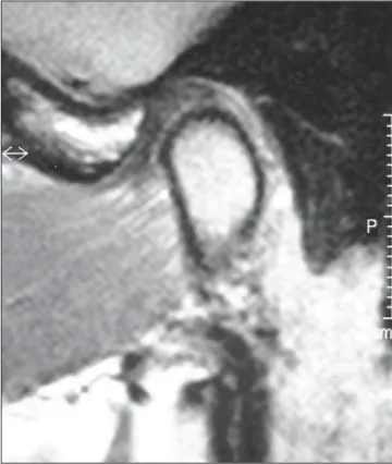
The MRI studies were independently in-terpreted by two experienced radiologists who elaborated the diagnostic reports ac-cording to the criteria established by Nebbe et al.(2) (Table 1; Figures 1 and 2). In case
of disagreement between these diagnostic reports, the final diagnosis was achieved by consensus between both radiologists.
The disease indicators consisted in a questionnaire developed by the American Academy of Orofacial Pain – Guidelines for Assessment, Diagnosis and
Manage-Table 1 Criteria established by Nebbe et al.(2).
a. Normal positioning of the articular disk.
b. Mild anterior articular disk displacement with opening reduction.
c. Moderate anterior articular disk displacement with opening reduction.
d. Total anterior articular disk displacement with opening reduction.
e. Total anterior articular disk displacement without opening reduction.
f. Other categories of articular disk positioning alteration.
Figure 1. MRI sagittal image of closed mouth demonstrating a normal disk positioning.
Figure 2. MRI sagittal image of closed mouth demonstrating anterior disk displacement.
ment of temporomandibular Joint Disor-der(3). The main complaint was obtained by
means of an interview as the initial step of the clinical evaluation. Then, the presence of articular pain was evaluated by means of both lateral and posterior palpation of the TMJ and auscultation for articular sounds (clicking and crepitus). The presence of muscular pain was evaluated by bilateral, extraoral palpation of the following muscles or regions: superficial masseter, deep masseter, temporal anterior, temporal posterior, frontal, vertex, posterior cervical regions, digastric and sternocleidomastoid; and intraoral palpation of temporalis, ptery-goid and lateral pteryptery-goid.
According to previous studies(4–6),
tem-poromandibular joint clicking or popping sounds are brief noises which occur in
some points during opening, closing or lat-eral movements; and crepitus is a mildly perceptible grating sound, suggestive of subchondral sclerosis.
the whole extent of the mandibular move-ment, with dynamic video images. All the images were acquired with the patient in dorsal decubitus.
Descriptive statistics was utilized for summarizing the data regarding sex and age range, facial pain, articular sounds and disk positioning at MRI. The correlation between MRI and facial pain and articular sounds was based on MRI versus clinical findings respectively on the right and left sides. The statistical significance of the clinical as-sessment of patients with TMJD and MRI findings was evaluated with the kappa test for determining the correlation level.
RESULTS
A female predominance was observed (24 patients), with a women/men ratio equal to 3.2:1, and higher incidence in the age range between 18 and 29 years (41.7% of the patients). As regards painful symp-toms, 14 patients (46.7%) presented bilat-eral pain, 4 (13.3%), right-sided pain, 3 (10.0%), left-sided pain, and 9 patients (30.0%) had no pain-related complaint.
As regards the presence or absence of articular sounds, the right side was affected in 7 patients (23.3%), the left side in 7 (23.3%), both sides in 6 (20.0%), and ab-sence of this symptom was observed in 10 (33.3%). The incidence of the different articular sounds was the following: on the left side — clicking in 12 patients (40.0%), crepitus in 1 (3.3%), and absence in 17 (56.7%); on the right side — clicking was observed in 12 patients (40.0%), crepitus in 1 (3.3%) and absence in 17 (56.7%).
Table 2 shows the frequency of articu-lar alterations and distribution related to their side according to the MRI diagnostic reports. Tables 3 and 4 present the fre-quency of articular alterations as well as the incidence in both sides of each type of al-teration according to the MRI diagnostic reports. Table 5 shows the relation between the clinical and the MRI diagnosis of left-sided disk displacement while the right-sided findings are shown on Table 6.
DISCUSSION
An attempt was made to combine the utilization of MRI (the method of choice
for evaluating TMJ) with a clinical ques-tionnaire. MRI is considered as the method of choice for evaluating the TMJ function-ing, because of its non-invasiveness and absence of collateral effects, besides the high accuracy comparable to arthrography for visualizing functioning structures. However, the association of clinical and imaging findings is essential for an
accu-rate diagnosis and prognostic evaluation of TMJD(4). Data collected by means of the
anamnesis and clinical examination of the patients constitutes the basis for a correct diagnosis of TMJD(7).
A high interobserver agreement was ob-served in the evaluation of TMJD by MRI, corroborating the acceptance and reliabil-ity of this diagnostic method(8–12). Many
Table 2 Distribution in relation to frequency and side of articular alterations.
Variable
Normal
Right-sided MRI
Left-sided MRI
Both sides
Total
Frequency
9
3
5
13
30
Percentage
30.0%
10.0%
16.7%
43.3%
100.0%
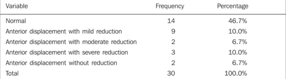
Table 3 Distribution of left-sided MRI results.
Variable
Normal
Anterior displacement with mild reduction
Anterior displacement with moderate reduction
Anterior displacement with severe reduction
Anterior displacement without reduction
Total
Frequency
14
9
2
3
2
30
Percentage
46.7%
10.0%
6.7%
10.0%
6.7%
100.0%
Table 4 Distribution of right-sided MRI results.
Variable
Normal
Anterior displacement with mild reduction
Anterior displacement with moderate reduction
Anterior displacement with severe reduction
Anterior displacement without reduction
Total
Frequency
12
10
1
4
3
30
Percentage
40.0%
33.3%
3.3%
13.3%
10.0%
100.0%
Table 5 Distribution of the diagnosis of left-sided displacement by MRI versus clinical findings.
Variables
MRI
Total
Normal
MRI – displacement
Clinical – displacement
With displacement MRI – displacement
Clinical – displacement
MRI – displacement
Clinical – displacement
Clinical diagnosis of left-sided displacement
Normal
4 28.6%
57.1%
3 18.8%
42.9%
7 23.3%
100.0%
With displacement
10 71.4%
43.5%
13 81.3%
56.5%
23 76.7%
100.0%
Total
14 100.0%
46.7%
16 100.0%
53.3%
30 100.0%
100.0%
times, the clinicians are not aware of the actual nature of TMJD, as their diagnosis is based only on clinical findings(13).
How-ever, they should be aware of the imaging methods both for recommending and inter-preting them.
The female prevalence observed in this group is similar to the one demonstrated in other study about TMJD(14), which has
evaluated 73 patients (56 women and 17 men). The patients were evaluated accord-ing to the distribution of the facial pain, and the most frequent type of facial pain oc-curred in both sides para 14 patients (46.7%), similarly to data described in other casuistics of TMJD(15). A study about
morphological alterations of the styloid process in patients with TMJD demon-strated their presence in 74 female and 9 male patients concentrated in the age range between 41 and 50 years (32.5%)(16).
A descriptive analysis demonstrated that the presence of clicking was the most fre-quent clinical finding — 20 cases (63.3%), 14 (46.6%) of them unilateral and six (20%) bilateral — while another study(17)
with 98 patients demonstrated unilateral articular sound in 60% of cases and bilat-eral in 40%..
On Table 2, it can be observed that, most frequently, articular alterations occurred in both sides in a total of 13 patients, similarly to the results of another study(18). In 34
cases (70%) some type of TMJ alteration was found at MRI, the most frequent one being anterior displacement with mild re-duction in 19 cases (Tables 3 and 4). In another study (19) 37.3% of the patients
were diagnosed with mild disk
displace-ment by MRI, while 74.4% presented a severe disk displacement. The correlation between right-sided MRI results and clini-cal diagnosis of right-sided disk displace-ment (Tables 5 and 6) demonstrated that of 12 patients (100%) who had normal MRI studies, nine (83.3%) had clinical diagno-sis of disk displacement. Also on the right side, 18 patients (100%) were diagnosed with disk displacement by MRI, 15 of them (83.3%) with clinical diagnosis of disk dis-placement. Interobserver agreement for the right side was 56.7% (kappa = 0 and p = 1). On the left side, of 14 patients (100%) who had a normal MRI study, 10 (71.4%) were clinically diagnosed with disk dis-placement, and, of 16 patients (100%) with MRI results positive for disk displacement, 13 (81.3%) were clinically diagnosed with disk displacement. The interobserver agreement for the left side was 56.7% (kappa = 0.1 and p = 0,526). Similar results have been obtained in a study of 46 patients with disk displacement with reduction compared with clinical assessment, with an interobserver agreement of 40.7% and kappa = 0.2(20).
Based on these results, it can be ob-served that the presence of MRI findings does not correspond to the presence of painful symptoms and vice-versa.
Pain is an extremely individualized ex-perience, whose threshold is quite variable among patients. The type or site of the pain may correspond to different etiological fac-tors. In the case of TMJD, several factors may lead to painful symptoms. Notwith-standing, a high incidence of asymptomatic patients affected by disk displacement with
and without reduction (33%) is observed. But there is a prevalence of symptomatic patients, representing up to 77% of cases with disk displacement(21,22).
The present study demonstrated a high incidence of patients symptomatic for TMJD (19 TMJs) with no MRI finding. In-dividual observation of the MRI studies demonstrated the following situations: pa-tients with clicking, but with MRI showing a normal disk positioning without displace-ment; and patients with clicking, and with MRI findings of anterior displacement with or without reduction. In another series, correlation has not been observed between the degree of disk displacement and pain at palpation of masticatory muscles, articu-lar sounds or occlusal findings(23) neither
between symptoms severity and degree of disk displacement(24). However, another
series shows a significant relationship be-tween MRI images and clinical evalua-tion(25). Another study of patients
submit-ted to MRI(26) has correlated clicking with
normal disk positioning in 36% of TMJs, and with anterior displacement with reduc-tion in 82%, concluding that the clinical diagnosis of clicking cannot be considered as a rule for determining the presence and type of disk displacement.
Although this is a frequent finding in patients with suspicion for TMJD, clicking should not be considered as a pathogno-monic sign of disk displacement, consid-ering that it was found in only 53% of these patients, with only 7% of crepitus(27). The
present study demonstrated that the pres-ence of clinical signs, clicking or crepitus is not sufficient for the diagnosis of ante-rior disk displacement.
REFERENCES
1. Ramos ACA, Sarmento VA, Campos PSF, et al. Articulação temporomandibular – aspectos nor-mais e deslocamentos de disco: imagem por res-sonância magnética. Radiol Bras. 2004;37:449– 54.
2. Nebbe B, Brooks SL, Hatcher D, et al. Magnetic resonance imaging of the temporomandibular joint: interobserver agreement in subjective clas-sification of disk status. Oral Surg Oral Med Oral Pathol Oral Radiol Endod. 2000;90:102–7.
3. Dworkin SF, LeResche L. Research diagnostic criteria for temporomandibular disorders: review, criteria, examinations and specifications, critique. J Craniomandib Disord. 1992;6:301–55.
4. Manfredini D, Tognini F, Zampa V, et al. Predic-tive value of clinical findings for
temporoman-Table 6 Distribution of the diagnosis of right-sided displacement by MRI versus clinical findings.
Variables
MRI
Total
Normal
MRI – displacement
Clinical – displacement
With displacement MRI – displacement
Clinical – displacement
MRI – displacement
Clinical – displacement
Clinical diagnosis of right-sided displacement
Normal
3 16.7%
40.0%
3 16.7%
60.0%
6 20.0%
100.0%
With displacement
9 83.3%
40.0%
15 83.3%
60.0%
24 80.0%
100.0%
Total
12 100.0%
40.0%
18 100.0%
60.0%
30 100.0%
100.0%
dibular joint effusion. Oral Surg Oral Med Oral Pathol Oral Radiol Endod. 2003;96:521–6. 5. Lobo LFL, Nunes LJ. ATM: diagnóstico e
trata-mento. São Paulo: Pancast; 2000.
6. Prado SD, Pereira HP, Gonçalves A. Ruídos arti-culares: métodos de detecção e tratamento. Rev Serv ATM. 2003;3:60–5.
7. Brasileiro CB, Cardoso VN, Ruckert B, et al. Ava-liação de processos inflamatórios na articulação temporomandibular empregando leucócitos autó-logos marcados com tecnécio-99m em modelo animal. Radiol Bras. 2006;39:283–6.
8. Brandlmaier I, Grüner S, Rudisch A, et al. Vali-dation of the clinical diagnostic criteria for tem-poromandibular disorders for the diagnostic sub-group of degenerative joint disease. J Oral Reha-bil. 2003;30:401–6.
9. Taskaya-Yilmaz N, Ogütcen-Toller M. Clinical correlation of MRI findings of internal derange-ments of the temporomandibular joints. Br J Oral Maxillofac Surg. 2002;40:317–21.
10. Liedberg J, Panmekiate S, Petersson A, et al. Evi-dence-based evaluation of three imaging methods for the temporomandibular disc. Dentomaxillofac Radiol. 1996;25:234–41.
11. Raustia AM, Pyhtinen J, Tervonen O. Clinical and MRI findings of the temporomandibular joint in relation to occlusion in young adults. Cranio. 1995;13:99–104.
12. Sano T, Westesson PL. Magnetic resonance im-aging of the temporomandibular joint. Increased T2 signal in the retrodiskal tissue of painful joints. Oral Surg Oral Med Oral Pathol Oral Radiol Endod. 1995;79:511–6.
13. Pharoah MJ. The prescription of diagnostic im-ages for temporomandibular joint disorders. J Orofac Pain. 1999;13:251–4.
14. Taskaya-Yilmaz N, Ogütcen-Toller M. Magnetic resonance imaging evaluation of temporoman-dibular joint disc deformities in relation to type of disc displacement. J Oral Maxillofac Surg. 2001;59:860–6.
15. Tanaka EE. Análise dos efeitos da terapia com placas estabilizadoras em pacientes com disfun-ções têmporo-mandibularespor meio da resso-nância magnética. (Tese de Doutorado). São Pau-lo: Universidade de São Paulo; 2000. 16. Guimarães SMR, Carvalho ACP, Guimarães JP,
et al. Prevalência de alteração morfológica do pro-cesso estilóide em pacientes com desordem tem-poromandibular. Radiol Bras. 2006;39:407–11. 17. Milano V, Desiate A, Bellino R, et al. Magnetic
resonance imaging of temporomandibular disor-ders: classification, prevalence and interpretation of disc displacement and deformation. Dentoma-xillofac Radiol. 2000;29:352–61.
18. Barclay P, Hollender LG, Maravilla KR, et al. Comparison of clinical and magnetic resonance imaging diagnosis in patients with disk displace-ment in the temporomandibular joint. Oral Surg Oral Med Oral Pathol Oral Radiol Endod. 1999; 88:37–43.
19. Takaku S, Sano T, Yoshida M, et al. A compari-son between magnetic recompari-sonance imaging and pathologic findings in patients with disc displace-ment. J Oral Maxillofac Surg. 1998;56:171–7.
20. Emshoff R, Innerhofer K, Rudisch A, et al. Clini-cal versus magnetic resonance imaging findings
with internal derangement of the temporoman-dibular joint: an evaluation of anterior disc dis-placement without reduction. J Oral Maxillofac Surg. 2002;60:36–43.
21. Katzberg RW, Westesson PL, Tallents RH, et al. Anatomic disorders of the temporomandibular joint disc in asymptomatic subjects. J Oral Ma-xillofac Surg. 1996;54:147–55.
22. Kurita H, Ohtsuka A, Kobayashi H, et al. Is the morphology of the articular eminence of the tem-poromandibular joint a predisposing factor for disc displacement? Dentomaxillofac Radiol. 2000;29:159–62.
23. Augthun M, Müller-Leisse C, Bauer W, et al. An-terior disk displacement of the temporomandibu-lar joint. Significance of clinical signs and symp-toms in the diagnosis. J Orofac Orthop. 1998;59: 39–46.
24. Tenenbaum HC, Freeman BV, Psutka DJ, et al. Temporomandibular disorders: disc displace-ments. J Orofac Pain. 1999;13:285–90.
25. Toyama M, Kurita K, Koga K, et al. Magnetic resonance arthrography of the temporomandibu-lar joint. J Oral Maxillofac Surg. 2000;58:978– 84.
26. Bell KA, Miller KD, Jones JP. Cine magnetic resonance imaging of the temporomandibular joint. Cranio. 1992;10:313–7.