J. Braz. Chem. Soc., Vol. 14, No. 3, 475-478, 2003. Printed in Brazil - ©2003 Sociedade Brasileira de Química 0103 - 5053 $6.00+0.00
S
ho
rt
R
e
p
o
rt
* e-mail: [email protected]
A New Tormentic Acid Derivative from
Luehea divaricata
Mart. (Tiliaceae)
Júlio C. A. Tanaka, Gentil J. Vidotti and Cleuza C. da Silva*
Departamento de Química, Universidade Estadual de Maringá, Avenida Colombo, 5790, 87020-900 Maringá - PR, Brazil
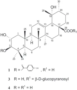
Um extrato metanólico das folhas de Luehea divaricata (Tiliaceae), vulgarmente conhecida no Brasil como “açoita-cavalo”, forneceu um novo triterpeno caracterizado como ácido 3β-p -hidroxibenzoiloxitormentico [ácido 3β-(p-hidroxibenzoiloxi)-2α-hidroxiurs-12-en-28-óico], juntamente com uma mistura contendo o ácido maslínico, um triterpeno conhecido. As estruturas dos compostos foram estabelecidas por métodos espectroscópicos.
A methanolic extract from leaves of Luehea divaricata (Tiliaceae), known in Brazil as “açoita-cavalo”, yielded two triterpene: a novel characterized as 3β-p-hydroxybenzoyloxytormentic acid [3β-(p-hydroxybenzoyloxy)-2α-hydroxyurs-12-en-28-oic acid] and a mixture containing the maslinic acid. The new compound’s structure was established by spectroscopic methods.
Keywords: Luehea divaricata, 3β-p-hydroxybenzoyloxytormentic acid, maslinic acid
Introduction
The Tiliaceae family has not been extensively studied yet. α-Amyrin derivatives have already been isolated from the genus Corchorus.1 There is no studies about the
chemical composition of this plant and no reports were found on the genus Luehea as well. Luehea divaricata
Mart. (Tiliaceae), known in Brazil as “açoita-cavalo”, is a tree which grows in Brazil, Argentina and Paraguay.2, 3 The L. divaricata is used in Brazilian folk medicine for different purposes: the leaves are used as diuretic, the stems as anti-inflammatory,4 the bark and aerial parts are used for healing
skin wounds, pimples, and for vaginal washes.2 Also the L. divaricata was assayed for antifungal properties and exhibited a broad spectrum of activity against dermatophytes.2 The aqueous extract of L. divaricata
presented genotoxic activity in the Ames test (Salmonella/ microsome) with microsomal activation.5 However, a
phytochemical screening of L.divaricata reported the presence of flavonoids, tannins and saponins.4 In this paper
we report the isolation and the structure elucidation of a new α-amyrin derivative, which was characterized as
3β-p-hydroxybenzoyloxytormentic acid and a mixture containing the maslinic acid.
Results and Discussion
Structural elucidation and NMR signal assignments of 3β-p-hydroxybenzoyloxytormentic acid (1)
Compound 1 was isolated as white crystals. Its EIMS spectrum showed a molecular peak at m/z 608 in agreement with C37H52O7 molecular formula. It also showed a base peak at m/z 121 attributed to p-hydroxybenzoyl cation and also the peaks at m/z 563 (10.8%, [M - COOH]+) and
562 (17.0%, [M- HCOOH]+ • ). The peak at
m/z 146 (61.9%, [C11H14]+ •) is a characteristic of a tertiary hydroxyl function
presence at C-19 in the urs-12-ene skeleton.6 Other
important peaks were noticed at m/z 246 (14.0%), 219 (7.9%), 218 (14.0%) and 201 (5.8%). Some low relative abundance peaks at m/z 179 and 264 were detected, as previously related to triterpenes from ursane class.6 The 1H
chemical shifts of 1 (CD3OD), in the range of δ 0.82-1.37, showed six singlets from methyl groups and one doublet, partly superposed by the singlets in agreement with an ursane type compound. The 1H NMR spectrum of an
oleanane type compound has seven methyl singlets and no doublets.7 The 1H NMR spectrum also showed two
476 Tanaka et al. J. Braz. Chem. Soc.
tormentic acid,8 and a strong agreement was observed,
ex-cept the chemical shift corresponding to H-3. The differ-ence (∆δH = 1.79 ppm)of the H-3 chemical shift (δH 4.70) in respect to tormentic acid8 was justified by the presence of a p-hydroxybenzoyl group linked to the oxygen of C-3 in compound 1, which induces an electron density reduction by inductive and resonance withdrawal effects of the p -hydroxybenzoyloxy group. The 1H and 13C NMR spectra’s
compound had signals for a p-hydroxybenzoate group [δ 7.92 (2H, dd, J 9.0 Hz, 2.1 Hz) and δ6.83 (2H, dd, J 9.0 Hz, 2.1 Hz), δ 168.7 (C-7’), 163.6, (C-4’), 133.0 (2’ and CH-6’), 123.0 (C-1’), 116.2 (CH-3’ and CH-5’)]. The presence of this group was supported by the observation of a strong peak in the mass spectrum at m/z 121 corresponding to [C7H5O2] + •. The correlation between H-3 and C-7’ observed
in HMBC spectrum was very important because it was pos-sible to establish a link between the p-hydroxybenzoyl group and the triterpene skeleton from the structure 1 in the C-3 position. Initially, compound 113C NMR spectrum was run
in CD3OD, however it was also necessary to run in C5D5N to confirm the presence of the signals that were superposed by the solvent signal in the range of δ 48-50 (Table 1). The 13C
NMR spectrum of 1 confirmed that is a triterpene skeleton with an ursolic acid type (C-12 and C-13 at δ 129.3 and 140.4). The 13C NMR spectral data of 1 were compared with
those from tormenticacid ester glucoside.9 The coupling
constant (J2,3) of 10.3 Hz is typical to an antiperiplanar (axial-axial) relationship between H-2 and H-3 (Table 1). The NOE difference NMR experiment was also performed to confirm the p-hydroxybenzoyloxy group orientation at C-3. Irra-diation of H-2β signal at δ 3.90 produced an enhancement in the methyl hydrogens resonance at δ 1.01 (3H-24) and 1.08 (3H-25) which showed a coaxial relationship between 3H-24 and 3H-25. In the same irradiation, it wasn’t observed
NOE enhancement at H-3 or H-5, showing an antiperiplanar relationship between H-2 / H-3 and H-2 / H-5.
The complete and unequivocal 1H and 13C chemical
shifts assignments of 1 were assisted by DEPT, COSY (1H x 1H), HSQC (13C x 1H) and HMBC (13C x 1H) spectra (Table 1).
Assignments of maslinic acid(2)
The 1D 1H NMR from the mixture showed signals for
methyl groups (δ0.80–1.16), for two carbinolic methine hydrogens at δ2.90 (d, J 9.9Hz, H-3α) and 3.61 (ddd, J
9.9 Hz, 9.9 Hz, 3.9 Hz, H-2β) and for one olefinic hydrogen at δ 5.24 (t, J 3.6 Hz). These data suggested 2 to be a triterpene with an oleanane skeleton.8 The 13C NMR
spectrum showed the signals at δ145.5, 123.6, 84.5 and 69.5 confirming that is olean-12-ene-2α, 3β -diol.10 Some
aspects of the maslinic acid structure was made by comparison of its 1H NMR and 13C NMR data with those
proposed to a similar compound.8,10,11
Experimental
General
EIMS: 70 eV; 1H (300 or 500 MHz) and 13C (75.5 or 125
MHz) NMR spectra were obtained in pyridine-d5 or
methanol-d4 with TMS as internal reference; column chromatography (CC): silica gel 60 (70-230 mesh); thin-layer chromatography (TLC): silica gel F254 (0.25 mm in thickness).
477 A New Tormentic Acid Derivative from Luehea divaricata Mart. (Tiliaceae)
Vol. 14, No. 3, 2003
Plant material
The plant was collected in April 1999, Mandacaru stream, Maringá city, State of Paraná, Brazil and identified by Dr. Maria Conceição de Souza, Universidade Estadual de Maringá. A voucher specimen (HUM 9057) was kept at the herbarium of the Biological Department of Universidade Estadual de Maringá.
Isolation
Air-dried and powdered leaves (600 g) of L. divaricata
Mart., were extracted with MeOH at room temp. The MeOH extract was concentrated in vacuum and yielded 74 g of crude methanolic extract. Part of the crude methanolic
extract (38 g) was partitioned with n-hexane (600 mL), chloroform (600 mL), ethyl acetate (600 mL) and methanol (100 mL), yielding 5.8 g (15.3%), 2.9 g (7.6%), 4.9 g (12.9%), 24 g (63.2%) respectively. The choroformic fraction was subjected to CC on silica gel (70 g) and eluted with different rations of n-hexane, CHCl3 and MeOH. The appropriate frs (monitored by TLC analysis) were combined resulting in 22 frs. Fr 14 (160 mg), eluted with chloroform-methanol (90:10), was subjected to repeated CC on silica gel, eluted with n-hexane, chloroform and methanol mixts of increasing polarity to give 3β-p -hydroxybenzoyloxytormentic acid (1) (2.8 mg-1.8%) and a mixture containing maslinic acid (2) (4.3 mg-2.7%).
3β-p-hydroxybenzoyloxytormentic acid (1). White crystals. EIMS m/z (rel. int.): [M]+ • 608, 563 (10.8), 562
Table 1.1H, 13C and 2D NMR spectral data for 3β-p-hydroxybenzoyloxytormentic acid (1), 13C NMR data for tormentic acid ester glucoside (3)9 and for tormentic acid (4),81H NMR in CD
3OD [1 (300 MHz) and 4 (400 MHz)] and
13C NMR in CD
3OD [1 (75.5 MHz)] and C5D5N [3 (50 MHz)]a
C (DEPT) δ13C δ1H COSY (1H x 1H) HMBC (13C x1H)
1 (1b, 3) 1 4 1 (2J and3J) 1 (2J, 3Jand4J)
11 (CH2) 48.6 (48.4, 48.0) 1.10 / 2.04 nd H-1b / H-1a; H-2 H-5; H-25
12 (CH) 67.7 (66.3, 68.6) 3.90 td 3.62 ddd H-1; H-3 H-3; H-25
(10.3, 10.3, 4.0) (9.8; 9.8; 3.5)
13 (CH) 85.9 (85.2, 83.8) 4.70 d (10.3) 2.91 d (9.8) H-2 H-23; H-24
14 (C) 41.1 (40.2, 38.5) H-3; H-5; H-23; H-24
15 (CH) 56.5 (55.4, 56.0) 1.04 nd H-6 H-23; H-24; H-25
16 (CH2) 19.5 (18.6, 19.1) 1.59 nd H-5; H-7 H-5; H-25
17 (CH2) 34.0 (33.1, 33.5) 1.37 / 1.64 nd H-6 H-26
18 (C) 40.8 (40.2, 40.6) H-27
19 (CH) 48.6 (47.5, 47.9) 1.82 nd H-11 H-5; H-25; H-26
10 (C) 39.2 (38.3, 39.9) H-25
11 (CH2) 24.7 (23.9, 24.2) 2.04 nd H-12; H-9
12 (CH) 129.3 (127.7, 128.2) 5.30 br s 5.28 t (3.2) H-11 H-18
13 (C) 140.4 (140.1, 139.5) H-18; H-27
14 (C) 42.7 (42.2, 42.3) H-27; H-26
15 (CH2) 29.6 (29.1, 29.3) 1.02 / 1.85 nd H-15b / H-15a; H-16 H-27 16 (CH2) 27.3 (26.7, 26.8) 1.28 / 1.75 nd H-16b / H-16a; H-15 H-18
17 (C) 48.6 (48.1, 48.6) H-18
18 (CH) 55.1 (54.5, 54.4) 2.51 s 2.50 s H-29
19 (C) 73.7 (72.6, 72.6) H-18; H-29; H-30
20 (CH) 43.1 (42.0, 42.2) 1.37 nd H-30 H-30; H-29; H-18
21 (CH2) 26.6 (26.2, 26.1) 1.54 / 2.59 nd H-21b / H-21a H-30 22 (CH2) 39.0 (38.2, 37.8) 1.75 nd
23 (CH3) 29.2 (28.8, 29.5) 0.90 s H-3; H-24
24 (CH3) 18.3 (16.5, 16.8) 1.01 s H-3; H-23
25 (CH3) 17.0 (16.6, 17.1) 1.08 s 26 (CH3) 17.5 (18.1, 17.8) 0.82 s 27 (CH3) 24.8 (24.5, 24.6) 1.37 s
28 (C) 182.7 (180.8, 176.9) H-18
29 (CH3) 27.0 (26.9, 27.0) 1.20 s
30 (CH3) 16.5 (17.0, 17.5) 0.93 d ( 6.6) H-20
1’ (C) 123.0 (122.7, - ) H-3’; H-5’
2’, 6’ (CH) 133.0 (132.5, - ) 7.92 dd (9.0, 2.1) H-3’, H-5’ H-6’, H-2’ 3’, 5’ (CH) 116.2 (116.0, - ) 6.83 dd (9.0, 2.1) H-2’, H-6’ H-5’, H-3’
4’ (C) 163.6 (163.4, - ) H-3’; H-5’; H-2’; H-6’
7’ (C) 168.7 (167.0, - ) H-3; H-2’; H-6’
a Values are in ppm (δ). Coupling constants (J), in parentheses, are in Hz; b13C NMR in C
478 Tanaka et al. J. Braz. Chem. Soc.
(17.0), 264 (< 3.0), 246 (14.0), 219 (7.3), 218 (14.1), 201 (5.8), 189 (31.8), 187 (15.6), 179 (< 3.0), 146 (61.9), 121 (100).
Maslinic acid(2). White crystals.13C NMR (75.5 MHz,
CD3OD): 48.1 (C-1), 69.5 (C-2), 84.5 (C-3), 40.5 (C-4), 56.7 (C-5), 19.5 (C-6), 33.9 (C-7), 39.2 (C-8), 49.0 (C-9), 39.2 (C-10), 24.0 (C-11), 123.6 (C-12), 145.5 (C-13), 42.6 (C-14), 28.8 (C-15), 24.0 (C-16), 47.7 (C-17), 42.7 (C-18), 47.2 (C-19), 31.6 (C-20), 34.9 (C-21), 33.8 (C-22), 29.3 (C-23), 17.0 (C-24), 17.1 (C-25), 17.4 (C-26), 23.9 (C-27), 180.0 (C-28), 33.5 (C-29), 23.9 (C-30).
Acknowledgements
The authors thanks CAPES for scholarships and CNPq for financial support. We also thanks Dr. M. C. de Souza for support in the plant collection and for the identification of the plant material, Dr. A. J. Marsaioli (Unicamp) for running the HSQC and HMBC spectra.
References
1. Khan, M. S. Y.; Javed, K.; Khan, M. H.; Shamsi, M. A.; Siddiqui, A. A.; Phytochemistry 1991, 30, 1989.
2. Zacchino, S.; Santecchia, C.; Lopez, S.; Gattuso, S.; Muñoz, J. de D.; Cruañes, A.; Vivot, E.; Cruañes, M. del C.; Salinas, A.; Ruiz, R. E. de; Ruiz, S.; Phytomedicine 1998, 5, 389. 3. Lorenzi, H.; Árvores Brasileiras: Manual de Identificação e
Cultivo de Plantas Arbóreas Nativas do Brasil / Harri Lorenzi,
2th ed., Plantarum: Nova Odessa, 1998.
4. Alice, C. B.; Silva, G. A. A. B.; Cad. Farm.1985, 1, 83. 5. Vargas, V. M. F.; Guidobono, R. R.; Henriques, J. A. P.; Mem.
I. Oswaldo Cruz1991, 86, 67.
6. Delgado, G.; Hernández, J.; Pereda-Miranda, R.;
Phyto-chemistry 1989, 28, 1483.
7. Biessels, H. W. A.; Hoof, A. C. K.; Bosch, J. J. K.; Salemink, C.
A.; Phytochemistry 1974, 13, 203.
8. Yamagishi, T.; Zhang, D-C.; Chang, J-J.; McPhail, D. R.; McPhail, A. T.; Lee, K-H.; Phytochemistry 1988, 27, 3213. 9. Gopalsamy, N.; Vargas, D.; Guého, J.; Ricaud, C.; Hostettmann,
K.; Phytochemistry 1988, 27, 3593.
10. Bilia, A. R.; Mendez, J.; Morelli, I.; Acta Pharm. Helv.1996,
71, 191.
11. Zucaro, Z.; Yasmin, L.; Compagnone, R.S.; Hess, S.C.; Delle Monache, F.; J. Braz. Chem. Soc.2000, 11, 241.
![Table 1. 1 H, 13 C and 2D NMR spectral data for 3β-p-hydroxybenzoyloxytormentic acid (1), 13 C NMR data for tormentic acid ester glucoside (3) 9 and for tormentic acid (4), 8 1 H NMR in CD 3 OD [1 (300 MHz) and 4 (400 MHz)] and 13 C NMR in CD 3 OD [1 (](https://thumb-eu.123doks.com/thumbv2/123dok_br/18989775.460208/3.892.94.826.163.754/table-nmr-spectral-hydroxybenzoyloxytormentic-tormentic-ester-glucoside-tormentic.webp)
