Article
Printed in Brazil - ©2017 Sociedade Brasileira de Química 0103 - 5053 $6.00+0.00
*e-mail: [email protected] #These authors contributed equally to this work.
Laccase-Assisted Rapid Synthesis of Colloidal Gold Nanoparticles for the Catalytic
Reduction of 4-Nitrophenol
Fang Li,a,# Zheng Li,b,c,# Chang Zengc,# and Yonggang Hu*,b,c
aCollege of Food and Bioengineering, Henan University of Science and Technology, 471023
Luoyang, China
bState Key Laboratory of Agricultural Microbiology and cCollege of Life Science and Technology,
Huazhong Agricultural University, 430070 Wuhan, China
A green method for the rapid preparation of uniform-sized colloidal gold nanoparticles under ambient conditions was presented and validated using laccase as a reduction agent in alkaline medium. UV-Vis spectrophotometry,field-emission high-resolution transmission electron microscopy, X-ray diffraction, selected area electron diffraction, energy dispersive X-ray analysis, zetasizer, and Fouriertransform infrared spectroscopy were used to characterize the gold nanoparticles.Thegold nanoparticles were spherical, crystalline, uniform, and monodisperse with an average size of 12.24 nm. Thesenanoparticles were successfully used to reduce 4-nitrophenol in the presence of NaBH4 and exhibited an excellent catalytic activity.
Keywords: colloidal gold nanoparticle, laccase, catalysis, 4-nitrophenol
Introduction
Over the past decade, gold nanoparticles (GNPs) have attracted considerable attention for their synthesis and application. Given their outstanding physicochemical and
optoelectronic properties,1 GNPs have been widely used in
spectroscopy,2 electrochemistry,3 catalysis,4 biomedicine,5
and environmental biotechnology.6 Synthesizing GNPs
with a tight control on their size and shape is crucial because these parameters determine material properties at
the nanoscale.7-10 Various chemical and physical methods
for the preparation of GNPs with controlled size and shape have been reported.4,10-15 However, the synthesis procedures
for GNPs usually either involve the use of hazardous chemicals or the requirement of rigorous conditions, such
as elevated temperature.14,15 A method employing enzymes
as a template for the biosynthesis of GNPs16-26 has recently
attracted increasing attention because of its simplicity1,26
and requirement for non-toxic chemicals. Moreover, enzymes can act as reducers and stabilizers because of their chemical, biochemical, and biological functions. For
example, Sharma et al.19 used glucose oxidase (GOx) as
the reducing agent to synthesize GNP-GOx composites
and GNP-polyaniline nanocomposites. Venkatpurwar and
Pokharkar20 reported a green method to prepare GNPs
by using the therapeutic enzyme serratiopeptidase and demonstrated that the enzyme capped on the as-prepared GNPs retains its therapeutic activity. Zou et al.21 directly
employed trypsin as the linking and reducing agent to synthesize gold nanoparticle-nanocluster composites.
Laccases (p-diphenol: dioxygen oxidoreductase,
EC 1.10.3.2) are blue multicopper oxidases that have been widely distributed in many eukaryotes [e.g., plants,27
insects,28 and fungi29 and prokaryotes (e.g., bacteria)30] since
their discovery more than one century ago in the Japanese tree Rhus venicifera. Laccases generally show a high stability in the extracellular environment, and their purification is easy probably because most laccases are extracellular
enzymes.31 As environmentally benign enzymes, laccases
exhibit limitless applications in many fields, from textile to food industries, and from bioremediation processes to green synthesis.31-33 The use of laccase to synthesize GNPs
was also studied. For example, Faramarzi and Forootanfar24
used laccase from Paraconiothyrium variabile to synthesize GNPs at 70 °C and obtained GNPs with sizes varying
from 71 to 266 nm. Guo et al.25 reported the use of
laccase for the in situ synthesis of GNPs with an average
size of 3 ± 2 nm at 4 °C for 48 h. Considering that the
accelerated in the presence of NaOH, we proposed a simple, rapid, and eco-friendly method for the biosynthesis of monodispersed GNPs by using commercial laccase
from Trametes versicolor. The synthesized GNPs were
thoroughlycharacterized via UV-Vis spectrophotometry,
field-emission high-resolution transmission electron microscopy (HRTEM), X-ray diffraction (XRD), selected area electron diffraction (SAED), energy dispersive X-ray
analysis (EDXA), zetasizer, and Fouriertransform infrared
spectroscopy (FTIR). Furthermore, the Au-catalyzed
reduction of 4-nitrophenol in the presence of NaBH4 was
used as a model system to demonstrate that the GNPs are a promising catalyst.
Experimental
Materials
Laccase from T. versicolor was acquired from
Sigma-Aldrich (St. Louis, MO, USA). HAuCl4·3H2O powder and
NaOH were purchased from Sinopharm Chemical Reagent Co., Ltd. (Shanghai, China). All chemical reagents were of analytical grade.
Purification of laccase
Laccase was purified according to our previously
reported procedures.34 In brief, 10.0 mg of laccase was
dissolved in 1.0 mL of 10.0 mmol L–1 sodium acetate
buffer (pH 5.5) and centrifuged using CR21G II high speed refrigerated centrifuge (Hitachi Ltd., Tokyo, Japan) at 14,000 rpm for 1 hour at 4 °C. The supernatant was subjected to a DEAE-SephadexA-50 (BioMan, Shanghai, China) anion-exchange column (previously equilibrated with buffer) at 4 °C and washed with buffer to remove
unbound material. Laccase was eluted with 60.0 mmol L–1
ammonium sulfate, then desalted into 20.0 mmol L–1
sodium acetate buffer (pH 5.5), and stored at 4 °C. The purified enzyme was applied for biosynthesis of GNPs.
Synthesis of GNPs
Purified laccase solution (180.0 µL, 0.575 mg mL–1) was
added to a solution containing 164.6 µL of 24.3 mmol L–1
HAuCl4 and 50.0 µL of 1.0 mol L–1 NaOH, and the volume
was adjusted to 1.0 mL with ultrapure water. The mixture was incubated for 60.0 min under ambient conditions with mild stirring. Finally, the synthesized GNPs were purified via double centrifugation at 14,000 rpm for 45 min at 4 °C to remove unbound laccase. Then, the GNPs were resuspended in ultrapure water for further characterization.
Characterization of GNPs
The UV-Vis spectra of the GNPs were recorded on an UV-Vis spectrometer (Ocean Optical QE65000, Ocean Optical Co., Ltd., Dunedin, USA). The The FTIR spectra of laccase and as-prepared GNPs were collected using an FTIR instrument (330 FTIR, Thermo Fisher Scientific, Waltham, MA, USA). Dried specimen was crushed with KBr in a mortar at 1:100 ratio. The pressed pellet was recovered with a clip and immediately analyzed in the
4000-400 cm–1 region over 500 scans in the FTIR instrument.
The morphology of GNPs was recorded using a JEM-2100 HRTEM instrument (HR, JEOL Ltd., Tokyo, Japan) with an accelerating voltage of 200 kV. The atomic percentage was determined using the JEM-2100 HRTEM instrument equipped with an EDX system. The SAED patterns of the prepared TEM samples were also determined. Powder X-ray diffraction was operated on an X-ray diffractometer (D/MAX-RB, Rigaku, Tokyo, Japan) at 40 kV and 40 mA
with CuKα radiation. Size distribution was analyzed by
Zetasizer (Nano-ZS90, Malvern Instrument Ltd., Malvern, Worcestershire, UK).
Catalytic reduction of 4-nitrophenol
The catalytic activity of the synthesized GNPs was researched by reducing 4-nitrophenol to 4-aminophenol
with NaBH4 as the reducing agent. In brief, a reaction
mixture of water (1.38 mL), aqueous 4-nitrophenol
solution (50.0 µL, 3.0 mmol L–1), and GNPs (20.0 µL,
0.096 mmol L–1) was first collected in a quartz cuvette.
The absorption spectrum was immediately recorded after the addition of NaBH4 (50.0 µL, 0.3 mol L–1).
Results and Discussion
Biosynthesis of GNPs
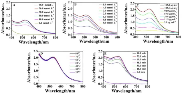
The parameters that affected the formation of GNPs by
reducing HAuCl4 with laccase were investigated through
UV-Vis spectroscopy. Alkaline condition was conducive to the synthesis and formation of GNPs. The concentrations
of NaOH ranging from 0 to 90.0 mmol L–1 were added to
adjust the reaction environment. As shown in Figure 1A, the absorption peak of the GNPs was not observed during 1 h in the absence of NaOH. Interestingly, an obvious surface plasmon resonance (SPR) peak corresponding to the GNPs emerged in the presence of NaOH. As expected, the intensities of the SPR band continuously increased with
increasing NaOH and peaked at 50.0 mmol L–1. However,
concentration was further increased. Thus, 50.0 mmol L–1
NaOH was selected for the subsequent synthesis. The GNPs
yield increased with the increase of AuCl4
−
concentration from 0-4.0 mmol L–1, and up to 4.0 mmol L–1 (Figure 1B).
Accordingly, 4.0 mmol L–1 was used for the subsequent
synthesis. The concentration of laccase was examined from 2.9 to 115.0 µg mL–1, and 103.5 µg mL–1 laccase was
selected to be the optimum concentration (Figure 1C). Results in Figure 1D indicate that the temperature in the range of 20 to 80 °C did not contribute to the change in SPR band. In other words, the size and yield of GNPs affected by the temperature is not obvious. As a result, the GNPs could be easily synthesized at room temperature (25 °C). In addition, the reaction time was also investigated in the range of 0.0 to 90.0 min. As shown in Figure 1E, the yield of GNPs increased with time, peaked at 60.0 min, and then plateaued.
Thus, the reaction solution containing 50.0 mmol L–1
NaOH, 4.0 mmol L–1 AuCl
4
−
, and 103.5 µg mL–1 laccase
incubated at ambient temperature for 60.0 min was selected for the biosynthesis of GNPs. As shown in Table 1, the present method was quick and facile in comparison with other chemical synthesis and biosynthesis methods. This result indicates that the developed method is a promising candidate for the rapid preparation of GNPs.
Characterization
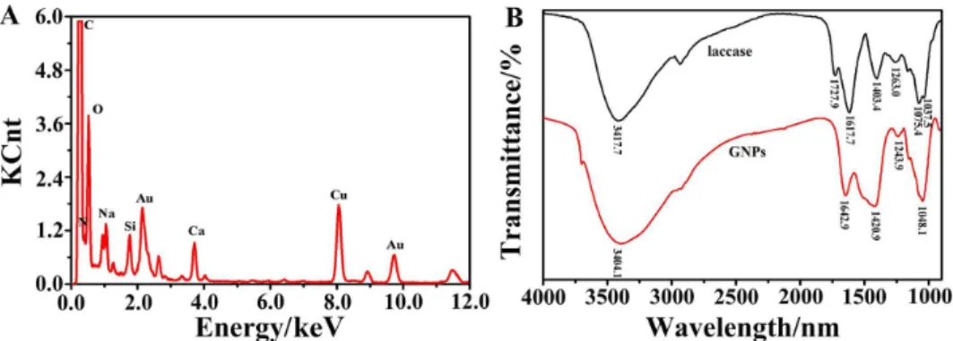
UV-Vis spectroscopy is a primary technique for the characterization of metallic nanoparticles largely attributed
to SPR phenomenon. The color of the as-prepared nanoparticles changed from yellow to deep wine because of the collective oscillation of free electrons induced by an interacting electromagnetic field in GNPs, indicating the formation of GNPs. Figure 2A shows that the synthesized GNPs displayed a single but strong SPR band at 518 nm. The XRD pattern (Figure 2B) consists of four prominent Bragg reflections at 2θ values of 38.1, 44.3, 64.5, and 77.6°,
which corresponded to the (111), (200), (220), and (311) lattice planes of the face-centered cubic (fcc) structure of gold, implying the formation of crystalline gold in the specimen. The TEM (Figure 2C) and HRTEM (Figure 2D) images of the GNPs clearly indicate that the composites were uniform and had a spherical morphology with good dispersion. The clear lattice fringes in the GNPs were observed in the HRTEM image, and the interfringe spacing was 0.23 nm, which is close to the interplane distance of the (111) plane in the fcc gold. The SAED pattern of GNPs (Figure 2E) further confirmed that the GNPs were nanocrystalline in nature and that the structure was fcc type, which was in accordance with the XRD result. The particle size distribution of the GNPs was analyzed using a Zetasizer (Figure 2F) and found to range from 10.10 to 13.54 nm with an average of approximately 12.24 nm.
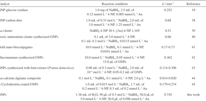
The elemental composition of the as-prepared GNPs was determined through EDXA analysis. As shown in Figure 3A, two characteristic peaks of gold atoms at approximately 2.195 and 9.700 keV showed the presence of gold. The C, N, and O signals may originate from the
Figure 1. Effect of experimental conditions on the biosynthesis of GNPs. (A) Effect of NaOH concentration, the solution containing 1.0 mmol L–1 HAuCl 4, 23.0 µg mL–1 laccase, and NaOH was incubated at 30 °C for 60.0 min; (B) effect of HAuCl
4 concentration, the solution containing HAuCl4, 23.0 µg mL–1 laccase, and 50.0 mmol L–1 NaOH was incubated at 30 °C for 60.0 min; (C) effect of laccase concentration, the solution containing 4.0 mmol L–1 HAuCl
laccases bound to the surface of GNPs, whereas the Cu and some of the C in the EDS spectrum might be attributed to the TEM grid background.
FTIR analysis of laccase and GNPs was further carried out (Figure 3B). The FTIR spectrum of laccase showed NH
and OH bands at 3417.7 cm–1, COO– stretching at 1727.9
and 1403.4 cm–1, amide I band at 1617.7 cm–1, amide III
band at 1263.0 cm–1, P−OH at 1075.4 cm–1,and P−O−C at
1037.5 cm–1 (black line). The adsorption band of P−OH and
P−O−C stretching signified the presence of protein phosphate
groups and the adsorption band of the COO– derived from
the carboxyl side groups in the amino acid residues. The spectrum of GNPs (red line) has similar FTIR bands to that of laccase. However, the wavenumber corresponding to the
asymmetric stretching mode of the COO−
group shifted from
1727.9 to 1420.9 cm–1, whereas that corresponding to the
Table 1. Comparison of reaction conditions for the preparation of GNPs
Reductant Reaction condition Reference
Trisodium citrate the solution containing 0.02% HAuCl4 + 0.08% trisodium citrate was microwave-assisted heated with 120 W power for 15.0 min
4
NaBH4 the solution containing 0.02 g mL–1 PVP, 2.0 mL of 0.024 mol L–1 and 1.0 mL of 0.0167 g NaBH4 was incubated at 170 °C for 3.0 h
14
URAK the solution containing 1.0 mg mL–1 URAK and 1.0 mmol L–1 HAuCl
4 in 10.0 mmol L–1 Tris-HCl buffer (pH = 9.0) was incubated at 37 °C for 60.0 h
17
Glucose oxidase the solution containing 6 × 10⁻⁴ mol L−1 HAuCl
4 and 3.0 mL of 0.7 mg mL–1 glucose oxidase in 10.0 mmol L–1 PBS (pH = 7.0) was incubated at 37 °C for 36.0 h
19
Trypsin the solution containing 4.0 mmol L–1 HAuCl
4 and 20.0 mg mL–1 trypsin (pH = 12.0) was incubated at 37 °C for 510.0 min
21
Laccase the solution containing 0.5 mL of 8.0 mg mL–1 laccase and 2.5 mL of 0.6 mol L–1 HAuCl
4 was incubated at 70 °C for 20.0 min
24
Laccase the solution containing 0.125 mg mL–1 laccase and 0.5 mmol L–1 HAuCl
4 in PB buffer (pH = 6.0) was incubated at 4 °C for 48 h
25
α-Amylase the solution containing 4.5 × 10⁻⁴ mol L–1 HAuCl
4 and 1.0 mg mL–1α-amylase in 10.0 mmol L–1 PBS (pH = 7.0) was incubated at 37 °C for 48 h
35
Laccase the solution containing 50.0 mmol L–1 NaOH, 4.0 mmol L–1 AuCl 4
− and 103.5 µg mL–1 laccase was incubated at 25 °C for 60.0 min
this work
Figure 2. (A) UV-Vis absorption spectra of GNPs synthesized by laccase. Inset shows (a) the control sample (the solution containing 50.0 mmol L-1 NaOH, 4.0 mmol L-1 AuCl
NH stretching vibration shifted from 3417.7 to 3404.1 cm–1.
The adsorption band of amide I changed from 1617.7 to
1642.9 cm–1. Meanwhile, the adsorption bands of P−OH
and P−O−C disappeared while a new band appeared at
1048.1 cm–1. Figure 3 indicates that GNP-laccase composites
were formed during our experiments.
Catalytic reduction of 4-nitrophenol
To evaluate the catalytic activity, the reduction of
4-nitrophenol to 4-aminophenol in the presence of NaBH4
was selected as the model system.36 The SPR band of
4-nitrophenol immediately shifted from 317 to 400 upon
the addition of fleshly prepared NaBH4 solution with an
associated color change from light yellow to bright yellow, implying the formation of 4-nitrophenolate ions. The addition of excess NaBH4 solution exerted no change on the
SPR band at 400 nm. Interestingly, the color gradually faded away with the addition of GNPs, and this process lasted for approximately 5 min. Similarly, this phenomenon was clearly observed by the gradual disappearance of the SPR band at 400 nm after the addition of 4.82 × 10⁻⁶ mmol L–1
GNPs with a concurrent appearance of a new peak at 300 nm, which corresponded to the formation of 4-aminophenol (Figure 4A).
Furthermore, the kinetics of the GNP-catalyzed reaction of 4-nitrophenol was investigated. Since the amount of NaBH4 largely exceeded in the reaction, the reduction rate
can be assumed to be independent of NaBH4 concentration.
Thus, the catalytic reaction could be treated as pseudo-first-order kinetics with respect to 4-nitrophenol concentration. The values of the apparent reaction rate constant k of these catalytic reactions in the presence of different doses of the as-prepared nanocomposites were calculated from the plot
of ln A vs. time (Figure 4B), where A is the absorbance
at 400 nm of 4-nitrophenolate ions. The rate constant k
gradually increased with increasing GNPs and reached 0.745 min–1 after using 7.712 × 10⁻⁶ mmol L–1 catalyst,
which was found to be significantly superior to those of most relevant literature reports for the reduction of 4-nitrophenol (Table 2). The GNPs could be stably stored in aqueous medium for as long as 3 months, and remained well dispersive without aggregation after reaction. The excellent stability and high catalytic activity of the GNPs probably attributed to the formation of GNP-laccase composites (Figure 3). The laccase, as a stabilizer, could resist the aggregation of GNPs to bulk materials, which leads to the loss of the catalytic activity of nanoparticles.37 Moreover,
the abundant function groups (–OH, –COOH, and –C=O) derived from laccase can increase the adsorption capacity,
Figure 4. (A) Time-dependent absorption spectra for the catalytic reduction of 4-nitrophenol by NaBH4 in the presence of GNPs synthesized by laccase; (B) plot of ln A (A = absorbance at 400 nm of 4-nitrophenolate ion) versus time.
and accelerate the reduction rate, eventually enhance the
catalytic acitivity.38 These results demonstrate that the
GNPs prepared by the proposed method are the promising catalyst.
Conclusions
We developed a simple, quick, and eco-friendly method to biosynthesize well-dispersed GNPs by using laccase as both reducing and stabilizing reagents. The synthesis was carried out for 60 min in alkaline solution under ambient conditions. The prepared GNPs exhibited an uniform spherical shape, narrow size distribution, and excellent storage stability. The obtained GNPs served as an efficient catalyst in the borohydride reduction of 4-nitrophenol. In consideration of the huge potential of nano-composite materials in various applications, the development of the biogenic route using laccase as a template under ambient conditions will offer a green alternative toward the formation of gold nanoparticles.
Acknowledgments
This work was supported by the National Natural Science Foundation of China (Grant No. 31501563), the State Key Laboratory of Agricultural Microbiology, Huazhong Agricultural University (Grant No. AMLKF201405) and the Opening Foundation of State Key Laboratory of Agricultural Microbiology (Grant No. AMLKF201508).
References
1. Panda, T.; Deepa, K.; J. Nanosci. Nanotechnol.2011, 11, 10279. 2. Schmit, V. L.; Martoglio, R.; Scott, B.; Strickland, A. D.; Carron,
K. T.; J. Am. Chem. Soc.2012, 134, 59.
3. Du, L.; Jiang, H.; Liu, X.; Wang, E.; Electrochem. Commun.
2007, 9, 1165.
4. Du, J.; Xia, Z.; Anal. Methods2013, 5, 1991.
5. Dykman, L.; Khlebtsov, N.; Chem. Soc. Rev.2012, 41, 2256. 6. Das, S. K.; Das, A. R.; Guha, A. K.; Langmuir 2009, 25, 8192. 7. Daniel, M.-C.; Astruc, D.; Chem. Rev.2004, 104, 293.
8. Campelo, J. M.; Luna, D.; Luque, R.; Marinas, J. M.; Romero, A. A.; ChemSusChem2009, 2, 18.
9. White, R. J.; Budarin, V. L.; Clark, J. H.; ChemSusChem2008, 1, 408.
10. Sánchez-Iglesias, A.; Pastoriza-Santos, I.; Pérez-Juste, J.; Rodríguez-González, B.; García de Abajo, F. J.; Liz-Marzán, L. M.; Adv. Mater. 2006, 18, 2529.
11. Teranishi, T.; Hasegawa, S.; Shimizu, T.; Miyake, M.; Adv. Mater.2001, 13, 1699.
12. Ruan, Q.; Shao, L.; Shu, Y.; Wang, J.; Wu, H.; Adv. Opt. Mater.
2014, 2, 65.
13. Zhang, L.; Xia, K.; Lu, Z.; Li, G.; Chen, J.; Deng, Y.; Li, S.; Zhou, F.; He, N.; Chem. Mater.2014, 26, 1794.
14. Zhao, F.; Yang, Y.; J. Dispersion Sci. Technol.2010, 31, 471. 15. Brust, M.; Walker, M.; Bethell, D.; Schiffrin, D. J.; Whyman,
R.; J. Chem. Soc., Chem. Commun.1994, 801.
16. Sharma, B.; Mandani, S.; Sarma, T. K.; Sci. Rep. 2013, 3, 2601.
Table 2. A comparison between the proposed system and literature reports for the room-temperature reduction of 4-nitrophenol
Catalyst Reaction condition k / min–1 Reference
GNP-glucose oxidase 1.0 mg of NaBH4, 2.5 mL of
0.12 mmol L–1 4-NP, 0.005 mmol L–1 Au
0.252 19
GNP-carbon dots 1.0 mL of 0.33 mol L–1 NaBH
4, 2.0 mL of 3.0 mmol L–1 4-NP, 1.25 mmol L–1 Au
0.68 38
Au cluster NaBH4:4-NP 30:1; [Au]:4-NP 1:105 0.51 39
Ferric ammonium citrate-synthesized GNPs 0.1 mL of 3.0 mmol L–1 4-NP, 0.1 mL 0.3 mol L–1 NaBH
4, 0.0115 mmol L–1 Au
0.66 40
Gold nano-bioconjugates 10.0 mmol L–1 NaBH
4, 0.1 mmol L–1 4-NP, 0.0101 mmol L–1 Au
0.17-0.73 41
Glucomannan-synthesized GNPs 10.0 mmol L–1 NaBH
4, 0.05 mmol L–1 4-NP, 15.0 µL of GNPs
0.362 42
GNPs synthesized with fruit extract (Prunus domestica) 0.08 mL of 0.3 mol L–1 NaBH
4, 2.0 mL of 10–4 mol L–1 4-NP, 0.05-0.2 mL of GNPs
0.114-0.306 43
Au-calcium alginate composite 0.1 mol L–1 NaBH
4, 0.1 mmol L–1 4-NP, 2.0 g L–1 Au 0.014-0.020 44
α-Cyclodextrin-coated GNPs 1.0 mL of 0.015 mol L–1 NaBH
4, 1.7 mL of 0.2 mmol L–1 4-NP, 0.3 mL of 0.2 mmol L–1 Au
0.179-0.274 45
GNPs 1.38 mL of H2O, 50 µL of 0.3 mol L–1 NaBH4, 50.0 µLof 3.0 mmol L–1 4-NP, 20.0 µL of 0.096 mmol L–1 Au
0.745 this work
17. Deepak, V.; Umamaheshwaran, P. S.; Guhan, K.; Nanthini, R. A.; Krithiga, B.; Jaithoon, N. M.; Gurunathan, S.; Colloids Surf., B2011, 86, 353.
18. Govindaraju, K.; Kiruthiga, V.; Manikandan, R.; Ashokkumar, T.; Singaravelu, G.; Mater. Lett.2011, 65, 256.
19. Sharma, B.; Mandani, S.; Sarma, T. K.; J. Mater. Chem. B2014, 2, 4072.
20. Venkatpurwar, V. P.; Pokharkar, V. B.; J. Biomed. Nanotechnol.
2010, 6, 667.
21. Zou, L.; Qi, W.; Huang, R.; Su, R.; Wang, M.; He, Z.; ACS Sustainable Chem. Eng.2013, 1, 1398.
22. Bharde, A.; Kulkarni, A.; Rao, M.; Prabhune, A.; Sastry, M.; J. Nanosci. Nanotechnol.2007, 7, 4369.
23. Sun, J.; Ge, J.; Liu, W.; Lan, M.; Zhang, H.; Wang, P.; Wang, Y.; Niu, Z.; Nanoscale2014, 6, 255.
24. Faramarzi, M. A.; Forootanfar, H.; Colloids Surf., B2011, 87, 23.
25. Guo, S.; Li, H.; Liu, J.; Yang, Y.; Kong, W.; Qiao, S.; Huang, H.; Liu, Y.; ACS Appl. Mater. Interfaces 2015, 37, 20937. 26. Tikariha, S.; Singh, S.; Banerjee, S.; Vidyarthi, A. S.; Int. J.
Pharm. Sci. Res.2012, 3, 1603.
27. McCaig, B. C.; Meagher, R. B.; Dean, J. F.; Planta2005, 221, 619.
28. Dittmer, N. T.; Suderman, R. J.; Jiang, H.; Zhu, Y. C.; Gorman, M. J.; Kramer, K. J.; Kanost, M. R.; Insect Biochem. Mol. Biol.
2004, 34, 29.
29. Dwivedi, U. N.; Singh, P.; Pandey, V. P.; Kumar, A.; J. Mol. Catal. B: Enzym.2011, 68, 117.
30. Alexandre, G.; Zhulin, I. B.; Trends Biotechnol.2000, 18, 41. 31. Rodgers, C. J.; Blanford, C. F.; Giddens, S. R.; Skamnioti, P.;
Armstrong, F. A.; Gurr, S. J.; Trends Biotechnol.2010, 28, 63.
32. Riva, S.; Trends Biotechnol.2006, 24, 219.
33. Alcalde, M.; Ferrer, M.; Plou, F. J.; Biocatal. Biotransform.
2007, 25, 113.
34. Zhang, Y.; Tan, C.; Fei, R.; Liu, X.; Zhou, Y.; Chen, J.; Chen, H.; Zhou, R.; Hu, Y.; Anal. Chem.2014, 86, 1115.
35. Rangnekar, A.; Sarma, T. K.; Singh, A. K.; Deka, J.; Ramesh, A.; Chattopadhyay, A.; Langmuir 2007, 23, 5700.
36. Al-Naji, M.; Balu, A. M.; Roibu, A.; Goepel, M.; Einicke, W.; Luque, R.; Glaser, R.; Catal. Sci. Technol.2015, 5, 2085. 37. Watt, J.; Cheong, S.; Toney, M. F.; Ingham, B.; Cookson, J.;
Bishop, P. T.; Tilley, R. D.; ACS Nano2010, 4, 396.
38. Liu, R.; Liu, J.; Kong, W.; Huang, H.; Han, X.; Zhang, X.; Liu, Y.; Kang, Z.; Dalton Trans.2014, 43, 10920.
39. Shivhare, A.; Ambrose, S. J.; Zhang, H.; Purves, R. W.; Scott, R. W.; Chem. Commun. 2013, 49, 276.
40. Rashid, M. H.; Mandal, T. K.; Adv. Funct. Mater.2008, 18, 2261.
41. Das, S. K.; Dickinson, C.; Lafir, F.; Brougham, D. F.; Marsili, E.; Green Chem.2012, 14, 1322.
42. Gao, Z.; Su, R.; Huang, R.; Qi, W.; He, Z.; Nanoscale Res. Lett.
2014, 9, 404.
43. Dauthal, P.; Mukhopadhyay, M.; Ind. Eng. Chem. Res.2012, 51, 13014.
44. Saha, S.; Pal, A.; Kundu, S.; Basu, S.; Pal, T.; Langmuir2010, 26, 2885.
45. Huang, T.; Meng, F.; Qi, L.; J. Phys. Chem. C2009, 113, 13636.
Submitted: April 20, 2016