A Further Investigation of the Effects of
Extremely Low Frequency Magnetic Fields on
Alkaline Phosphatase and
Acetylcholinesterase
Gary Silkstone*, Michael T. Wilson
School of Biology, University of Essex, Wivenhoe Park, Colchester, Essex, CO4 3SQ, United Kingdom
Abstract
Using a custom build spectrophotometer equipped with Helmholtz coils and designed to study the effects of magnetic fields on enzyme reactions in real-time we have investigated the influence of fields, from 100μT to 10 mT and at a variety of field frequencies, on the membrane bound enzymes alkaline phosphatase and acetylcholinesterase. We have also employed other methods to apply a magnetic field, e.g. Biostim. In contrast to earlier reports we have been unable to detect any field effects on these enzymes under any field/frequency regime. We discuss possible reasons for the discrepancy between this and earlier work and note the particularly complex influence of small temperature changes that may confound analysis.
Introduction
Over many years, and especially more recently, there has been considerable interest in the effects of magnetic fields on biological systems [1–10]. Much of this interest has focused on the effects of low intensity fields on the reactivity of light induced radicals. The reason for this is that there is good experimental evidence that navigation during the migration of some birds and insects is partly based on sensing the earth’s magnetic field [6–10] and that photoactivated cryptochrome, a flavoprotein located in the eye of the organism, is the magnetosensor [6,11–
13]. Furthermore, there is a well established theory that can provide a mechanism through which low strength magnetic fields may influence the temporal behavior of correlated radical pairs [6,11], such as may be formed in cryptochrome during photoreduction or, possibly, dur-ing re-oxidation of the photoreduced protein by oxygen [14].
Alternative radical based mechanisms through which biological systems may be influenced by magnetic fields have also been advanced. For example, Buchachenko and co-workers have proposed a mechanism based upon the fact that one of the isotopes of Mg, namely25Mg, pos-sesses a magnetic nucleus [15]. The proposal is that Mg participates in the mechanism of Mg dependent kinases through phosphate-to-Mg electron transfer forming a radical-ion-pair and OPEN ACCESS
Citation:Silkstone G, Wilson MT (2016) A Further Investigation of the Effects of Extremely Low Frequency Magnetic Fields on Alkaline Phosphatase and Acetylcholinesterase. PLoS ONE 11(3): e0148369. doi:10.1371/journal.pone.0148369
Editor:Maria Rosaria Scarfi, National Research Council, ITALY
Received:September 8, 2014
Accepted:January 18, 2016
Published:March 10, 2016
Copyright:© 2016 Silkstone, Wilson. This is an open access article distributed under the terms of the Creative Commons Attribution License, which permits unrestricted use, distribution, and reproduction in any medium, provided the original author and source are credited.
Data Availability Statement:All relevant data are contained within the paper.
Funding:This study was funded by the "Electromagnetic Field Research Trust" (EMF Research Trust) grant code number XLE51 1123718.
that the two spin states of this pair, singlet and triplet, contribute differently to ATP synthesis with the triplet state being the more efficient. Because of coupling between the unpaired elec-tron and the magnetic nucleus,25Mg was proposed to enhance the activity of kinases by induc-ing a higher population of the triplet state and to make the enzyme highly sensitive to applied magnetic fields. Buchachenkoet al. reported an increase in the rate of ATP synthesis catalysed by creatine kinase (V.Xanthiavenom) in the presence25Mg and an increase by 50% at ~50 mT field strength. Recent attempts to reproduce these results have, however, failed [16].
In 2005, Morelliet al. (2005) studied the effects of extremely low frequency (ELF) electro-magnetic fields (EMF’s) on several (seven in total) membrane-associated enzymes [17]. They claimed that the activities of three of these (alkaline phosphatase, acetylchlinesterase, and phos-phoglycerate kinase) exhibited a decrease in activity (about 50%) when exposed to ELF EMF’s and that this effect disappeared when the enzymes were dissolved in a buffer containing the detergent Triton x-100, suggesting that fields may modify the membrane organization and structure by acting directly on the anisotropy of diamagnetic susceptibility of membrane phos-pholipids. In subsequent studies on acetylcholinesterase activity in cerebellum synaptosomal membranes, Raveraet al. (2010) reported the effects of magnetic fields (50 Hertz sinusoidal) exhibiting a sharp threshold, having no effect below a certain field strength and a significant effect above this [18]. This behaviour is strongly reminiscent of a phase change in the mem-brane lipid and this is a possible mechanism through which magnetic field influence memmem-brane bound enzymes. Previous physico-chemical studies by Caseli and co-workers support the find-ings of Morelliet al., where lipid-anchored alkaline phosphatase incorporated into artificial phospholipid monolayers showed a substantial decrease in enzyme activity following a phase transition on applied increases in pressure [19]. Indeed, observed magnetic field effects on the enzyme acetylcholinesterase are quite numerous [18,19–21], as they are for a few other mem-brane bound enzymes [5]. Most of these reports focus on the effects of magnetic fields on these enzymes being attributed to the diamagnetic anisotropic properties of the membrane phos-pholpids to which they are bound [5].
The intriguing findings of Morelliet al. have important implications for biology and indeed human health, and prompted us to further investigate the effects of magnetic fields on the activity of alkaline phosphatase (from a microsomal preparation from fresh bovine liver) and acetylcholinesterase (from ghost cell membranes from fresh erythrocytes). We have adhered rigidly to the protocols reported by Morelliet al. for enzyme preparation (ensuring that the enzymes remain membrane bound) and for the assay systems. In addition we have employed a specially designed spectrometer equipped with paired Helmholtz coils capable of real-time spectroscopic assay [16]. For consistency with the earlier work we have also applied magnetic fields using the Biostim equipment (seeMaterials and Methods), that was used by Morelliet al. The Biostim is commercially available and has been used clinically to aid bone healing follow-ing fracture and the findfollow-ings that this device decreases the activity of specific membrane-asso-ciated enzymes may therefore have important implications for our understanding of the proposed beneficial effects of magnetic fields. With this in mind we consider it warranted to revisit these studies given their possible medical importance and the use of the Biostim in clini-cal contexts.
Materials and Methods
The blood samples used in this study were sourced from the blood bank based at Colchester Gen-eral Hospital,Turner Road, Colchester, Essex, CO4 5JL, U.K. (URLwww.colchesterhospital.nhs. uk/pathology). These samples were of anonymous origin and is standard procedure. These sam-ples were of course suitably screened by the Hospital's Pathology/Haemotology Departmant for the presence of the main blood bourne diseases such as HIV, HBV, HCV, HTLV, Syphilis, Malaria, to name but a few, to make them safe and so available for patient donation. Our labora-tory based at the University of Essex and where this work was conducted, is one that specialises in handling blood samples like those of donated blood obtained from Hospitals, as we work pre-dominantly on the biochemical mechanisms of how haemoglobin functions.
Magnetic field production
Dual Helmholtz coil/spectrometer system. We have constructed a dual beam spectro-photometer with sample and reference compartments each between paired Helmholtz coils. The coils surrounding the sample apply a field, static or at a selected frequency, while those of the reference are counter wound so that no field is produced but all heating and vibration effects are equivalent to the sample coils. The generator system supplies a sinusoidal wave shape on application of a desired frequency. The dual Helmholtz coils were placed approxi-mately 1 m apart to avoid any mutual stray fields. Care was taken to ensure that no metal (espe-cially iron) containing materials were in the local vicinity of the apparatus, and all the
equipment was arranged on a wooden bench and away from electrical wires. These procedures ensured that no unwanted fields were detected in the vicinity of the apparatus on mapping the area using a Hall probe. The spectrophotometer can follow the activity of enzyme reactions in real time. The enzyme reaction mixture is split, one portion placed in each coil, and both sam-ples are monitored. The apparatus has been fully described previously [16].
The temperature was maintained at a 25°C throughout all experiments and, most impor-tantly, any temperature difference between the two reaction mixtures one where a field could be applied (sample) and the other not (reference) was kept below 0.1°C to avoid any confound-ing influence arisconfound-ing from temperature dependent activity differences between the two reaction mixtures. The apparatus was housed in a constant temperature room and the optical cuvettes placed in custom built Perspex water jackets connected to a temperature controlled water bath. The coils were equipped with individually shaped aluminium cooling fins to ensure that their temperatures were identical. In addition the temperature was monitored periodically using thermometers (accurate to 0.1°C) placed in the two reaction mixture cells, sample and refer-ence. The specific temperature of 25°C that we used was identical to that reported by Morelli et al. for their studies.
Biostim system. ELF EMF’s were also produced by an apparatus (Biostim Igea, Modena, Italy) used mainly for clinical applications (e.g. accelerate the healing of bone fractures). The generator system supplies a square wave with a maximal applied tension of 180 V, a period of 13.3 ms (75 Hz of frequency), a duty cycle of 10%, to a couple of Helmoltz coils (each with 1000 turns of copper wire of 0.2 mm of diameter) with internal and external diameter of 72.5 and 82.5 mm, respectively. Measurements of the maximal intensity of the magnetic field with a gaussmeter, showed that it was fairly constant midway between the coils, giving values of about 2.5 mT at a distance of 3 cm.
Sample preparations
containing 0.25 M sucrose. The homogenate was centrifuged for 10 minutes at 1000g. The supernatant was collected and centrifuged for 20 min at 20,000g. The resulting supernatant containing cytosol and microsomes was collected and centrifuged at 100,000g for 1 hour. The pellet containing microsomes was stored at -80°C.
Ghost cell membrane preparation from human erythrocytes. Starting from 20 ml human blood treated with 1 mg/ml EDTA (to avoid coagulation). The sample was centrifuged at 3000g for 15 minutes at 4°C and the pellet containing erythrocytes was collected and re-sus-pended in 150 mM KCl and 20 mM Tris–HCl, pH 7.4 and centrifuged/washed three to five times depending at 5000g for 15 minutes. To obtain the erythrocyte membranes, the last pellet was re-suspended in hemolysis buffer containing 1 mM EDTA and 10 mM Tris–HCl, pH 7.4 for. . .and then centrifuged at 20,000g for 30 minutes. The pellet containing erythrocyte mem-branes was re-suspended in a few milliliters of distilled water. Samples were stored at -80°C.
Enzyme activity measurements
Alkaline phosphatase activity. Alkaline phosphatase activity was measured by using 4-nitrophenylphosphate as a substrate and monitoring 4-nitrophenol formation. The substrate concentration range studied was from 0.07 to 5 mM. Microsomes from liver were added to a reaction mixture containing 4-nitrophenylphosphate, 0.1 M glycine buffer (pH 10.5), 0.1 mM ZnCl2, and 1 mM MgCl2. The increase in absorbance at 405 nm (ε= 1.85 mmol-1cm-1) is
pro-portional to the nitrophenol produced. Microsome concentrations were varied in order to opti-mize reaction times.
Acetylcholinesterase activity. Acetylcholinesterase activity was measured by using acet-ylthiocholine chloride as substrate and monitoring the thiocoline production. The substrate concentration range studied was from 0.025 to 0.40 mM. Ghost erythrocyte membrane prepa-rations were added to a reaction mixture containing acetylthiocholine in 50 mM phosphate buffer, pH 7.2. DTNB (0.5 mM) was added. Thiocoline reaction with DTNB formed thionitro-benzoate (ε405= 1.33 mM-1cm-1) which was monitored spectrophotometrically by following
the rise in absorbance at 405 nm. Ghost erythrocyte membrane concentrations were varied in order to optimize reaction times.
Determination of protein concentrations. The activities of the enzymes alkaline phos-phatase and acetylcholinesterase in the work of Morelliet al. were expressed in U/mg (defined as n/moles of substrate transformed/min/mg protein). Therefore, the total amount of protein in our enzyme samples where microsomes (for alkaline phosphatase) and ghost erythrocyte membranes (for acetylcholinesterse) were being used was calculated. This was achieved by car-rying out Biuret (protein determination) assays using known amounts of microsomal and ghost erythrocyte membrane samples [22]. A standard curve was made by adding known amounts of bovine serum albumin (BSA) (10 mg/ml stock) to Biuret reagent, the concentration range of BSA being 0–0.5 mg/ml final in 0.1 mg/ml increments, and leaving to stand for 15 mins at room temperature. An increase in absorbance (540–460 nm) was observed on increas-ing [BSA], and a line of best fit plotted to the data points. Suitable known dilutions of micro-somes and ghost erythrocyte membranes were made such that following the Biuret reaction as described for the standards, the final absorbance value obtained was within the range of the standard curve. All experiments were carried out in duplicate.
Outline of MFE exposures we and Morelli
’
s group used
Results
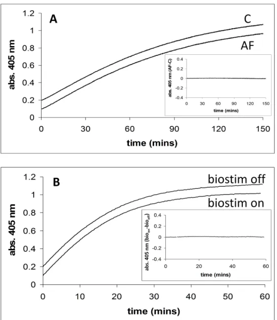
Fig 1Ashows the time courses for the release of 4-nitrophenol from 4-nitrophenlyphosphate catalysed by alkaline phosphatase, in the absence and presence of a magnetic field (2.5 mT/75 Hz) applied using the dual Helmholtz/spectrophotometer system. TheinsettoFig 1Ashows a plot of the time course with applied field (AF) minus that in the absence of the applied field (C).Fig 1Bshows similar time courses in the presence of the field applied by the Biostim appa-ratus. The difference between Biostim on (bioon) and Biostim off (biooff) is also shown, see inset. The activity of alkaline phosphatase (AF-C) was studied at five different substrate con-centrations (0.07, 0.1, 0.25, 1, and 5 mM) and at each concentration experiments were carried out three times (5x3 = 15 experiments in total).Table 2shows the mean difference in activity with standard deviation between the two field conditions. The activity of alkaline phosphatase (bioon-biooff) was also studied at one substrate concentration (0.07 mM), in triplicate. The
mean and standard deviation of the difference in activity, (absorbance/min) between the“field on”and“field off”conditions were calculated to be 3x10⁻⁵and 2x10⁻⁵ΔA405/min respectively.
This experiment using the Biostim has been designed to be as closely similar in methodology as possible to that carried out by Morelliet al. [17] in which they observed a marked decrease in enzyme activity.
InFig 2the activity of alkaline phosphatase in the presence and absence (“on”and“off”) of an applied magnetic field is shown. At the start of the assay all reactant mixtures were placed in the cuvette which was positioned in the monitoring beam. At t = ~5 mins an aliquot of
enzyme/microsomes was added (denoted 1x enzyme). It can be clearly seen that prior to this first addition of enzyme, no reaction was observed. A further aliquot of enzyme (2x total) was added at time = ~9 mins, and on this addition it can be seen that the rate of product formation doubles. TheinsettoFig 2shows an expanded region of the linear time course of the reaction in which the field (2.5 mT/75 Hz) was turned“on”and then“off”. A straight line of best fit is shown plotted through all the data points that start one minute before the field was applied to one minute after the field was switched“off”(R2=>0.99), and this shows there is no change in
slope during the time the field was applied. Further more detailed analysis was carried out on these experiments, where in a single experiment straight lines were fitted to the data points one minute before the field was turned“on”to one minute after the field remained“on”and, one minute after the field remained“on”to one minute after the field was turned“off”. This analy-sis revealed that there was no change in the slopes on the field being turned either“on”or“off”
(R2values =>0.99). These experiments were carried out five times in total, and all the lines of
best fit were analysed to ascertain if there was any deflection from linearity due to the field. No significant effect of the field was observed in any of the experiments.
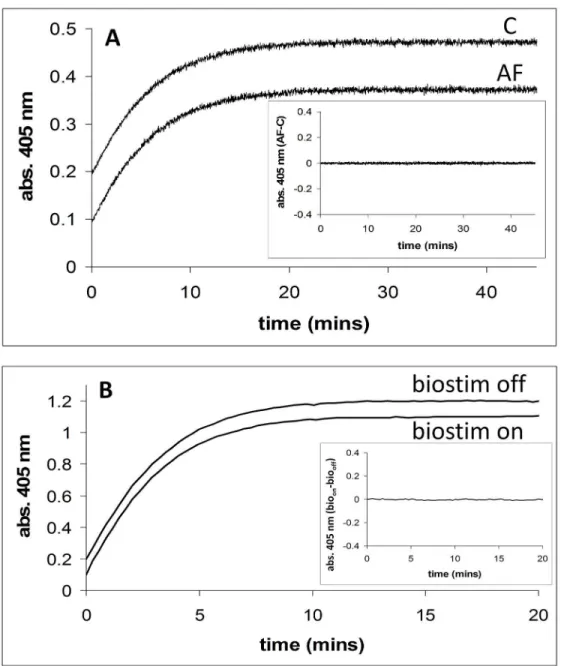
InFig 3A, the activity (thionitrobenzoate formation, abs. 405 nm) of the enzyme acetylcho-linesterase in the presence of an externally applied magnetic field (2.5 mT/75 Hz) is shown. The same reaction is also shown where no external field was applied. TheinsettoFig 3Ashows a plot of the applied field (AF) time course minus the no applied field (C) time course.Fig 3B
shows the activity of acetylcholinesterase in the presence and absence of an externally applied magnetic field using the Biostim apparatus. The Biostim on (bioon) minus Biostim off (biooff)
Table 1. Different MF conditions are denoted as numbers 1, 2 and 3.T = tested and NT = not tested.
MF conditions Biostim Dual Helmholtz/ spectrophotometer
12.5 mT, 75 Hz, square wave, 10% on/off duty cycle T Morelli and Silkstone NT
22.5 mT, 75 Hz, sinusoidal wave NT T Silkstone
32.5 mT staticfield NT T Silkstone
plot is also shown for this experiment (seeinset). Using the dual Helmholtz/spectrophotometer system, the activity of acetylcholinesterase (AF-C) was studied at five different substrate con-centrations (0.025, 0.05, 0.1, 0.2, and 0.5 mM) and at each concentration experiments were car-ried in triplicate (5x3 = 15 experiments in total).Table 3shows the results of these
experiments, with the mean and standard deviation for the difference in activity given. Using the Biostim apparatus and spectrometer system, the activity of acetylcholinesterase (bioon-bio
-off) was studied, in triplicate at each substrate concentration, at the same concentrations as for Fig 1. Alkaline phosphatase activity in the presence and absence of applied magnetic fields. A. Alkaline phosphatase activity in the presence (2.5 mT/75 Hz) and absence of an applied magnetic field using the dual Helmholtz/spectrophotometer system. Enzyme activity is assayed by measuring 4-nitrophenol formation (abs. 405 nm) with time.Inset. Shows a plot of the applied field (AF) time course minus the no applied field (C) time course. Conditions: [substrate] = 0.07 mM.B. Alkaline phosphatase activity in the presence (2.5 mT/75 Hz) and absence of an applied magnetic field using the Biostim system.Inset. Shows a plot of the applied field (biostim on,bioon) time course minus the no applied field (biostim off,biooff) time
course. Conditions: [substrate] = 0.07 mM.
the dual Helmholtz coil/spectrometer system (5x3 = 15 experiments in total). These results are reported inTable 4. This experiment using the Biostim has been designed to be as closely simi-lar in methodology as possible to that carried out by Morelliet al. [17] in which they observed a marked decrease in enzyme activity.
InFig 4, the activity of acetylcholinesterase is shown in the presence and absence of an applied magnetic field on the same sample, the field being switched“on”and the“off”during the time course (indicated by arrows). At the start of this assay, the reaction mixture was placed in the cuvette, positioned in the monitoring beam, and at t = ~7 mins an aliquot of enzyme/ ghost cell membranes was added. Prior to this first addition of enzyme no reaction was observed. TheinsettoFig 4shows an expanded region of the time course of the reaction where this was linear and the field (2.5 mT/75 Hz) was turned“on”and then“off”. A straight line of
Table 2. For alkaline phosphatase using the dual Helmholtz coil/spectrometer system, the mean (mean) value of the slopes fitted for AF-C for each substrate concentration is given. The mean value for each substrate concentration used was calculated from 3 separate experiments (n = 3), the slope for each experimentAF-Cwas fitted using a straight line of best fit with R2values>0.98 in all cases. The standard deviation (sd) is also given. Similar (less
exten-sive) data is also given for the alkaline phosphatase using the Biostim system (seeresults).
[substrate] mM 0.07 0.10 0.25 1.00 5.00
mean (x10-5) -0.33 1.00 0.33 6.00 -1.67
sd (x10-5) 8.33 7.00 6.11 3.46 8.14
doi:10.1371/journal.pone.0148369.t002
Fig 2. Alkaline phosphatase activity in the presence of an applied magnetic field (2.5 mT/75 Hz, field on/field off as indicated by arrows) using the dual Helmholtz/spectrophotometer system.At the start of the assay no enzyme is present in the reaction mixture. At time = ~5 mins an aliquot of enzyme/microsomes is added (denoted 1x enzyme), and at time = ~9 mins another aliquot of enzyme/microsomes is added (denoted 2x enzyme).Inset. A very good straight line fit of the data points for the region 1 min either side of where the field was applied in an on/off manner (field applied at 12 mins and switched off at 14 mins).
best fit is shown plotted through all the data points that start one minute before the field was applied to one minute after the field was switched“off”(R2=>0.99), and this shows there is
no change in slope during the time the field was applied. Further more detailed analysis was carried out on these experiments, where in a single experiment straight lines were fitted to the data points one minute before the field was turned“on”to one minute after the field remained
“on”and, one minute after the field remained“on”to one minute after the field was turned
Fig 3. Acetylcholinesterase activity in the presence and absence of applied magnetic fields. A. Acetylcholinesterase activity in the presence (2.5 mT/75 Hz) and absence of an applied magnetic field using the dual Helmholtz/spectrophotometer system. Enzyme activity is assayed by measuring thionitrobenzoate formation (abs. 405 nm) with time.Inset. Shows a plot of the applied field (AF) time course minus the no applied field (C) time course. Conditions: [substrate] = 0.025 mM.B. Acetylcholinesterase activity in the presence (2.5 mT/75 Hz) and absence of an applied magnetic field using the Biostim system.Inset. Shows a plot of the applied field (biostim on,bioon) time course minus the no applied field (biostim off,biooff) time
course. Conditions: [substrate] = 0.10 mM.
“off”. This analysis revealed that there was no change in the slopes on the field being turned either“on”or“off”(R2values =>0.99). These experiments were carried out five times in total,
and all the lines of best fit were analysed to ascertain if there was any deflection from linearity due to the field. No significant effect of the field was observed in any of the experiments.
InFig 5Aand 5B, Lineweaver-Burk plots have been calculated from the Michaelis-Menten kinetics displayed by acetylcholinesterase activity of ghost cell membranes.Fig 5Ashows these plots obtained using the dual Helmholtz coil/spectrometer system andFig 5Bthose for the Biostim spectrometer system. The data points shown for both plots are the mean values of three experiments at each substrate concentrations and the error bars are the standard devia-tion. InTable 5the Vmaxand KMvalues in the presence and absence of externally applied
mag-netic fields are given. The values are very similar to those reported by Morrelliet al. in the absence of field but in our experiments the Vmaxdid not exhibit an approximately 50%
diminu-tion in the presence of a field. The analysis/presentadiminu-tion of this experiment using the Biostim, is almost identical to that carried out by Morelliet al. [17].
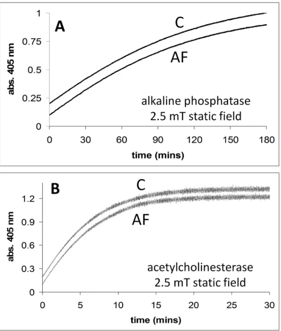
InFig 6Aand 6B, the activities of alkaline phosphatase and acetylcholinesterase are shown in the absence and presence of a 2.5 mT static field. InFig 6Athe time courses for the release of 4-nitrophenol from 4-nitrophenlyphosphate catalysed by alkaline phosphatase is shown, in the absence and presence of a magnetic field (2.5 mT static field) applied using the dual Helm-holtz/spectrophotometer system. InFig 6Bthe activity (thionitrobenzoate formation, abs. 405 nm) of the enzyme acetylcholinesterase in the absence and presence of an externally applied magnetic field (2.5 mT static field) is shown. We do not discuss these results, but show them for a more complete data set.
Discussion
In contrast to the reports by Morelliet al. [17], we have been unable to detect magnetic field effects on the steady state kinetics of either acetylcholinesterase or alkaline phosphatase. This discrepancy cannot be explained in terms of the assay conditions as we have paid particular attention to reproducing the conditions used by these authors, although Morelliet al. [17] do not report the temperature at which their experiments were performed. We also followed closely the protocols that were reported by Morelliet al. for the preparation of the membrane bound enzymes alkaline phosphatase and acetylcholinesterase. In addition, to be sure that there were no differences in the way in which the field was applied, we undertook experiments both with the Helmholtz coil system and with the Biostim apparatus used in the earlier studies.
Table 3. For acetylcholinesterase using the dual Helmholtz coil/spectrometer system, the mean (mean) value of the slopes fitted for AF-C for each substrate concentration is given. The mean value for each substrate concentration used was calculated from 3 separate experiments (n = 3), the slope for each experimentAF-Cwas fitted using a straight line of best fit with R2values>0.98 in all cases. The standard deviation (sd) is also given.
[substrate] mM 0.025 0.05 0.10 0.20 0.40
mean (x10-5) 0.00 -2.00 0.67 -1.00 3.00
sd (x10-5) 2.65 4.36 4.16 2.65 5.57
doi:10.1371/journal.pone.0148369.t003
Table 4. For acetylcholinesterase using the dual Helmholtz coil/spectrometer system, the mean (mean) value of the slopes fitted for bioon-bioofffor
each substrate concentration is given.The mean value for each substrate concentration used was calculated from 3 separate experiments (n = 3), the slope for each experimentbioon-biooffwas fitted using a straight line of best fit with R2values>0.98 in all cases. The standard deviation (sd) is also given.
[substrate] mM 0.025 0.05 0.10 0.20 0.40
mean (x10-5) -1.33 1.67 -13.0 4.00 2.67
sd (x10-5) 5.51 5.13 7.37 5.00 7.51
One clear difference in which our method of assay is distinct from the previous investigators is in regard to the method used to monitor activity, the earlier reports employing fixed-time assays while we have used real-time spectral assays. It is difficult to see how this methodological difference can account for the discrepancy between the studies.
The mechanism proposed by Morelliet al. to account for the magnetic field effect is that the activities of the enzymes depend upon the fluidity of the phospholipid membrane in which they are embedded. The field, acting through the diamagnetic susceptibility of the membrane, is suggested to orient the lipid and restrain its thermal motion and thus affect the enzyme’s activities. In support of this proposal Morelliet al. show that in the presence of Triton x-100, a detergent that dissolves the membrane, the effect of the field is abolished. Although this expla-nation is feasible in principle the fields that were used in the investigation make this unlikely on energetic grounds. Given a field of ~10 mT the low magnetic susceptibility for diamagnetic materials such as lipid molecules indicate an aligning energy of ~10−27J. This may be
com-pared with thermal energy at 300°K (~kT, where k is the Boltzmann constant) of ~10−21J, i.e. a
factor of 106fold greater. Thus one may presume that fields of ~1 mT or less will have unde-tectable influence on membrane fluidity (see for example [5]). Studies that have demonstrated the effects of magnetic fields on phospholipids membranes have typically used field of the order of 1 T or greater [23]. Water soluble proteins or protein domains tethered to membranes can be affected by magnetic fields through the field-induced orientation/phase change in the associated membrane e.g. bacterial purple membrane, but again at high field~10 T [24]. Effects at lower field strengths have, however, been reported in membranes containing ion channels.
Fig 4. Acetylcholinesterase activity in the presence of an applied magnetic field (2.5 mT/75 Hz, field on/field off as indicated by arrows) using the dual Helmholtz/spectrophotometer system.At the start of the assay no enzyme is present in the reaction mixture. At time = ~6.5 mins an aliquot of enzyme/ghost cell membranes is added.Inset. A very good straight line fit of the data points for the region 1 min either side of where the field was applied in an on/off manner (field applied at 8 mins and switched off at 10 mins).
Fig 5. Lineweaver-Burk plots calculated from the Michaelis-Menten kinetics of acetylcholinesterase from ghost cell membranes. A. Plots for acetylcholinesterase activity in the presence and absence of an applied magnetic field using the dual Helmholtz/spectrometer system (2.5 mT/75 Hz).B. Plots for
acetylcholinesterase activity using the Biostim apparatus. For each experiment using either apparatus, at any given substrate concentration, one sample was placed in the counter wound coil where no field could be applied and the other in the coil or Biostim where a field could be applied, and reactions were followed simultaneously. Data obtained in the absence of a field is denoted bycirclesand those in the presence of field bydiamonds. The enzymatic activity V was measured in U/mg, which is defined as nmoles of substrate converted per minute per mg of protein (nmoles/minute/mg). The protein concentration in mg/ml was calculated using the Biuret method [19]), and these calculated values were used to establish the total protein concentration present in the assays.
doi:10.1371/journal.pone.0148369.g005
Table 5. For acetylcholinestersase the Vmaxand KMvalues in the presence and absence of externally applied magnetic fields for both apparatus
systems are calculated and given.The control coil in the dual Helmholtz system had no applied field administered in all experiments.
Helmholtz coils Helmholtz coils Biostim
field coil (no appliedfield) control coil field coil (2.5 mT/75Hz) control coil ON OFF
KM(mM) 0.121 0.116 0.130 0.111 0.109 0.114
Vmax(U/mg) 2.917 2.791 2.985 2.632 2.618 2.667
For example miniature endplate potentials recorded in the isolated murine presynaptic neuro-muscular junction were inhibited at relatively low field and exhibited an absolute flux density threshold of 39.7 mT [25] The authors postulate that this effect is mediated through reorienta-tion of diamagnetic molecular domains within the membrane. This behaviour has some simi-larity to that reported by Raveraet al. [18], and one may surmise that if the synaptosomal membrane associated acetylcholinesterase could also be associated with ion channels within the membrane that reorientation of these may influence the esterase activity.
Alternatively, small changes in temperature, especially in the region of a phase transition, may alter membrane fluidity and induce measurable effects in enzyme activity [26]. The field
Fig 6. A. Alkaline phosphatase activity in the presence (2.5 mT static field) and absence of applied magnetic fields. Conditions: [substrate] = 0.07 mM.B. Acetylcholinesterase activity in the presence (2.5 mT static field) and absence of an applied magnetic field. Conditions: [substrate] = 0.025 mM. All reactions were carried out in the dual Helmholtz/spectrophotometer system. The applied field (AF) and no applied field (C) time courses are shown.
effects reported by Morelliet al. for alkaline phosphatase exhibits a remarkably sharp transition between 130μT and 150μT the activity halving over this 20μT range. It is difficult to explain
such behaviour of system in response to a magnetic field without recourse to a mechanism involving correlated radical pairs. It is more reminiscent of phase transition that is coupled to temperature, for example the phase transition found in phospholipids lipid membrane that leads to enhanced fluidity [26]. The temperature dependence of acetycholinesterase, at least, is complex with clear inflection in the Arhenius plot at around 30°C. indicating a sharp change in activation energy. Furthermore if enzymes are not assayed at saturating substrate concentra-tion an increase in temperature will not only increase the rate (and hence Vmax) it is also likely
to alter KMand lead to a change in observed velocity which may be positive or negative.
During the course of our investigations we became aware of the need for extremely close control of temperature and continuous monitoring of the temperature of sample and reference reaction mixtures. In our hands differences between sample and reference of<0.1°C (at 25°C)
lead to measurable differences in these membrane bound enzyme’s activities. We suggest, therefore, that the complex and confounding influences off temperature on these complex physical systems, comprising mixed phospholipid membrane in which is embedded a dynamic protein possessing catalytic activities involving multi-step mechanisms, may account for why Morelliet al. observed magnetic field effects while we did not.
Although we have attempted to reproduce closely the experimental protocols and experi-mental conditions of Morrelli and Ravera there may be some important differences of which we are unaware. For example we do not know the phase transition temperature of the mem-branes we used. One may suppose that any magnetic effect mediated by a reorientation of lipid or protein within the membrane must depend strongly on temperature. Such orientation would be energetically easier close to the membrane transition temperature and more difficult below this temperature. If for example the lipid compositions of the membranes were different between the preparations we used and those the group based in Italy used, a not unreasonable proposition given the influence of nutritional differences on membrane composition, then a temperature of 25°C may or close or relatively far from the membrane transition temperature. This being the case parallel experiments at this temperature may yield conflicting results.
Acknowledgments
We would like to thank the EMF Biological Trust for their support.
Author Contributions
Conceived and designed the experiments: GS MTW. Performed the experiments: GS. Analyzed the data: GS MTW. Contributed reagents/materials/analysis tools: GS MTW. Wrote the paper: GS MTW.
References
1. Kirschvink JL, Kobayashi-Kirschvink A, Diaz-Ricci JC, Kirschvink SJ. Magnetite in human tissues: a mechanism for the biological effects of weak ELF magnetic fields. Bioelectromagnetics Supplement 1: . 1992: 101–113.
2. Barnes F. Mechanisms for electric and magnetic field effects on biological cells. IEEE Trans. Magn. 2005; 11: 4219–4224.
3. Havas M. Biological Effects of Low Frequency Electromagnetic Fields. In: Clements-Croome D (Ed.). Electromagnetic Environments and Health in Buildings. Spon Press, London. 2004: 535.
4. Labes MM. doi:10.1038/211968a0Letts Nature. 1966; 11(968).
6. Ritz T, Adem S, Schulten K. A model for photoreceptor-based magnetoreception in birds. Biophysical Journal. 2000; 78: 707–718. PMID:10653784
7. Schulten K, Swenberg CE, Weller A. A biomagnetic sensory mechanism based on magnetic field mod-ulated coherent electron spin motion. Zeitschrift fur Physikalische Chemie. 1978; 111: 1–5.
8. Ball P. Physics of life: The dawn of quantum biology. Nature. 2011 June 15. doi:10.1038/474272a 2011; 474: 272–274. PMID:21677723
9. Wiltschko W, Wiltschko R. Magnetic Orientation in Birds. The Journal of Experimental Biology. 1996; 199: 29–38. PMID:9317275
10. Kobayashi A, Kirschvink J. Magnetoreception and Electromagnetic Field Effects: Sensory Perception of the Geomagnetic Field in Animals and Humans. Magnetoreception and EMF Effects. 1995; 21: 367– 394.
11. Rogers CT, Hore PJ. Chemical magnetoreception in birds: The radical pair mechanism. PNAS. 2008; 106(2): 353–360.
12. Solov’yov IA, Schulten K. Reaction kinetics and mechanism of magnetic field effects in cryptochrome. Journal of Physical Chemistry B. 2012; 116: 1089–1099.
13. Solov’yov IA, Domratcheva T, Shahi ARM, Schulten K. JACS. 2012; 134: 8046–1852.
14. Muller P, Ahmad M. Light-activated cryptochrome reacts with molecular oxygen to form a flavin-super-oxide radical pair consistent with magnetoreception. JBC. 2011; 286: 21033–21040.
15. Buchachenko AL, Kusnetsov DA. Magnetic field affects enzymatic ATP synthesis. J. Am. Chem. Soc. 2008; 130: 12868–12869. doi:10.1021/ja804819kPMID:18774801
16. Crotty D, Silkstone G, Poddar S, Ranson R, Prina-Mello A, Wilson MT, Coey JMD. Reexamination of magnetic isotope and field effects on adenosine triphosphate production by creatine kinase. PNAS. 2012; 109(5): 1437–1442. doi:10.1073/pnas.1117840108PMID:22198842
17. Morelli A, Ravera S, Panfoli I, Pepe IM. Effects of extremely low frequency electromagnetic fields on membrane-associated enzymes. Archives of Biochemistry and Biophysics. 2005; 441: 191–198. PMID:16126157
18. Ravera S., Bianco B., Cugnoli C., Panfoli I., Calzia D., Morelli A., Pepe I. M. Sinusoidal ELF magnetic fields affect acetylcholinesterase activity in cerebellum synaptosomal membranes. Bioelectromag-netics. 2010; 31(4): 270–276. doi:10.1002/bem.20563PMID:20041436
19. Caseli L, Oliveira RG, Masui DCM, Furriel RP, Leone FA, Maggio B, Zaniquelli MED. Influence of a sta-tionary magnetic field on acetylcholinesterase in murine bone marrow cells. Langmuir. 2005; 21: 4090– 4095. PMID:15835979
20. Stegemann S, Altman KI, Muhlensiepen H, Feinendegen LE. Influence of a stationary magnetic field on acetylcholinesterase in murine bone marrow cells. Radiat. Environ. Biophys. 1993; 1: 65–72.
21. Hamdy HA, Mahmoud BF, Shalaby TE, El-Sharkawy AM, Farag IMF. Effect of static and alternating magnetic fields on acetylcholinesterase and monoamine oxidase activities in the brain of mice. J. Invest. Biochem. 2013. doi:10.5455/jib.201307160759202013; 3(4): 133–137.
22. Fenk CJ, Kaufman N, Gerbig DG. Protein detection and measurement. J. Chem. Educ. 2007; 84: 1676–1678.
23. Orchard AF. Magnetochemistry (Publisher: OUP Oxford, 2003, ISBN 9780198792789).
24. Polk C, Postow E. Handbook of Biological effects of electromagnetic field, Second Edition, CRC Press 1996. Chapter 3“Biological effects of static magnetic fields”by Frankel RB and Liburdy RP: 159–162. 25. Rosen D. Studies on the Effect of Static Magnetic Fields on Biological Systems. BBA (Biomembranes).
1994; 1193: 62–66.