Panic-like behaviors in Carioca High-and Low-conditioned
Freezing rats
Bruno de Oliveira Galvão
1, Vitor de Castro Gomes
1, Silvia Maisonnette
1, and J.
Landeira-Fernandez
1,21 - Pontifícia Universidade Católica do Rio de Janeiro, Rio de Janeiro, RJ, Brazil
2 - Universidade Estácio de Sá, Rio de Janeiro, RJ, Brazil
Abstract
Panic disorder involves both recurrent unexpected panic attacks and persistent concern about having additional attacks. Electrical stimulation of the dorsal periaqueductal gray (dPAG) is an animal model of both panic attack and panic disorder, whereas contextual fear conditioning represents a model of anticipatory anxiety. Previous research indicated that anxiety has an inhibitory effect on panic attack-like behavior. However, still unclear is the role that anticipatory anxiety plays in panic disorder-like behaviors. This issue was investigated with two lines of animals selectively bred for high (Carioca High-Freezing) and low (Carioca Low-Freezing) freezing in response to contextual cues associated with footshock. The results suggest that although anticipatory anxiety might exert an inhibitory effect on the expression of panic attack, it might also facilitate the pathogenesis of panic disorder. Keywords: breeding lines, dorsal periaqueductal gray, contextual fear conditioning, freezing behavior, escape behavior, panic attack, panic disorder.
Received 28 January 2011; received in revised form 1 June 2011; accepted 3 June 2011. Available on line 10 October 2011
Bruno de Oliveira Galvão, Vitor de Castro Gomes, and Silvia Maisonnette, Department of Psychology, Pontifícia Universidade Católica do Rio de Janeiro, Rio de Janeiro, RJ, Brazil. J. Landeira-Fernandez, Pontifícia Universidade Católica do Rio de Janeiro, Rio de Janeiro, RJ, Brazil AND curso de Psicologia, Universidade Estácio de Sá, Rio de Janeiro, RJ, Brazil Correspondence concerning this article should be addressed to J. Landeira-Fernandez, Departamento de Psicologia, Pontifícia Universidade Católica do Rio de Janeiro, Ed. Cardeal Leme, Sala 201L, Rua Marquês de São Vicente 225, Gávea, RJ-Brazil, 22451-900. Email: [email protected]
Introduction
Panic disorder is a complex anxiety disorder that involves both recurrent, unexpected panic attacks and persistent concern about having additional attacks (American Psychiatric Association, [APA], 1994). A panic attack is a sudden surge of overwhelming fear or terror that occurs without warning or any obvious reason. Symptoms are accompanied by major neurovegetative
changes such as palpitation, hypertension, dificulty
with deep breathing, sweating, an urge to void the
bladder, increased peristalsis, the desire to lee, and a
feeling of imminent death or losing control. An attack is an acute condition with an abrupt onset that reaches a peak within 10 min.
Although the occurrence of a panic attack is a hallmark of panic disorder, the chronic conditioning
of this anxiety disorder is defined by the constant and persistent fear of experiencing further attacks or worry about the possible consequences of a panic attack. Patients suffering from panic disorder may live in a nearly constant state of apprehension. The frightening prospect of experiencing another panic attack might be so extreme that the patient can develop agoraphobia (i.e., a fear of being in places or situations in which escape might be difficult or help might not be available in the event of a panic attack or panic-like symptoms).
Brandão, & Landeira-Fernandez, 2001). Freezing and escape responses triggered by dPAG stimulation are mediated by the same neurochemical processes and represent a model of panic attack, whereas dPAG post-stimulation freezing at the aversive escape threshold is mediated by a distinct neuronal substrate and appears to be a model of panic disorder (for review, see Brandão, Zanoveli, Ruiz-Martinez, Oliveira, & Landeira-Fernandez, 2008).
Considerable evidence also indicates that the freezing response to contextual cues previously associated with electrical footshock is one of the most reliable animal models of anticipatory anxiety (Brandão et al., 2008). In a typical experiment, a rat is exposed to a novel chamber, and a brief unsignaled footshock is presented several minutes later. Some time later (i.e., the next day), the animal freezes when returned to the same chamber in the absence of footshock (Landeira-Fernandez, 1996). This defensive freezing response differs from the one triggered by dPAG stimulation because no piloerection or exophthalmus is observed. Moreover, freezing in response to contextual cues previously paired with footshock involves an initial active motor component with the purpose of withdrawing to a safe and hidden location next to an object (thigmotaxis), such as a corner or a wall.
Previous studies indicated that rats exposed to contextual cues previously associated with electrical footshock exhibited a robust defensive freezing response and exhibited a higher dPAG electrical stimulation threshold to induce escape responses compared with control animals that were not exposed to contextual fear conditioning (Magierek, Ramos, da Silveira-Filho, Nogueira, & Landeira-Fernandez, 2003). This pattern of results has been replicated recently (Galvão, Larrubia, Hommes, Cardenas, & Cruz, 2010) and indicates that anticipatory anxiety might play an inhibitory role on the occurrence of panic attack-like behavior. However, still unclear
is the extent to which anxiety might inluence the
development of panic disorder.
Two new lines of Wistar rats, termed Carioca High- and Low-Freezing (CHF, CLF), were selectively bred for high and low levels of freezing in response to contextual cues previously associated with footshock (Gomes & Landeira-Fernandez, 2008 ). After three generations of breeding, CHF rats were considered to naturally have a greater propensity for exhibiting higher freezing responses compared with CLF animals. A recent study indicated isomorphism between CHF rats and anticipatory anxiety (Dias, Bevilaqua, Silveira, Landeira-Fernandez, & Gardino, 2009). Therefore, these lines of animals may be an important tool for investigating the relationship between anxiety with panic attack and panic disorder. Accordingly, the purpose of
the present study was to investigate whether CHF and CLF animals exhibit different patterns of panic attack- and panic disorder-like behaviors induced by electrical stimulation of the dPAG.
Method
Subjects
Experimental animals selectively bred for high (CHF) and low (CLF) contextual fear conditioning were obtained according to a procedure described in a previous study (Gomes & Landeira-Fernandez, 2008).
Briely, albino Wistar rats were placed in an observation
chamber. Three minutes later, three unsignaled electrical footshocks (0.7 mA, 1 s duration) were delivered 20 s apart. Three minutes after the last shock, the animal was returned to its home cage. Approximately 24 h after the training session, the animal was returned to the same observation chamber for an 8-min test session in the absence of any stimulation. The present study employed male CHF and CLF rats from the ninth generation with the highest (CHF) and lowest (CLF) conditioned freezing scores. All animals were employed as breeders in our ongoing selective breeding program before the beginning of the experiment.
Animals were housed in groups of ive to seven,
according to their respective lines, in polycarbonate cages measuring 18 × 31 × 38 cm3, with food and water available
ad libitum. The room temperature was controlled (24 ± 1°C), with a 12 h/12 h light/dark cycle (07:00–19:00 h). The experiment was conducted during the light phase of the cycle. Animals were 6 months old at the beginning of the experiment. The experimental procedures were performed in accordance with the guidelines for experimental animal research established by the Brazilian Society of Neuroscience and Behavior, which are based on the United States National Institutes of Health Guide for the Care and Use of Laboratory Animals (revised 1996).
Surgery
All animals were implanted with a stainless steel unilateral guide-cannula aimed at the dPAG. Under tribromoethanol anesthesia (250 mg/kg, i.p.), each
animal was ixed in a Kopf stereotaxic frame and injected
Material
The experiment was conducted in the same observation chamber (25 × 20 × 20 cm3) where CHF
and CLF animals were phenotyped for contextual fear conditioning. The observation chamber was placed inside a sound-attenuating box. A red light bulb (25 W) was placed inside the box, and a video camera mounted on the back of the observation chamber was used to observe the animal’s behavior on a monitor placed outside the experimental room. A ventilation fan attached to the box supplied 78 dB background noise.
Procedure
Seven days after surgery, each animal was placed inside the observation chamber. Five minutes later, freezing and escape aversive thresholds were determined using electrical stimuli (alternating current, 60 Hz, 20 s) presented through a removable electrode connected to the guide-cannula aimed at the dPAG. Brain stimulation was presented at 20-s intervals, with the current intensity beginning at 5 µA and increasing by 5 µA steps. The freezing threshold was
operationally deined as the lowest current intensity that
produced an absence of movement, with the exception of respiration, accompanied by at least two of the following autonomic reactions: urination, defecation, piloerection, or exophthalmia. The current intensity producing running or jumping was considered to be the escape threshold. The dPAG electrical stimulation was stopped when the threshold for eliciting an escape was reached. To investigate freezing behavior that occurred after cessation of dPAG stimulation applied at the escape threshold, the animals remained in the observation chamber for an additional 8 min without any stimulation. During this period, freezing was scored using a time-sample procedure. Every 2 s, the animal’s freezing behavior was scored by a well-trained observer.
After the experiment, animals were sacriiced under
deep anesthesia with chloral hydrate. The brain was perfused through the heart with saline solution (0.9%) followed by 10% formalin solution, removed, and
postixed in 10% formalin. Frozen 55-µm sections were
cut using a microtome to localize the positions of the electrode tips according to the atlas of Paxinos and Watson (1986). Only data from rats with electrode tips located inside the dPAG were included in the statistical analysis.
Results
Data are expressed as mean ± SEM. Two-way repeated-measures analysis of variance (ANOVA) was used to evaluate differences in aversive threshold between CHF and CLF animals. The breeding line (CHF and CLF) was considered the between-subjects factor, and aversive thresholds (freezing and escape) were considered
the within-subjects factors. Two-way repeated-measures ANOVA was also used for dPAG-evoked post-stimulation freezing analysis, with breeding line (CHF and CLF) the between-subjects factor and time (min) the within-subjects
factor. Signiicant effects in the ANOVA were followed by the Newman-Keuls post hoc test. Values of p < 0.05 were
considered statistically signiicant.
Histological examination of the brain slices indicated that all electrode tips were located inside the
dPAG. The inal group samples were the following:
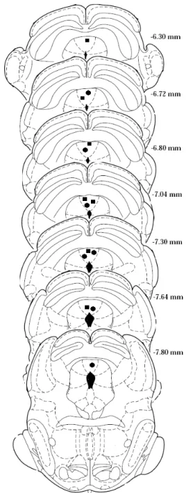
CHF, n = 7; CLF, n = 7. The representative sites of the dPAG stimulation are shown in Figure 1.
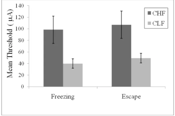
As reported previously (Oliveira, Nobre, Brandão, & Landeira-Fernandez, 2004), freezing and escape responses induced by dPAG electrical stimulation occurred in a stepwise fashion. As the intensity of the current applied to the dPAG increased, the animals suddenly stopped, became immobile, and often urinated and defecated. With higher intensities, this freezing behavior was followed by vigorous running and jumping reactions. The escape response stopped as soon as the dPAG electrical stimulation was switched off. Figure 2 shows the mean (± SEM) of the electrical current threshold required to trigger freezing and escape behaviors in CHF and CLF animals. Two-way ANOVA revealed main effects of breeding line (F[1,12] = 5.42, p < 0.05) and aversive threshold (F[1,12] = 234.37, p < 0.0001). No breeding line ´ aversive threshold interaction was found (F[1,12] = 0.37, p > 0.5). Post hoc analysis indicated that CHF animals presented higher aversive freezing and escape thresholds than CFL animals (both p < 0.05).
Figure 3 shows the mean (± SEM) percentage of time that CHF and CLF animals spent freezing following dPAG stimulation at the escape threshold. Two-way
repeated-measures ANOVA showed a signiicant effect
of breeding line (F[1,12] = 12.10, p < 0.005). No main effect of time (F[1,12] = 2.18, p > 0.1) or interaction between lines of animals during the 8-min dPAG post-stimulation period (F [1,12] = 0.34, p > 0.5) was found. Post hoc analysis indicated that CHF animals expressed more dPAG-evoked post-stimulation freezing behavior compared with CLF animals during the 8-min test period (all p < 0.05). Because CHF animals required a higher threshold current than CLF animals to elicit an escape response it is possible that differences between these two groups in freezing behavior immediately after dPAG electrical stimulation at the escape threshold might be due to differences in the threshold current between CHF and CLF. In order to test this possibility, an analysis of
covariance (ANCOVA) using the escape dPAG electrical stimulation threshold parameter as covariant factor was
performed. Results from this analysis indicated conirmed signiicant main effect of breeding line (F [1,12] = 9.34, p = 0.01). A main effect of time (F [1,12] = 8.47, p = 0.01) and no interaction between lines of animals during the 8-min dPAG post-stimulation period (F [1,12] = 0.55, p > 0.4) was also detected.
Discussion
The current understanding of anxiety disorders departed from an earlier concept of a unitary process and evolved into a more recent view that suggests a group of different but interrelated nosological categories. A major shift in this recent view of anxiety
disorder occurred with Klein’s pioneering work (1962;
1964), which showed that imipramine had a selective effect in the treatment of panic attacks. Since then, a qualitative distinction between anticipatory anxiety and spontaneous panic attack has been repeatedly observed in clinical settings (Battaglia & Ogliari, 2005). However, the relationship between anticipatory anxiety with panic attack and the development of panic disorder remains a subject of intense debate.
Experimental research employing rats selectively bred for high or low levels of emotionality represents an important and powerful tool for investigating the relationships between different aspects of anxiety disorders. Rats in this study were selectively bred to exhibit high (CHF) or low (CFL) levels of freezing in response to contextual cues previously associated with footshock (Gomes & Landeira-Fernandez, 2008). Although freezing appears to be the main conditioned response observed during fear conditioning, active escape responses have also been suggested to be present during this aversive learning situation (Tarpley, Shlifer, Halladay, & Blair, 2010). This is a particularly important issue because it might challenge the view that the CHF
Figure 2. Mean (± SEM) freezing and escape thresholds determined with the procedure of the dPAG electrical stimulation in the two breeding lines selected according to their emotional reactivity.
phenotype is associated with more anxiety-like behavior than the CLF phenotype. Thus, CLF rats may freeze less not because they are less “afraid,” but because they are more “frightened” and thus more prone to exhibit active escape responses than defensive freezing behavior.
Much evidence appears to exclude this possibility. For example, conditioned freezing is a direct function of footstock intensity (Morris & Bouton, 2008) and has been pharmacologically validated as an animal model of anticipatory anxiety. Accordingly, benzodiazepine receptor agonists such as diazepam and midazolam reduced the amount of conditioned freezing, whereas
the benzodiazepine inverse agonist
dimethoxy-β-carboline produced freezing behavior similar to that elicited by context fear conditioning (Fanselow, 1991). Consistent with this, anxiolytic-like substances such as 5-HT1A receptor agonists, selective serotonin reuptake inhibitors, and monoamine oxidase inhibitors with
veriied clinical eficacy in the treatment of anxiety
symptoms, attenuated conditioned behavior in rats, indicating considerable construct and face validity of this paradigm to human anxiety (Conti, Maciver, Ferkany, & Abreu, 1990; Maki et al., 2000). Moreover, mice selectively bred for high and low levels of freezing in response to contextual cues previously associated with footshock also presented, respectively, higher and lower levels of anticipatory anxiety in the fear-potentiated startle test (Ponder et al., 2007). Finally, previous results from our laboratory with different models of anxiety, such as the elevated plus maze and social interaction
test, indicated that CHF animals exhibited signiicantly
more anxiety-like behavior than control rats (Dias et al., 2008) Therefore, the CHF line appears to represent a robust animal model of anticipatory anxiety.
The results of the present study indicated that CHF animals had a higher dPAG electrical stimulation aversive threshold for producing freezing and escape reactions than CLF animals. This result is consistent with several other studies, which indicated that contextual fear conditioning can inhibit defensive responses to aversive
proximal or painful stimuli such as the tail-lick response
to radiant heat, complex and elaborated nociceptive responses elicited in the formalin test, vigorous running and jumping triggered by footshock (Fanselow, 1982)
and shock-induced defensive ight reactions (Bolles and
Collier, 1976). Moreover, contextual fear conditioning can also inhibit vigorous escape responses induced by N-methyl-D-aspartate (Galvão et al., 2010) or electrical stimulation (Magierek et al.,2003) of the dPAG. Much evidence indicates that the amygdaloid complex and its projections to the ventral portion of the PAG are critically involved in the regulation of contextual fear conditioning. Malfunctioning of this system might be associated with pathological forms of anticipatory anxiety (e.g., generalized anxiety disorder). Descending inhibitory projections from the amygdaloid complex might reach
the dPAG, which in turn might inhibit defensive reactions triggered by this structure. Therefore, activation of the neural circuitry involved in anxiety might indeed inhibit the occurrence of panic attack-like behavior associated with neurons located within the dPAG.
The present results also indicated that CHF animals displayed more freezing behavior immediately after dPAG electrical stimulation at the escape threshold compared with CLF animals. This difference might be attributable to the fact that CHF animals required a higher threshold current than CLF animals to elicit an escape response. An ANCOVA contested this hypothesis indicating that although CHF animals were more resistant to the expression of escape behavior in response to dPAG stimulation, they were more prone to freezing after the occurrence of the dPAG aversive stimulation compared with CLF animals.
The dPAG post-stimulation freezing is not fear-conditioning in response to contextual cues associated with the dPAG electrical stimulation. Previous studies employed a context shift procedure and indicated that freezing after dPAG stimulation persisted when animals were placed in a different context immediately after the dPAG stimulation (Vianna et al., 2001). Moreover, several studies indicated that freezing observed after dPAG stimulation has a different neural mechanism from freezing and escape responses elicited by dPAG electrical stimulation. For example, electrolytic lesions or muscimol-induced inactivation of the amygdaloid complex reduced dPAG post-stimulation freezing but did not affect freezing or escape responses induced by dPAG electrical stimulation (Oliveira et al. 2004; Ruiz-Martinez, de Oliveira, & Brandão, 2006). Indeed, dPAG post-stimulation freezing appears to be mediated by ascending projections, possibly relayed through the thalamus to forebrain structures related to the sensory
processing of aversive stimuli. These indings suggest
the possibility that dPAG post-stimulation freezing might represent an animal model of panic disorder. Therefore, the fact that CHF animals expressed more dPAG post-stimulation freezing than CLF animals might indicate that anticipatory anxiety could enhance the development of panic disorder triggered by panic attacks.
reactions that are present in panic disorder might also recruit some forebrain structures related to anticipatory anxiety. Further studies are needed to elucidate whether activation of neural circuitries associated with the amygdaloid complex might play an inhibitory role in the occurrence of panic attack and excitatory modulation of structures associated with panic disorders.
Acknowledgment
This research was supported by Brazilian National Research Council CNPq, 522720/95-10 awarded to J. Landeira-Fernandez.
References
American Psychiatric Association. (1994) Diagnostic and statistical manual of mental disorders, (4th ed). Washington: American Psychiatric Association.
Battaglia, M., & Ogliari, A. (2005). Anxiety and panic: from human studies to animal research and back. Neuroscience Biobehavioral Reviews, 29, 169-79.
Bolles, RC., & Collier, A. C. (1976). The effect of predictive cues of freezing in rats. Animal Learning Behavior, 4, 6-8.
Brandão, M.L., de Aguiar, J.C., & Graeff, F.G. (1982) GABA mediation of the anti-aversive action of minor tranquilizers.
Pharmacology Biochemistry Behavior, 16, 397-402.
Brandão, M.L., Zanoveli, J.M., Ruiz-Martinez, R.C., Oliveira L.C., & Landeira-Fernandez J. (2008). Different patterns of freezing behavior organized in the periaqueductal gray of rats: association with different types of anxiety. Behavioral Brain Reviews, 188, 1-13. Conti, L.H., Maciver, C.R., Ferkany, J.W., & Abreu, M.E. (1990).
Footshock-induced freezing behavior in rats as a model for assessing anxiolytics. Psychopharmacology (Berl), 102, 492-497. de Carvalho, M.R., Dias, G.P., Cosci, F., de-Melo-Neto, V.L., Bevilaqua,
M.C., Gardino, P.F, & Nardi, A.E. (2010) Current indings of fMRI
in panic disorder: contributions for the fear neurocircuitry and CBT effects. Expert Reviews of Neurotherapeutics, 10, 291-303. Dias, G.P., Bevilaqua, M.C., Silveira, A.C., Landeira-Fernandez, J.,
& Gardino P.F. (2009). Behavioral proile and dorsal hippocampal
cells in carioca high-conditioned freezing rats. Behavioral Brain Research, 205, 342-8.
Fanselow, M.S. (1982). The post-shock activity burst. Animal Learning and Behavior, 10, 448-54.
Fanselow, M.S. (1991). Analgesia as a response to aversive Pavlovian
conditional stimuli: Cognitive and emotional mediators. In Denny M.R. Fear, avoidance, and phobias: A fundamental analysis, (pp. 61-86) Hillsdale, NJ: Lawrence Erlbaum.
Galvão, B., Larrubia, B., Hommes, W.J., Cardenas, F.P., Cruz, A.P.M., & Landeira-Fernandez, J. (2010). Effects of contextual fear conditioning and pentylenetetrazol on panic-like reactions induced by dorsal periaqueductal gray stimulation with N-methyl-D-aspartate. Psychology Neuroscience, 3, 67-72.
Gomes, V.C., & Landeira-Fernandez, J. (2008). Amygdaloid lesions produced similar contextual fear conditioning disruption in the Carioca high- and low-conditioned freezing rats. Brain Research,
1233, 137-45.
Klein, D.F. (1964). Delineation of two drug-responsive anxiety
syndromes. Psychopharmacologia, 5, 397-408.
Klein, D.F., & Fink, M. (1962). Psychiatric reaction patterns to
imipramine. American Journal of Psychiatry, 119, 432-8. Landeira-Fernandez, J. (1996). Context and Pavlovian conditioning.
Brazilian Journal ofMedical Biology Research, 29, 149-73. Magierek, V., Ramos, P.L., da Silveira-Filho, N.G., Nogueira, R.L., &
Landeira-Fernandez, J. (2003). Context fear conditioning inhibits panic-like behavior elicited by electrical stimulation of dorsal periaqueductal gray. Neuroreport, 14, 1641-4.
Maki, Y., Inoue, T., Izumi, T., Muraki, I., Ito, K., Kitaichi, Y., Li X., & Koyama T. (2000). Monoamine oxidase inhibitors reduce
conditioned fear stress-induced freezing behavior in rats. European Journal of Pharmacology,406, 411-8.
Morris, R.W., & Bouton, M.E. (2006). Effect of unconditioned stimulus magnitude on the emergence of conditioned responding. Journal of Experimental Psychology: Animal Behavior Processes, 32, 371-85. Oliveira, L.C., Nobre, M.J., Brandão, M.L., & Landeira-Fernandez,
J. (2004) Role of amygdala in conditioned and unconditioned fear generated in the periaqueductal gray. Neuroreport, 15, 2281-5. Paxinos G., & Watson C. (1986). The rat brain in stereotaxic
coordinates, (2nd ed). New York: Academic Press.
Ponder ,C.A., Kliethermes, C.L., Drew, M.R., Muller, J., Das, K.,
Risbrough, V.B., Crabbe, J.C., Gilliam, T.C., & Palmer, A.A. (2007). Selection for contextual fear conditioning affects anxiety-like behaviors and gene expression. Genes Brain and Behavior, 6, 736-49. Ruiz-Martinez, R.C., de Oliveira, A.R., & Brandão, M.L. (2006).
Conditioned and unconditioned fear organized in the periaqueductal gray are differentially sensitive to injections of muscimol into amygdaloid nuclei. Neurobiology Learning and Memory, 85, 58-65. Tarpley, J.W., Shlifer, I.G., Halladay, L.R., & Blair, HT. (2010).
Conditioned turning behavior: a Pavlovian fear response expressed during the post-encounter period following aversive stimulation.
Neuroscience, 169,1689-704.