Article
J. Braz. Chem. Soc., Vol. 26, No. 12, 2536-2544, 2015. Printed in Brazil - ©2015 Sociedade Brasileira de Química 0103 - 5053 $6.00+0.00
A
*e-mail: [email protected]
Luminescent Terbium Doped Aluminate Particles: Properties and Surface
Modification with Asparagine
José M. A. Caiut,*,a,b,c Younes Messaddeq,a Hervé Dexpert,b Marc Verelst,b
Sidney J. L. Ribeiroa and Jeannette Dexpert-Ghysb
aInstituto de Química, Universidade Estadual Paulista - UNESP, CP 355, 14801-970 Araraquara-SP, Brazil
bCentre for Materials Elaboration and Structural Studies/CNRS, BP 4347, 31055 Toulouse CEDEX 4, France
cDepartamento de Química, Faculdade de Filosofia, Ciências e Letras de Ribeirão Preto, Universidade de São Paulo, 14040-901 Ribeirão Preto-SP, Brazil
New luminescent organic-inorganic hybrid particles based on Tb-doped aluminates and asparagine (Asn) surface modifiers were investigated. The Tb3+ doped inorganic core was obtained
by spray pyrolysis, at 200 °C γ-AlOOH (BOE:Tbx%) or at 700 °C γ-Al2O3 (γTA:Tbx%). The
reaction of Asn with boehmite in water disaggregated the sub-micronic boehmite particles to give stable dispersion of surface modified nanoparticles Asn:BOE:Tbx% (x = 1 or 5). Concerning the Asn:γTA:Tbx% system, an Asn film wrapping alumina particles was observed. Photoluminescence spectra exhibited the bands assigned to Tb3+5D
4 → 7FJ = 6-3 transitions. A broad absorption band
(240 nm) was assigned to the host (aluminate) to ion (Tb3+) energy transfer. Efficient energy
transfer was observed when active ions are incorporated in the defect-spinel structure of γTA, whereas it was relatively weak for BOE:Tb where Tb3+ are bonded to the hydroxyls groups at
nanocrystals surface. It is noticeable that Asn strengthens the linkage of Tb3+ with the aluminate
matrix, enhancing the host to dopant energy transfer.
Keywords: hybrid, spray pyrolysis, alumina, rare earth, terbium luminescence
Introduction
“Alumina” is the general name encompassing a large number of products with different chemical formulas and/or structures. The thermodynamically stable phase at standard
temperature and pressure conditions is α-Al2O3, having
the hexagonal crystal structure of the mineral corundum.
Lanthanide ions doped alumina (α) provide extremely
versatile photoluminescent ceramics. The green emitter
Tb3+ was for instance successfully incorporated into a dense
alumina matrix, achieving a transparent light-emitting
ceramic.1 Transitions alumina’s (partially dehydrated
aluminum hydroxides) possess high surface areas which renders them appropriate for use as adsorbents, catalysts
and catalyst carriers. The “γ-Al2O3” transition alumina
is particularly important in catalysis and details of its structure and exact composition have been the subject of a
number of controversial works.2-7 Boehmite, γ-AlOOH, is
actually the most important precursor for the synthesis of transition aluminas following the transformation sequence
boehmite →γ → δ → θ → α. Crystallization degree,
morphology and surface properties are observed to be highly dependent on the structure.
Nanocrystalline boehmite can be obtained through a
sol-gel route as firstly described by Yoldas.8,9 Nanocrystalline
boehmite may be employed as sorbent, for instance by removing potentially toxic metal from polluted waste
water.10 The surface of boehmite particles is highly reactive
towards carboxylic acids in aqueous media. The adsorption of low molecular weight carboxylic acids, ubiquitous in natural environments, on positively charged mineral surfaces, e.g., aluminium oxihydroxides, has also been the
subject of many investigations.11
Material chemists have taken benefit of this high affinity to synthesize alumoxanes from the reaction of boehmite
carboxylate-substituted alumoxanes may be employed as precursors for a wide range of ceramics, including lanthanide-aluminium
mixed-metal oxides.13
Alumina-based nanoparticles or nano-structured objects with possible applications in the field of nano-bio-technology or nano-medicine have been described. Alumina membranes with highly ordered nanopores are proposed
for instance in smart implantable drug delivery systems.14
Although far less described in literature than mesoporous silica, powdered mesoporous (hydrated) alumina is a potentially available substrate for controlled drug release,
after suitable chemical functionalization of its surface.15
On the other hand, luminescent micro- or nano- particles are becoming of great importance in the field of bio-labeling. Several examples of crystalline lanthanide oxides as luminescent biolabels are also found in
literature. Eu3+ and Tb3+ containing nanomaterials have
also been reported as in nanocrystalline hydroxyapatite
or in lanthanum phosphate for example.16-18 Lanthanide
emitters (Eu3+, Tb3+) are particularly interesting for the
purpose of luminescent bio-labeling since the decay times
for the main intra 4fn transitions are much longer than the
decay times of the background fluorescence of biological samples. By employing well-suited experimental set-ups the photoluminescence signal from the optical probe may be discriminated from the background emission from all
non-probed species.19
Concerning preparation methods spray pyrolysis (SP) is an aerosol process commonly used to form a variety of materials in powder form, including transition aluminas or
α alumina.20-22 A protocol based on the rapid drying of an
aluminium alcoholate suspension was proposed in order to
synthesize nanocrystalline γ-AlOOH or γ-Al2O3 in a single
step, by the SP process.23 Moreover, by the addition of an
optically active lanthanide ion Eu3+ or Tb3+, luminescent
aluminates can be obtained in just one step.24-26
Due to its high affinity for the boehmite surface, the amino-acid asparagines (Asn) strengthens the attachment of the luminescent europium ions at the boehmite
nanoparticles after their suspension in water.24
On the basis of these preceding observations, we present here a systematic investigation of Asn:aluminates:Tb nanostructured particles. Terbium-doped aluminates, having the boehmite (BOE) or the gamma alumina structure were obtained by spray pyrolysis, and will
be noted BOE:Tbx% and γTA:Tbx%, respectively.
The luminescence properties of these powders will be discussed regarding the possible sites of terbium ions in the aluminates and the effects of matrix-dopant electronic interactions. The possibility of modifying the surface of these aluminates by the amino acid Asn was then explored.
Asparagine (Asn = H2N-CO-CH(NH3+)-COO− at pH = 7.0)
was chosen to render the oxide particles bio-compatibles, because the carboxylic end will presumably attach the aluminate surface, with the formation of alumoxanes, whereas the amide group will be let free for further reaction. Hereafter, composites with Asn will be compared with the corresponding un-modified aluminate, to highlight the organic-inorganic interactions. Finally, the possibility to achieve luminescent nano-labels in the investigated systems will be discussed.
Experimental
Hydrated alumina samples were prepared by spray pyrolysis. The precursor (boehmite sol) was obtained by the methodology established by Yoldas modified to obtain the
rare earth doped alumina.8,9 TbCl
3 (0.37 g, 0.001 mol) was
dissolved in water (300 mL) at 80 °C. Aluminum tri-sec-butoxide (25.93 g, 0.1 mol) was added to terbium aqueous solution under stirring at 80 °C. After 2 h stirring, nitric
acid was added as a peptizing agent, up to 0.07 HNO3/Al3+
(molar ratio). The sol (with concentration adjusted to
0.2 mol L-1 Al3+ by water dilution) was spray dried in an
experimental setup already described.23 In this setup, the
spray was generated by a piezoelectric pellet oscillating at
2.4 MHz. The aerosol, made of fine droplets (ɸ ca. 5 µm)
from the precursor solution in air, was then driven into two heating zones, the first one around 100 °C and the second one was the decomposition-densification zone where the temperature may be adjusted at a chosen temperature
(TSYN). The powder was separated from the gas phase by
an electrostatic collector. The drying and decomposition
were accomplished within about 10 s. TSYN was fixed at
200 °C for the synthesis of boehmite (BOE), and at 700 °C
for that of the “γ” transition alumina (γTA). Three dopant
percentages were investigated: 1, 5 and 10% expressed in
mol of Tb3+/mol of Al3+.
Amino acids
Aluminoxanes nanocomposites were obtained by dispersing the hydrated aluminas particles in Asn water solutions. Samples activated with Tb1% (BOE:Tb1% or
γTA:Tb1%) were dispersed in water (300 mg in 25 mL).
used for dynamic laser light scattering measurements
and also to complete the analysis of Tb3+ luminescence.
For the high Asn composition (0.5Asn:1BOE:Tb1%), the sample was observed to be composed off a mixture of nanoparticles and crystallized Asn; concerning the lower content (0.1 Asn), changes on alumina particles were not measureable. Therefore only results obtained for compositions 0.3Asn:1Al will be presented. Composite samples will be named Asn:BOE:Tb (%, mol Tb/Al) and
Asn:γTA:Tb (%, mol Tb/Al).
Characterization
The powders were characterized by X-ray diffraction (XRD), with a Seifert XRD3000 diffractometer. The coherent length was estimated with the Scherrer equation
(D = 0.9l (FWHM) cosθ) with the full width at half
maximum measured in (2θ) and expressed in radians.
The size distributions of powders dispersed in water were determined by dynamic laser light scattering (DLS) using a Malvern Instruments ZETASIZER 3000, model DTS5300. Samples were observed by transmission electron microscopy (TEM) with a Philips-CM120 and by scanning electron microscopy (SEM) with a SEM-JEOL 6700F. Fourier transform infrared (FTIR) spectra were recorded in KBr pellets using a model 2000 Perkin-Elmer spectrometer. Room temperature luminescence excitation and emission spectra were measured with a Hitachi-Fl100 spectrofluorimeter (at 2.5 nm spectral resolution) for solids
samples. The 5D
4 lifetime measurements were obtained by
coupling a phosphorimeter to a spectrofluorimeter SPEX Fluorolog, model F212I, monitoring the emission intensity at 543 nm. Finally, a Varian ECLIPSE (model El05123699) operating in time-resolved mode with excitation and emission band-passes of 10 nm was employed to record the spectra of particles in suspension.
Results and Discussion
XRD and FTIR: structural investigations
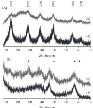
Investigations by X-ray diffraction (Figure 1) and FTIR spectroscopy (Figure 2) confirmed that powders
synthesized at 200 and 700 °C were boehmite γ-AlOOH
and γ-Al2O3, respectively, both being weakly crystallized
and with a high degree of hydration. Surface modification by Asn induced tenuous changes as discussed hereafter.
At TSYN = 200 °C the diffractogram matched with the
reference JCPD No. 21-1307. In a former paper, details of X-ray data of boehmite samples elaborated by SP have
been analyzed.24 The average nanocrystal size following the
orthorhombic axis b does not exceed one cell, so that half the hydroxyl groups are at the external surface of nanocrystals.
At TSYN = 700 °C, the diffractogram corresponded to
reference data JCPD No. 10-0425: with peaks labeled
(400) and (440) at 2θ = 46 and 66.5°, respectively, and a
very broad and asymmetrical band between 30 and 40°, which contains the badly resolved contributions from (220) and (311) planes. The peak positions matched with the gamma-alumina standard apart from the absence of (111) and (222), which should be observed in well crystallized
transition aluminas.2,5 The coherent length evaluated was
about 3 nm for the SP transition aluminas, but the important background observed under the peaks supports that the size of most nanocrystals in the samples were below this value. On reacting with asparagine, an additional broad band
appeared at 2θ ca. 20° for boehmite samples, assigned to
the organic counterpart of the composite. The reaction of γ
alumina with Asn has lead to the partial transformation of
γ-Al2O3 into γ-AlOOH, the diffraction peaks of γ-AlOOH
being marked by stars in Figure 1B.
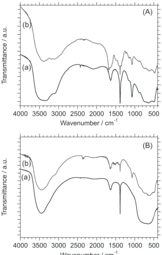
FTIR spectrum of boehmite γ-AlOOH has been
described in literature.27,28 In agreement with these previous
papers, the spectrum recorded for the powder TSYN = 200 °C
(Figure 2A) displayed vibrational modes of [AlO6] at 481,
632, 734 cm-1 and δ
OH at 1072 and 1157 cm-1. Samples were
highly hydrated, as were amorphous or nanocrystallized
boehmite described in references.28,29 The two δ
OH modes
at 3083 and 3310 cm-1 were partly obscured by the broad
10 20 30 40 50 60 70 80
10 20 30 40 50 60 70 80
(B)
(b) (b)
(a)
(2
5
1
)
(0
0
2)
(2
0
0
)
(0
3
1
)
(1
2
0)
2θ/degree
(0
2
0
)
(a)
*
(A)
*
*
2θ/degree
*
band around 3400 cm-1, this last one assigned to adsorbed
hydroxyls. The presence of adsorbed water molecules was
evidenced by the δHOH at 1630 cm-1. FTIR spectrum of
sample TSYN = 700 °C (Figure 2B) was quite in agreement,
in the wave number range 400-1400 cm-1, with the one of
γ-Al2O3,28 with relatively broad bands peaking at 635 cm-1
(stretching AlO6) and 785 cm-1 (stretching AlO4). Adsorbed
water and hydroxyls were attested at 1630 cm-1 and around
3400 cm-1.
Boehmite and aluminates from spray pyrolysis
displayed features around 1385 cm-1 from residual nitrates
but no trace of organics from the preparation mixture were
detected. The band at 1673 cm-1 (C=O) and CH vibrations
at 1360, 1316 and 1236 cm-1 were well isolated for sample
0.3Asn:BOE:Tb1%. Additional features were observed at
1500 cm-1 and superimposed with the hydroxyls modes in
the region 3000-3400 cm-1, but were not assigned. In the
spectrum of Asn:γTA:Tb1%, shown in Figure 2B, only
very weak effect from the organic part was detectable, as
a low intensity band at 1416, 1428 and 1511 cm-1δ CN,
υS CO2– and δ NH3+ vibration, respectively.30 Important
changes were observed in the aluminate response since
bands characteristic of boehmite were observed proving
the partial transformation of γ-Al2O3 into γ-AlOOH, also
evidenced by XRD.
Morphology and water dispersion
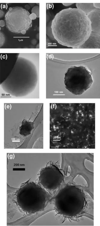
The particles prepared by spray pyrolysis were always spherical and sub-micrometric with a moderately dispersion in size, whatever their composition. The major fraction of hydrated alumina particles presents diameters between 100 and 500 nm. Some characteristic electron microscope images are gathered in Figure 3. SEM images recorded
on BOE:Tb1%, Figure 3a, and on γTA:Tb1%, Figure 3b,
evidence irregular surfaces, and spherical particles consisting of smaller (nanometric) sub-particles, aggregated in the spray pyrolysis processing. TEM observations, Figures 3c and 3d, give additional information since the spheres were homogeneously packed with sub-particles, and not hollow, although hollow particles can be obtained
by SP.22
In composite materials, the organic and inorganic counterparts could be well observed, the effect of the dispersant being more noticeable on boehmite than on transition alumina particles. Most of the boehmite spheres had disaggregated during reaction with Asn. Sample Asn:BOE:Tb1%, shows an organic film in which the nanoparticles appear dispersed. TEM image Figure 3e shows the composite film and one bigger particle not completely disaggregated. Aluminum was identified by local analysis (EDXS in TEM) throughout the film. Figure 3f was recorded on the film in dark field conditions: all bright parts are due to crystallized boehmite nanoparticles (a few nm in size), embedded in
the amorphous polymer. For sample Asn:γTA:Tb1% (TEM
image in Figure 3g), the organic film was observed to wrap the sub micrometric alumina particles.
Asn molecules were observed to interact differently
with boehmite and Al2O3. Boehmite23,24 micrometric spheres
obtained by SP had been spontaneously dispersed in water. The same remark was made here for BOE:Tb1% by DLS analysis. 60% of the particles fall in the range 25-40 nm, while higher values (up to 85 nm) were measured for the remaining 40%. Spherical sub-micrometric particles were disaggregated in water and nanometric sub-particles
remained very stable. At 5 or 10% Tb3+ particles were
noticeably bigger: higher contents in Ln3+ lead to more
tightly linked sub-particles. Once formed, these spherical microparticles had not been fragmented in water. In
addition, all particles synthesized at TSYN = 700 °C with
the γ-Al2O3 structure, remained stable in water. The
addition of Asn did not change the particles form and the
4000 3500 3000 2500 2000 1500 1000 500
4000 3500 3000 2500 2000 1500 1000 500
(b)
(b)
(a) (a)
(B)
Wavenumber / cm-1
(A)
Wavenumber / cm-1
T
ransmittance / a.u.
T
ransmittance / a.u.
final composite presented low concentration of Asn as corroborated by the low organic content detected by FTIR.
Photoluminescence
The two hydrated aluminates under consideration have quite different crystal structures, in particular when one
considers the way that bigger cations as Ln3+ could be
accommodated on the Al3+ sites. γ-Al
2O3 has a defect-spinel
structure with most of Al atoms in octahedral coordination
and the minority ones in tetrahedral coordination.3 At crystal
surface, more distorted tetrahedral Al sites may occur with
penta- and hepta-coordinated Al.31γ-Alumina has a lot of
structural defects or empty cationic sites, especially near the surface of nano-crystals in poorly crystallized sample, and
it has been admitted that Eu3+ ions may be accommodated
in γ-Al2O3, most probably at these defect sites.25,26,32 The
crystal structure of boehmite,33 is orthorhombic. The unit
cell consists of two double layers of aluminium-centered
distorted octahedral AlO4(OH)2. OH groups locate at the
outer surface of the double layers and interact to hold the
layers together. There is no substitution of Eu3+ on the
Al3+ octahedral sites in boehmite for Eu3+,24 but that partly
hydrated Eu3+ ions are directly bonded to OH groups at the
boehmite surface. It is highly probable that Tb3+ behave
the same way as Eu3+, hereafter we discuss characteristic
features from the terbium luminescence data that support these hypotheses.
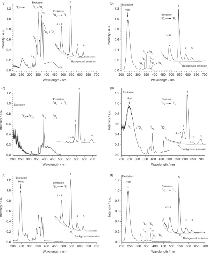
The photoluminescence emission spectra of samples
BOE:Tb1% and γTA:Tb1% are displayed in Figures 4a and
4b, respectively. Characteristic emission bands assigned to
the 5D
4→7FJ transitions (J = 6, 5, 4, 3) with the dominant
green band at 540 nm are observed. The shape of Tb3+
emission is generally little sensitive to the local environment and no additional information could be extracted from their comparison in the two hydrated aluminates. Considering photoluminescence excitation a number of narrow lines
assigned to Tb3+ (4f8 configuration) 7F
6→2S + 1LJ transitions
could be observed.34 Of particular interest is the broad
contribution observed at wavelengths 320-400 nm since it matches the excitation wavelengths most often encountered in laboratory fluorescence microscopes and supports the
consideration of Tb3+ as fluorescent bio-label. In addition,
an intense broad band at around 240 nm was observed for
γTA:Tb1%, but not for BOE:Tb1%. The strength to the way
terbium ions are connected with the host may be responsible for the differences. Two assignments are conceivable for
the band around 240 nm. The first assignment is the of Tb3+
interconfigurational parity-allowed absorption. From the
ground state 7F
6 (4f8), transitions occur to the multiplets
9D
J and 7DJ (4f7 5d1). The former transition (ending at 9DJ)
is spin forbidden and occurs at lower energy than the spin
allowed one ending at 7D
J.
Dorenbos35 initiated a shift model in the analysis of
5d levels of Tb3+. The spin-allowed transition energies
for Tb3+ in some mixed oxides (borates or silicates) are
actually reported at around 5.20 eV (238 nm). Consistently,
Zawadski et al.36 assigned the absorption band at 240 nm
observed in mixed oxides Al2O3-ZrO2:Tb3+ to (4f8) →7DJ
(4f7 5d1). All these values agree with the band at 240 nm
(5.17 eV) observed in γTA:Tb1%. Considering BOE:Tb1%, (Figure 4a) no strong absorption band could be observed, but rather the low energy tail of a stronger excitation truncated by inner filter effect. This would mean that the spin allowed band is shifted towards higher energies, as it
is in the aquo ion [Tb(OH)2]8 where absorption maximum is
at 5.71 eV (217 nm)37 supporting the existence of hydrated
terbium ions in boehmite.
The second possible assignment is the host sensitization, which may be described according to the Förster-Dexter energy transfer. A spectral overlap of the host emission with the acceptor absorption is necessary. The visible blue
photoluminescence of γ-Al2O3 nanoparticles has been
investigated in detail by Yu et al.38 An emission band peaking
at 405 nm, associated with excitations at 236, 245, 255
nm has been observed for γ-Al2O3 calcined at 500 °C, and
assigned to electron-hole recombination at oxygen defects in the transition alumina. Indeed, the emission background observed for our samples may be assigned to that of the aluminate matrices. The broad emission from alumina
overlaps the wavelengths of Tb3+ intra 4f8 absorption lines
(7F
6 →5G6, 5D3, 5L10, 5L8, 5D2, 5L8, 5H7), so that γ-Al2O3
to acceptor Tb3+ energy transfer would occur favorably.
Inversely, although a similar photoluminescene occurs
for boehmite,39,40 there would be no boehmite to terbium
energy transfer in BOE:Tb. This can be understood if the
bonding of Tb3+ with the boehmite is weaker than it is with
the transition alumina. So finally both mechanisms would easily explain our observations. An argument in favor of the second assignment that of host sensitization, is given by the
consideration of Eu3+ in same hosts. The photoluminescence
spectra from our preceding work24 are also shown in Figures
4c and 4d. At wavelengths below 300 nm, a broad absorption
band was detected for γTA:Eu1% but not for BOE:Eu1%.
This band was assigned to absorption in the localized LMCT
state (ligand to metal charge transfer),24 but it could also be
due to non radiative energy transfer between the host and
Eu3+, since the visible emission of aluminates matches the
group of absorption lines 7F
0→5D3, 5L6, 5L7, 5D4.
Also in line with terbium ions having a much more hydrated environment in boehmite than in transition
alumina, the 5D
4 emission lifetime was shortened due to
the enhanced probability of non-radiative de-excitation
by the OH vibrators. The 5D
4 luminescence decay curves
were fitted with first-order exponential functions and the decay time was found to be markedly lower in sample
BOE:Tb1% (1.00 ± 0.10 ms) than in sample γTA:Tb1%
(2.56 ± 0.26 ms). Ishizaka et al.41 have measured the 5D
4
decay times in Tb3+ doped alumina films prepared by
sol-gel method, then heat-treated to remove residual water molecules: our observations agree rather well with those
reported on films after heat treatment at 25 °C and at 500 °C, respectively.
Photoluminescence spectra of the nanocomposites
Asn:BOE:Tb1% and Asn:γTA:Tb1% are displayed in
Figures 4e and 4f, respectively. Concerning PLE spectra, the main difference between Asn modified or unmodified aluminates were the intensities ratios between the band at
240 nm and 7F
6 →2S + 1LJ lines. The broad band appeared
clearly in Asn:BOE:Tb1%, whereas it was absent in un-modified boehmite. This observation supports the assumption that host to terbium energy transfer is the cause of this excitation band: indeed, it may be understood that the amine acid strengthens the linkage of hydrated Tb ions with the boehmite surface and favors energy transfer.
Photoluminescence in aqueous medium
After the screening of various amine acid/aluminate/
terbium ratios it was established that compositions Tb3+/
BOE equal 1 or 5 percent and Asn/BOE equal 0.3/1 may be considered as luminescent labels in aqueous media since they remain well dispersed at the nanometric scale in solution. We have thus evaluated the possibility to record
the Tb3+ luminescence with the measurement conditions
usually employed in rare earth luminescent bio-assays: using proper excitation wavelength and optimized time-resolution conditions. Figure 5 shows the emission of
Asn:BOE:Tb1% at concentration 0.16 mol L-1 (in mol of
Al3+) under excitation at 250 or 351nm and with a signal
detection fixed from 0.1 milliseconds to 0.5 milliseconds after the exciting pulse.
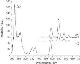
It is important to establish the lower concentration compatible with the unambiguous observation of time-resolved spectra. In that work, it was checked the
luminescence emission up to 0.1 mol L-1 for hybrid in
water medium, however the results let us to affirm at
the 0.05 mol L-1 as the low limit concentration to
time-resolution analysis. The water stability and luminescence behavior could enable the hybrid system to monitor
wastewater treatment, e.g., labeling Escherichia coli.42
Since, Escherichia coli and enterococci are used as
regulatory tools to monitor water and presence of potential enteric pathogens yet their source (human or animal) cannot
be determined with routine methods.43
Conclusions
The spray pyrolysis (SP) of aluminium tri-sec-butoxide
and TbCl3 in aqueous medium allowed synthesizing
nano-structured powders having the crystalline phase
(noted γTA). Particles were spherical and sub-micrometric, consisting of aggregated nanometric sub-particles. The photoluminescence emission spectra of samples
BOE:Tb1% and γTA:Tb1% exhibited the well-known
green emission 5D
4→7FJ = 6-3 of Tb3+.The strong near-UV
absorption (around 240 nm)is assigned to the host to ion
Figure 4. Photoluminescence of (a) BOE:Tb1%; (b) γTA:Tb1%; (c) BOE:Eu2%; (d) γTA:Eu2%; (e) Asn:BOE:Tb1%; (f) Asn:γTA:Tb1%. Excitation spectra monitored at λ emission = 543 nm (a, b, e, f) or at λ emission = 592 nm (c, d); emission spectra by excitation at 351 nm (a), 250 nm (host at b, e, f) and 394 nm (c, d).
200 250 300 350 400 450 500 550 600 650 700 0.0 0.2 0.4 0.6 0.8 1.0 1.2 Intensit y / a .u .
Wavelength / nm
7 F6 Excitation 3 4 5
J = 6 Emission 5 D4 7 FJ 5
G6+5
D3
5
L10 5
L9+ 5 D2 5 L8 5 H7 (a)
200 250 300 350 400 450 500 550 600 650 700 0.0 0.2 0.4 0.6 0.8 1.0 1.2 Excitation (b) Intensit y / a .u .
Wavelength / nm
Host
3 4 5
J = 6 Emission
5
D4 7
FJ
5
G6+ 5
D3 5
L10 5
L9+5
D2
5
L8 5
H7
200 250 300 350 400 450 500 550 600 650 700 0.0 0.2 0.4 0.6 0.8 1.0 1.2 Excitation (c) Intensit y / a .u .
Wavelength / nm
5 D 3 5 L 6 5 D 2 4 3 2 1
J = 0 Emission
5
D0 7
FJ
7
F0
200 250 300 350 400 450 500 550 600 650 700 0.0 0.2 0.4 0.6 0.8 1.0 1.2 Host 7 F0 (d) Intensit y / a .u .
Wavelength / nm Excitation 5 D 3 5 L 6 5 D 2 4 3 2 1
J = 0 Emission
5
D0 7
FJ
200 250 300 350 400 450 500 550 600 650 700 0.0 0.2 0.4 0.6 0.8 1.0 1.2 Host (e) Intensit y / a .u .
Wavelength / nm
3 4 5
J = 6 Emission
5
D4 7
FJ Excitation
200 250 300 350 400 450 500 550 600 650 700 0.0 0.2 0.4 0.6 0.8 1.0 1.2 Excitation (f) In te n s it y / a .u .
Wavelength / nm
3 4 5
J = 6 Emission 5 D4 7 FJ 5
G6+5
D3
5
L10 5
energy transfer. We associate its presence in γTA:Tb1% to
the incorporation of Tb3+ in the defect-spinel structure of
the γ-Al2O3 matrix. In BOE:Tb1%, in contrast, the energy
transfer was very weak, confirming our earlier hypothesis
that Ln3+ ions do not enter the aluminate network in
boehmite, but are bonded to the hydroxyls groups at boehmite surface.
The surface of luminescent terbium doped boehmite particles was modified by asparagine Asn. The particles, in (water + Asn), disaggregated into nanoparticles (hydrodynamic diameter from 5 to 100 nm). Asn is localized at the surface of the particles and at the same time the linkage of hydrated terbium ions with boehmite
was strengthened, enhancing the aluminate → Tb energy
transfer. The goal to get modified aluminate nanoparticles
with enhanced luminescence starting from the γTA:Tb1%
was not achieved since it was not possible to disaggregate the sub-micronic spheres in (water + Asn). Besides,
γ-Al2O3 hydrolysed partially into γ-AlOOH during the
reaction with Asn. When homogeneous suspensions in aqueous solution of nanoparticles were achieved, the limit of detection in time-resolution conditions analysis
was estimated to 0.05 mol L-1 AlOOH:Tb1%. Spray
pyrolysis is a cheap, fast and reliable production process. It is well adapted to the synthesis of hydrated aluminate particles. Moreover aluminates particles may be doped with any luminescent lanthanide Ln leading to strong photoluminescence in the visible, with a millisecond lifetime appropriated for efficient time gated detection. With a highly reactive surface, terbium-doped boehmite nanoparticles may be easily modified and therefore fulfill several of the key features for their actual use for in vitro or in vivo applications.
Acknowledgements
Authors thank Y. Kihn (CEMES-CNRS) and C. Galaup (SPCMIB-Université de Toulouse) for their support in electron microscopy and time-resolved luminescence investigations, respectively. The Brazilian agencies FAPESP (2007/55723-6, 2011/21551-0), CNPq, CAPES, and the CAPES-COFECUB Brazil-France cooperation program for the grant to J. M. A. C. are also acknowledged.
References
1. Penilla, E. H.; Kodera, Y.; Garay, J. E.; Adv. Funct. Mater. 2013,
23, 6036.
2. Zhou, R. S.; Snyder, R. L.; Acta Crystallogr. 1991, B47, 617.
3. Sohlberg, K.; Pennycook, S. J.; Pantelides, S. T.; J. Am. Chem. Soc. 1999, 121, 7493.
4. Pecharromán, C.; Sobrados, I.; Iglesias, J. E.; González-Carreño, T.; Sanz, J.; J. Phys. Chem. B 1999, 103, 6160.
5. Krokidis, X.; Raybaud, P.; Gobichon, A.-E.; Rebours, B.; Euzen, P.; Toulhoat, H.; J. Phys. Chem. B2001, 105, 5121. 6. Paglia, G.; Rohl, A. L.; Buckley, C. E.; Gale, J. D.; Phys. Rev. B
2005, 71, 224115.
7. Alvarez, L. J.; Leon, L. E.; Sanz, J. F.; Capitan, M. J.; Odriozola, J. A.; J. Phys. Chem.1995, 99, 17872.
8. Yoldas, B. E.; J. Mater. Sci. 1975, 10, 1856. 9. Yoldas, B. E.; Ceramic Bulletin1975, 54, 289.
10. Granados-Correa, F.; Jiménez-Becerril, J.; J. Hazard. Mater.
2009, 162, 1178.
11. Yoon, T. H.; Johnson, S. B.; Musgrave, C. B.; Brown Jr., G. E.;
Geochim. Cosmochim. Acta 2004, 68, 4505.
12. Landry, C. C.; Pappé, N.; Mason, M. R.; Apblett, A. W.; Tyler, A. N.; MacInnes, A. N.; Barron, A. R.; J. Mater. Chem. 1995,
5, 331.
13. Kareiva, A.; Harlan, C. J.; MacQueen, D. B.; Cook, R. L.; Barron, A. R.; Chem. Mater. 1996, 8, 2331.
14. Kang, H.-J.; Kim, D. J.; Park, S.-J.; Yoo, J.-B.; Ryu, Y. S.; Thin Solid Films 2007, 515, 5184.
15. Kapoor, S.; Hegde, R.; Bhattacharyya, A. J.; J. Control. Release
2009, 140, 34.
16. Al-Kattan, A.; Santran, V.; Dufour, P.; Dexpert-Ghys, J.; Drouet, C.; J. Biomater. Appl. 2014, 28, 697.
17. Li, L.; Liu, Y.; Tao, J.; Zhang, M.; Pan, H.; Xu, X.; Tang, R.;
J. Phys. Chem. C 2008, 112, 12219.
18. Meiser, F.; Cortez, C.; Caruso, F.; Angew. Chem. Int. Ed. 2004,
43, 5954.
19. Jin, D.; Connally, R.; Piper, J.; Cytom. Part A 2007, 71A, 783.
20. Okada, K.; Tanaka, A.; Hayashi, S.; Otsuka, N.; J. Mater. Sci. Lett. 1993, 12, 854.
Figure 5. Time resolved photoluminescence of 0.3Asn:BOE:Tb1% 0.16 mol L-1 in water (a) excitation spectra monitored at λ emission = 543 nm and emission spectra by excitation at (b) 250 nm; (c) 351 nm. Delay time at 0.1 ms, sample window as 0.4 ms.
200 250 300 350 400 450 500 550 600 650 700 0
20 40 60 80 100 120 140 160 180
(c) (b)
Wavelength / nm
Intensity / a.u.
21. Vallet-Regi, M.; Rodriguez-Lorenzo, L. M.; Ragel, C. V.; Salinas, A. J.; Gonzalez-Calbet, J. M.; Solid State Ionics 1997,
101-103, 197.
22. Kato, T.; J. Ceram. Soc. Jpn. 2002, 110, 146.
23. Caiut, J. M. A.; Dexpert-Ghys, J.; Kihn, Y.; Vérelst, M.; Dexpert, H.; Ribeiro, S. J. L.; Messaddeq, Y.; Powder Technol. 2009, 190, 95.
24. Caiut, J. M. A.; Ribeiro, S. J. L.; Messaddeq, Y.; Dexpert-Ghys, J.; Verelst, M.; Dexpert, H.; Nanotechnology 2007, 18, 455605. 25. Marques, R. F. C.; Caiut, J. M. A.; Paiva-Santos, C. O.; Ribeiro, S. J. L.; Messaddeq, Y.; Garcia, C.; Neumeyer, D.; Dexpert, H.; Verelst, M.; Dexpert-Ghys, J.; Braz. J. Phys. 2009, 39, 176. 26. Caiut, J. M. A.; Bazin, L.; Mauricot, R.; Dexpert, H.; Ribeiro,
S. J. L.; Dexpert-Ghys, J.; J. Non-Cryst. Solids 2008, 354, 4860. 27. Fripiat, J. J.; Bosmans, H.; Rouxhet, P. G.; J. Phys. Chem. 1967,
71, 1097.
28. Boumaza, A.; Favaro, L.; Lédion, J.; Sattonnay, G.; Brubach, J. B.; Berthet, P.; Huntz, A. M.; Roy, P.; Tétot, R.; J. Solid State Chem. 2009, 182, 1171.
29. Wang, S.-L.; Johnston, C. T.; Bish, D. L.; White, J. L.; Hem, S. L.; J. Colloid Interface Sci. 2003, 260, 26.
30. Cooper, S. J.; Langmuir 2002, 18, 3749.
31. Alvarez, L. J.; Leon, L. E.; Sanz, J. F.; Capitan, M. J.; Odriozola, J. A.; J. Phys. Chem. 1995, 99, 17872.
32. Monteiro, M. A. F.; Brito, H. F.; Felinto, M.; Brito, G. E. S.; Teotonio, E. E. S.; Vichi, F. M.; Stefani, R.; Micropor. Mesopor. Mat. 2008, 108, 237.
33. Milligan, W. O.; McAtee, J. L.; J. Phys. Chem. 1956, 60, 273. 34. Carnall, W. T.; Crosswhite, H.; Crosswhite, H. M.; Energy
Level Structure and Transition Probabilities in the Spectra of
the Trivalent Lanthanides in LaF3, 1st ed.; Argonne National
Laboratory: Argonne, USA, 1977.
35. Dorenbos, P.; J. Phys. Condens. Matter 2003, 15, 6249. 36. Zawadski, M.; Hreniak, D.; Wrzyszcz, J.; Mista, W.; Grabowska,
H.; Malta, O. M.; Strek, W.; Chem. Phys. 2003, 291, 275. 37. Carnall, W. T.; J. Chem. Phys. 1968, 49, 4412.
38. Yu, Z. Q.; Li, C.; Zhang, N.; J. Lumin. 2002, 99, 29.
39. Yu, Z. Q.; Wang, C. X.; Gu, X. T.; Li, C.; J. Lumin.2004, 106, 153.
40. Bai1, X.; Caputo, G.; Hao, Z.; Freitas, V. T.; Zhang, J.; Longo, R. L.; Malta, O. L.; Ferreira, R. A. S.; Pinna, N.; Nat. Commun. 2014, 5, 5702.
41. Ishizaka, T.; Nozaki, R.; Kurokawa, Y.; J. Phys. Chem. Solids
2002, 63, 613.
42. Duarte, A. P.; Mauline, L.; Gressier, M.; Dexpert-Ghys, J.; Roques, C.; Caiut, J. M. A.; Deffune, E.; Maia, D. C. G.; Carlos, I. Z.; Ferreira, A. A. P.; Ribeiro, S. J. L.; Menu, M.-J.; Langmuir
2013, 29, 5878.
43. Srinivasan, S.; Aslan, A.; Xagoraraki, I.; Alocilja, E.; Rose, J. B.; Water Res.2001, 45, 2561.
Submitted: June 17, 2015
Published online: August 25, 2015