Cop
yright
© ABE&M t
odos os dir
eit
os r
eser
vados
.
Long-term follow-up of an 8-year-old
boy with insulinoma as the irst
manifestation of a familial form of
multiple endocrine neoplasia type 1
Seguimento de longo prazo em um menino de 8 anos de idade com insulinoma como primeira manifestação de neoplasia endócrina múltipla tipo 1
Helena Campos Fabbri1, Maricilda Palandi de Mello1, Fernanda Caroline
Soardi1, Adriana Mangue Esquiaveto-Aun1,2, Daniel Minutti de Oliveira3,
Fernanda Canova Denardi3, Arnaldo Moura-Neto3, Heraldo Mendes Garmes3,
Maria Tereza Matias Baptista3, Patrícia Sabino de Matos4,Soia Helena Valente
de Lemos-Marini2, Lilia Freire Rodrigues D’Souza-Li2, Gil Guerra-Júnior2
SUMMARY
Multiple endocrine neoplasia type 1 (MEN1) is an autosomal dominant hereditary cancer syn-drome characterized mostly by parathyroid, enteropancreatic, and anterior pituitary tumors. We present a case of an 8-year-old boy referred because of hypoglycemic attacks. His diagnosis was pancreatic insulinoma. Paternal grandmother died due to repeated gastroduodenal ulcerations and a paternal aunt presented similar manifestations. At a first evaluation, the father presented only gastric ulceration but subsequently developed hyperparathyroidism and lung carcinoid tumor. During almost 15 years of follow-up, three brothers and the index case presented hyper-parathyroidism and hyperprolactinemia. Molecular study showed a G to A substitution in intron 4, at nine nucleotides upstream of the splicing acceptor site, causing a splicing mutation. All affected members of the family have the same mutation. Paternal grandmother and aunt were not studied and the mother does not carry any mutation. MEN1 is a rare condition that requires permanent medical assistance. Early clinical and genetic identification of affected individuals is essential for their own surveillance and also for genetic counseling. Arq Bras Endocrinol Metab. 2010;54(8):754-60
SUMÁRIO
A neoplasia endócrina múltipla tipo 1 (NEM1) é uma doença hereditária autossômica domi-nante, caracterizada principalmente por tumores de paratireoide, enteropancreáticos e adeno--hipofisários. Apresentamos o caso de um menino com 8 anos encaminhado por crises de hipoglicemia. Seu diagnóstico foi insulinoma pancreático. Sua avó paterna faleceu por úlceras gastroduodenais de repetição e a tia paterna tinha as mesmas manifestações. Na primeira avaliação, o pai apresentou apenas úlcera gástrica, porém com a evolução desenvolveu hiper-paratireoidismo e tumor carcinoide pulmonar. Durante cerca de 15 anos de seguimento, os três irmãos e o caso índice desenvolveram hiperparatireoidismo e hiperprolactinemia. O estudo molecular mostrou a substituição G por A no intron 4, a nove nucleotídeos do sítio aceptor de
splicing, criando um novo sítio de splicing. Todos os membros da família afetados e estudados tinham a mesma mutação. A NEM1 é uma condição rara que requer assistência médica per-manente. As identificações clínicas e genéticas precoces são essenciais para o tratamento e aconselhamento genético. Arq Bras Endocrinol Metab. 2010;54(8):754-60
1 Center for Molecular Biology and Genetic Engineering (CBMEG), Universidade Estadual de Campinas (Unicamp), Campinas, SP, Brazil 2 Department of Pediatrics, Faculdade de Ciências Médicas (FCM), Unicamp, Campinas, SP, Brazil
3 Department of Clinical Medicine, FCM-Unicamp, Campinas, SP, Brazil 4 Department of Pathology Anatomy, FCM-Unicamp, Campinas, SP, Brazil
Correspondence to: Gil Guerra-Júnior Departamento de Pediatria, FCM-Unicamp
13083-887 − Campinas, SP, Brazil [email protected]
Cop
yright
© ABE&M t
odos os dir
eit
os r
eser
vados
.
INTRODUCTION
M
ultiple endocrine neoplasia type 1 (MEN1 – OMIM 131100) is an autosomal dominant familial cancer syndrome characterized by primary hyperparathyroidism in association with endocrine en-teropancreatic tumors and anterior pituitary adenomas, but manifestations of carcinoid tumors, adrenal adeno-mas, and lipomas are also reported. The occurrence of two MEN1-related endocrine tumors is suficient to es-tablish the clinical diagnosis (1-3). This syndrome often occurs at a young age with equal sex distribution (3). Primary hyperparathyroidism is responsible for more than 90% of clinical cases and usually it is the irst clini-cal manifestation of MEN1. Enteropancreatic tumors (gastrinomas, glucagonomas, VIPomas, insulinomas, or non-functioning tumors) affect about 30%-80% of patients (4), while the incidence of pituitary adenomas (prolactinomas, somatotropinomas, corticotropino-mas, or non-functioning tumors) in MEN1 patients varies from 15% to 90% (5-6).The MEN1 gene, which is responsible for the dis-ease, is located on 11q13, occupies 9.8 kb of genomic DNA, and encodes a 610-aminoacid protein called me-nin, within 10 exons (7). It is considered to be a tumor suppressor gene by altering JunD-mediated transcrip-tion (8). It is involved in the regulatranscrip-tion of cell functranscrip-tions such as DNA replication and repair, and also in the transcriptional machinery (7,9,10). MEN1 mutations have a high degree of penetrance: more than 95% of pa-tients who carry a mutation will develop the disease by the ifth decade of life (3), however, the phenotype is highly variable therefore a direct genotype-phenotype correlation has been dificult to be established (11). Carriers of MEN1 gene mutations may beneit from periodic clinical evaluations (12). Generally, MEN1 germline mutations are identiied with a prevalence av-erage of 70% in the familial forms, whereas the sporadic cases, associated with de novo mutation in the MEN1 gene, represents about 5%-10% (13). More than 1,000 MEN1 mutations have been reported; about 25% are nonsense, 45% intragenic deletions, 15% insertions, 10% missense mutations, and less than 5% are splice site mutations (14).
Herein we report an unusual index case presenting with hypoglycemia in early age (8 years of age) within a MEN1 family. Long-term follow-up with clinical mani-festations and management of all affected relatives are also discussed.
CASE REPORT
Clinical data
An 8-year-old boy was admitted at our pediatric clinic because of seizures misdiagnosed as epilepsy (normal EEG and cerebral CT). He also had sweating, palpi-tation, tremulousness, hunger, anxiety, and a sudden weight gain (8 kg in 4 months). Laboratory data con-irmed hyperinsulinemic hypoglycemia (glycemia = 19 mg/dL – Normal range – NR = 60-99; insulinemia = 26.2 IU/mL – NR < 5; and C-peptide = 3.2 ng/mL – NR = 1.1-5.0). Pancreatic angiography and abdomen CT suggested nodules in the pancreas region. Upon intraoperative ultrasound two lesions in the distal body of the pancreas were observed (0.6 cm and 0.3 cm in size); they were removed by partial pancreatectomy. Histological analysis conirmed insulinoma, and immu-nohistochemistry examination was also strongly posi-tive for insulin but also slightly posiposi-tive for glucagon and gastrin (Figure 1 A-D).
To date, his family history includes a healthy mother and 3 brothers (an older boy and two younger twins). In addition, his paternal grandmother had repeated gastroduodenal ulceration and urolithiasis since the age of 38 and died at the age of 45 due to gastric ul-cer perforation. Autopsy showed peptic ulul-cer with gastrin-secreting duodenal mucosa tumor (gastrinoma – Zollinger-Ellison syndrome) and primary hyperpara-thyroidism. His paternal aunt also had repeated gas-troduodenal ulceration and urolithiasis, and his father presented only gastroduodenal ulceration when he was irst examined.
The patient had been clinically stable during sev-eral years of follow-up and showed normal growth and puberty. However, at the age of 16 he also presented elevated PTH (90.9 pg/mL, NR = 10.0-50.0) and cal-cium (11.1 mg/dL – NR = 8.5-10.5) leading to pri-mary hyperparathyroidism diagnosis and subsequently to subtotal parathyroidectomy. He is now 25 years old, without symptoms, MRI of the pituitary region is nor-mal and prolactin high (54.3 ng/mL – NR < 15); PTH and calcium serum concentrations have been main-tained in the upper normal range throughout 5 years; and, both glycemia and insulin are normal.
Cop
yright
© ABE&M t
odos os dir
eit
os r
eser
vados
.
Figure 1. (A)HE-50X: Thearrow indicates a hypercellular pancreatic nodule with thin capsule. (B)HE-400X: Polygonal cells in a solid, trabecular and glandular pattern, with no atypia or necrosis. (C)Immunohistochemical assay demonstrating intense and diffuse positivity for insulin. (D)Ultrastructural study showing neuroendocrine granules.
A
C
B
D
chemotherapy was performed after lobectomy. He is now 51 years old and is doing well with normal serum levels of PTH, calcium, glucose, insulin and prolactin.
A 29-year-old brother reported hyperprolactinemia due to a pituitary macroadenoma when he was 24 years old. Since then he has been successfully treated with cabergoline. One year later, he developed primary hy-perparathyroidism; therefore, he underwent subtotal parathyroidectomy. The two 23-year-old younger twin brothers presented different symptoms. One developed hyperinsulinemic hypoglycemia due to insulinoma at the age of 18 and underwent subtotal pancreatectomy. Histological analysis conirmed multiple pancreatic nodules and immunohistochemistry examination was positive for insulin and negative for glucagon and gas-trin. One year later, he developed diabetes and started insulin replacement. At the age of 20, he had hyperp-rolactinemia that was treated with cabergoline and after other year he showed elevated PTH and calcium indi-cating subtotal parathyroidectomy. The other twin had elevated PTH and calcium when he was 18 years old; therefore subtotal parathyroidectomy was performed.
Two years later he presented hyperprolactinemia treat-ed with cabergoline. Father and brothers remain un-der treatment and periodic screening, which consists of medical appointments taking place twice a year, labo-ratorial investigation performed annually and imaging studies conducted every two years (Table 1).
Molecular analysis
Blood specimens of the patient and his relatives were collected with approval by the appropriate institutional review board and a signed informed consent was ob-tained.
Cop
yright
© ABE&M t
odos os dir
eit
os r
eser
vados
.
Table 1. Clinical data, management and follow-up of MEN1 manifestations in all cases of the Brazilian family studied
Patient Manifestation diagnosisAge at Management Follow-up follow-upTime of
Index case Hypoglycemia (insulinoma) 8 Partial pancreatectomy Normal insulin and glycemia 17 Primary hyperparathyroidism 16 Subtotal parathyroidectomy Upper normal range of PTH and calcium Hyperprolactinemia (normal MRI) 25 Observation Moderate hyperprolactinemia Paternal grandmother Repeated gastroduodenal
ulceration and urolithiasis
38 NR Died at 45 years-old due to gastric perforation (gastrinoma)
-Paternal aunt Repeated gastroduodenal ulceration and urolithiasis
NR NR NR NR
Father Gastroduodenal ulceration 35 Clinical treatment Without symptoms 17 Primary hyperparathyroidism 45 Subtotal parathyroidectomy Normal PTH and calcium
Bronchial carcinoid 45 Lobectomy + chemotherapy Without symptoms Older brother Hyperprolactinemia
(macroadenoma)
24 Cabergoline treatment Normal prolactin 5
Primary hyperparathyroidism 25 Subtotal parathyroidectomy Normal PTH and calcium
Younger brother (twin A) Hypoglycemia (insulinoma) 18 Near-total pancreatectomy Insulin-dependent diabetes 5 Hyperprolactinemia (normal MRI) 20 Cabergoline treatment Normal prolactin
Primary hyperparathyroidism 21 Subtotal parathyroidectomy Normal PTH and calcium
Younger brother (twin B) Primary hyperparathyroidism 18 Subtotal parathyroidectomy Normal PTH and calcium 5 Hyperprolactinemia (normal MRI) 20 Cabergoline treatment Normal prolactin
NR: not reported.
Table 2. Primers designed for amplification and sequencing of MEN1 gene
Primer Sequence (5’-3’) Tm (ºC)1 Size (bp)2
5’UTRs CCCCGAGTCTGCAGGTAGTG 61.8 801
Ex 2as AGCTCGGCAGCAAACAGG 60.6 801
Int 1s AACCTTAGCGGACCCTGGG 63 705
Int 2as GCATATCATTTCCCCTCTCTGG 62.4 705
Int 2s CCCCATGTTAAAGCACAGAGG 61.6 720
Int 4as ACTAACCCATTTTTCCAGGAGG 61.6 720
Int 4s CTTTCCCTTCCTGAGCTTCAG 60.3 596
Int 6as ACACAAAGTGAGACTGGATGGG 60.4 596
Int 6s TGGGAGTGGAGATGGAGAGG 60.6 956
Int 8as ATCCCTAATCCCGTACATGCAG 58 956
Int 7s3 TCCTTTCTTCCCCTCCATCAG 62.4
Int 7as3 ACTGGATGGAAAGGGGATG 58
Int 8s CTGGCTATGGATTGGCTTTATA 59.4 486
Int 9as AGCAAGGTGAGAGCAAGGTTG 61.2 486
Int 9s TGGGTGGGATGGGATGG 60.1 711
3’UTRas AGGGTTTGGGTAGAGGTGAGG 61.6 711
Ex 10s3 ACTGGACAAGGGCCTGGG 61.3
Ex 10as3 AGTTGCAGCTTGATGGCG 58.7
1: annealing temperature used in PCR; 2: size of amplified fragments; 3: internal primers used for
sequencing purposes.
All coding regions and their adjacent sequences of the MEN1 gene from the four brothers and their parents were ampliied and sequenced. The molecular
analyses identiied the heterozygous G>A nucleotide change in the splice acceptor region of intron 4 (IVS4-9G>A) in all brothers and the father (Figure 2 A-C). The mother did not carry the mutation; and the pater-nal grandmother and aunt were not tested. Heterozy-gosis for single nucleotide polymorphisms (SNPs) were identiied as described in table 3.
DISCUSSION
Clinical manifestations of MEN1-associated diseases are very rare in childhood (14) and the overall preva-lence of insulinoma is almost 10% (3). Primary hyper-parathyroidism is the most common endocrinopathy in MEN1, reaching nearly 100% penetrance by the age of 50 years (3). Urolithiasis is usually the irst manifes-tation of hyperparathyroidism associated with MEN1 and it affects about 30% to 75% of the cases (16).
Cop
yright
© ABE&M t
odos os dir
eit
os r
eser
vados
.
A
B
C
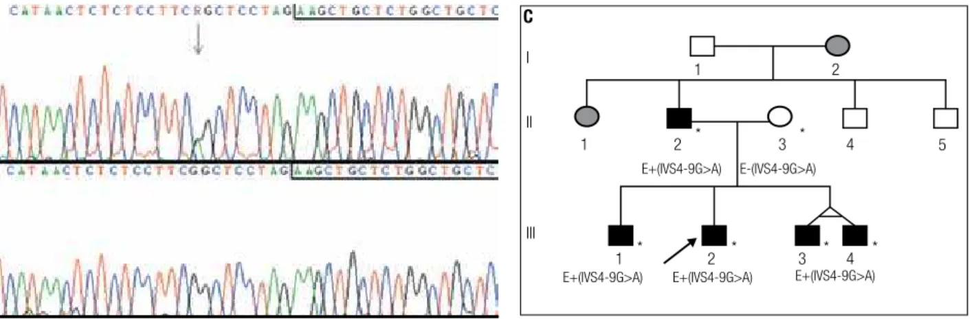
Figure 2.Sequence analysis showing the IVS4-9G>A mutation. Sequences boxed correspond to the first nucleotides of exon 5 and the arrow indicates the nucleotide change. (A) Heterozygous G to A substitution detected in blood DNA of the patient. (B) DNA from his normal mother. (C) Family pedigree. The arrow indicates the index-case. Asterisks indicate individuals that were personally analyzed. E+(IVS4-9G>A) and E-(IVS4-9G>A) denote that the individuals have been molecularly evaluated for the mutation and are, respectively, heterozygous and normal homozygous. Individuals I.2 and II.1 were not molecularly analyzed but clinical data indicated MEN1 manifestation.
all affected members showed variable clinical manifesta-tions, with early onset (from 8 to 24 years of age) in all the brothers.
Among enteropancreatic tumors, gastrinomas are the most common and the major cause of morbidity and mortality, as occurred in the paternal grandmother. They are often multiple and present high risk of me-tastasis. Conversely, insulinomas are usually benign and surgery tends to be curative (18). Non-functional enter-opancreatic tumors may appear in the irst two decades of life and present risk of malignancy. Medical conduct already recommends to begin screening for enteropan-creatic tumors, especially for insulinoma, at the age of 5 years (3,18). Nowadays, this recommendation has been extended to the non-functioning tumors as well (4).
To our knowledge, this is the second report describ-ing clinical symptoms and genetic testdescrib-ing in twins with MEN1. Similarly to the case reported by Rix and cols. (19), the twin brothers presented different phenotypes, with primary hyperparathyroidism and
hyperprolac-tinemia in both, but only one developed insulinoma. Variability in phenotypes may relect a random chance for a second mutation in susceptible cells with loss of heterozygosity, in accordance with Knudson’s “two hit” hypothesis (7,14). Some of the affected individuals described here are heterozygous for frequent SNPs in MEN1 gene. The p.D418D polymorphism caused by the c.1269C>T nucleotide change in exon 9 was found in the father and in individuals III-1 and III-4. This polymorphism has previously been associated with spo-radic hyperparathyroidism (20) and it was found to be more frequent in Brazilian patients than in those stud-ied in the USA and Europe (21). In the present study hyperparathyroidism was diagnosed in ive affected in-dividuals but only three carry the p.D418D polymor-phism, therefore it seems that hyperparathyroidism and p.D418D are not associated in those cases. Whereas, other polymorphisms have been identiied (Figure 1D), if combined to speciic haplotypes we found that the “TACC” haplotype (c.-35A>T>C, c.799-9G>A, Table 3. Heterozygosis for SNPs and c.799-9G>A mutation in MEN1 family
5’UTR Intron 4 Exon 9 Intron 9
SNP access number rs679946 mutation rs2071313 rs654440
cDNA position c.-35A>C>T c.799-9G>A c.1269C>T c.1365+103C>G
II-2 A/T G/A C/T C/G
II-3 T/T G/G C/C C/C
III-1 T/C G/A C/T C/G
III-2 T/T G/A C/C C/C
III-3 T/T G/A C/C C/C
III-4 T/C G/A C/T C/G
I
II
III
1 2
1 2 * *
* * *
*
3 4
4 3 2
1
E+(IVS4-9G>A) E+(IVS4-9G>A) E+(IVS4-9G>A) E-(IVS4-9G>A)
E+(IVS4-9G>A)
Cop
yright
© ABE&M t
odos os dir
eit
os r
eser
vados
.
c.1269C>T, and c.1365+103C>G) is present in the individuals who developed insulinoma. Two other hap-lotypes were identiied: “CAGT” in two brothers and probably “TATG” in the father. Since all siblings carry the IVS4-9G>A mutation inherited from the father, it is very dificult to ind an explanation for such dif-ferences within the MEN1 gene. Possible mechanisms might involve double independent events generating germline mosaicism.
In late onset heritable disorders, the beneits of genetic diagnosis must be balanced with the psycho-logical effects of the screening procedure. As no cu-rative treatment can be offered to carriers of MEN1 gene mutations, early detection will be positive for the possibility of initiating adequate treatment at a pres-ymptomatic stage, which may decrease morbidity and mortality (12,21,22). Therefore, parents within fami-lies with a clinical diagnosis of MEN1 should receive genetic counseling in order to be able to decide about genetically testing their children.
The earliest presentation of an endocrine tumor in MEN1 reported in the literature was an aggressive pro-lactin and GH-secreting pituitary macroadenoma in a 5-year-old boy (23). Pancreatic insulinoma has been described in a 7-year-old boy (24), followed by primary hyperparathyroidism at the age of 8 years (3). Our in-dex patient showed an insulinoma when he was 8 years old, reinforcing the proposition that a clinical screening program for children carrying MEN1 gene mutations should start at the age of 10-15 years (21,22,25,26). These children should be evaluated with annual blood tests by measuring serum calcium, pancreatic polypep-tide and gastrin, prolactin and insulin-like growth fac-tor-1. Annual pancreatic ultrasound, and pancreatic and pituitary MRI every 3-5 years, should also be per-formed. A consensus statement from 2001 (3) recom-mended that screening should begin at 5 years of age as mentioned above.
Several germline MEN1 gene mutations have been described in Brazilian patients (21,27) and they are mainly frameshift, nonsense or missense mutations. The G>A substitution at 9 nucleotides upstream from the splicing junction of the MEN1 gene in intron 4, described before in European and Japanese patients (28,29), is reported here for the irst time in a Brazil-ian family. The IVS4-9G>A that was irst described as a polymorphism with heterozygosity frequency of 3% (30), however, it was further demonstrated to produce two different transcripts. The most abundant transcript
presented an insertion of seven nucleotides in the exon 4 and 5 junction, and the less abundant had a normal mRNA sequence (29). Therefore, it was associated to MEN1 phenotype in an European adult patient with adrenocortical adenoma and lung carcinoid tumor (28) and also in three Japanese adult patients from two un-related families, but with different phenotypes. The different phenotypes were: one with hyperparathyroid-ism, repeated gastroduodenal ulceration, subcutaneous lipoma and insulinoma; the other two sisters presented hyperparathyroidism, whereas only one had insulinoma (29). Our index case also had insulinoma and hyper-parathyroidism, however, the manifestations were ob-served earlier than those previously reported. In all cas-es this alteration was dcas-escribed as a germline mutation.
In conclusion, the early diagnose and the long-term follow-up of the MEN1 family described in the present paper reinforces that careful family history with regards to familial MEN1 should always be investigated in pa-tients presenting with insulinoma in childhood. Addi-tionally, the possibility of conirming MEN1 diagnosis by genetic testing should be offered to the children and their families.
Acknowledgements: The authors would like to thank Dr. Márcio José da Silva from the sequencing facility. We also thank Dr. Lu-ciana de Campos Leite Medeiros and Mrs. Mara Sanches Guarag-na for reading the manuscript.
Disclosure: no potential conlict of interest relevant to this article was reported.
REFERENCES
1. Carling T. Multiple endocrine neoplasia syndrome: genetic basis for clinical management. Curr Opin Oncol. 2005;17:7-12. 2. Guang-Wen Z, Yao W, Xi C, Xiao-Hua J, Xiao-Ying L, Guang N, et
al. Diagnosis and surgical treatment of multiple endocrine neo-plasia. Chin Med J. 2009;122:1495-500.
3. Brandi ML, Gagel RF, Angell A, Bilczikian JP, Beck-Peccoz P, Bordi C, et al. Consensus: guidelines for diagnosis and therapy of MEN type 1 and type 2. J Clin Endocrinol Metab. 2001;86:5658-71. 4. Newey PJ, Jeyabalan J, Walls GV, Christie PT, Gleeson FV, Gould
S, et al. Nonfunctioning tumors in children with MEN1. J Clin En-docrinol Metab. 2009;94:3640-6.
5. Corbetta S, Pizzocaro A, Peracchim, Beck-Peccoz P, Faglia G, Spada A. Multiple endocrine neoplasia type 1 in patients with recognized pituitary tumours of different types. Clin Endocrinol. 1997;47:507-12.
6. Yoshimoto K, Saito S. Clinical characteristics in multiple endo-crine neoplasia type 1 in Japan: a review of 106 patients. Nippon Naibunpi Gakkai Zasshi. 1991;67:764-74.
Cop
yright
© ABE&M t
odos os dir
eit
os r
eser
vados
.
8. Chandrasckharappa SC, Guru SC, Manickan P, Olufemi SE, Collins PS, Emmert-Buck MR, et al. Positional cloning of the gene for multiple endocrine neoplasia-type 1. Science. 1997;276:404-7. 9. Franchi GM, Villa VV, Carrera P, Sartorio SM, Maffi P, Bosi E, et al.
A new mutation in the MEN1 gene. Cancer Genet Cytogenet. 2009;192:199-01.
10. Kim H, Lee JR, Cho EJ, Liu JO, Youn HD. Menin, a tumor suppres-sor, represses JunD-mediated transcriptional activity by asso-ciation with mSln3A-histonic deacctylase complex. Cancer Res. 2003;63:6135-9.
11. Falchetti A, Marini F, Brandi ML. Multiple endocrine neoplasia. Ge-neReviews 2010. Available at: http://www.ncbi.nlm.nih.gov/book-shelf/br.fcgi?book=gene&part=men1. Accessed at: 27 Jul 2010. 12. Geerdink BAM, Van der Luijt RB, Lips CJM. Do patients with
mul-tiple endocrine neoplasia syndrome type 1 benefit from periodi-cal screening? Eur J Endocrinol. 2003;149:577-82.
13. Falchetti A, Brandi ML. Multiple endocrine neoplasia type I va-riants and phenocopies: more than a nosological issue? J Clin Endocrinol Metab. 2009;94:1518-20.
14. Marx SJ, Nieman LK. Aggressive pituitary tumours in MEN1: do they refute the two-hit model of tmorigenesis? J Clin Endocrinol Metab. 2002;87:453-6.
15. Sambrook J, Fritsch EF, Maniatis TE. Molecular cloning, a labora-tory manual. New York: Cold Spring Harbor; 1989.
16. Lourenço Jr DM, Coutinho FL, Toledo RA, Montenegro FL, Cor-reia-Deur JE, Toledo SP. Early-onset, progressive, frequent, ex-tensive, and severe bone mineral and renal complications in multiple endocrine neoplasia type 1-associated primary hyperpa-rathyroidism. J Bone Miner Res. 2010;25(11):2382-91.
17. Ruszniewski P, Fave GD, Cadiot G, Komminoth P, Chung D, Kos--Kudla B. Well-differentiated gastric tumors/carcinomas. Neuro-endocrinology. 2006;84:158-64.
18. Schussheim DH, Skarulis MC, Agarwal SK, Simonds WF, Burns AL, Spiegel AM, et al. Multiple endocrine neoplasia type 1: new cli-nical and basic findings. Trends Endocrinol Metab. 2001;12:173-8. 19. Rix M, Hertel NT, Nielsen FC, Jacobsen BB, Hoejberg AS, Brixen
K, et al. Cushing’s disease in childhood as the first manifestation of multiple endocrine neoplasia syndrome type 1. Eur J Endocri-nol. 2004;151:709-15.
20. Correa P, Lundgren E, Rastad J, Åkerström G, Westin G, Carling T. Multiple endocrine neoplasia type 1 polymorphism D418D is associated with sporadic primary hyperparathyroidism. Surgery. 2002;132:450-5.
21. Toledo RA, Lourenço DM, Coutinho FL, Quedas E, Mackowiack I, Machado MC, et al. Novel MEN1 germline mutations in Brazilian families with multiple endocrine neoplasia type 1. Clin Endocri-nol. 2007;67:377-84.
22. Lourenço Jr DM, Toledo RA, Coutinho FL, Margarido LC, Siqueira SA, dos Santos MA, et al. The impact of clinical and genetic scre-enings on the management of the multiple endocrine neoplasia type 1. Clinics (Sao Paulo). 2007;62:465-76.
23. Stratakis CA, Shussheim DH, Freedman SM, Keil MF, Pack SD, Agarwal SK, et al. Pituitary macroadenoma in 5-year-old: an early expression of multiple endocrine neoplasia type 1. J Clin Endocri-nol Metab. 2000;85:4776-80.
24. Kontogeorgos G, Kapranos N, Tzavara I, Thalassinos N, Rologis D. Monossomy of chromosome 11 in pituitary adenoma in a patient with familial multiple endocrine neoplasia type 1. Clin Endocrinol. 2001;54:117-20.
25. Johnston LB, Chew SL, Trainer PJ, Reznck R, Grossman AB, Bes-ser GM, et al. Screening children at risk of developing inherited endocrine neoplasia syndromes. Clin Endocrinol. 2000;52:127-36. 26. Johnston LB, Chew SL, Lowe D, Reznck R, Monsom JP, Savage
MO. Investigating familial endocrine neoplasia syndromes in chil-dren. Horm Res. 2001;55:31-5.
27. Lourenço Jr DM, Toledo RA, Mackowiak II, Coutinho FL, Caval-canti MG, Correia-Deur JE, et al. Multiple endocrine neoplasia type 1 in Brazil: MEN1 founding mutation, clinical features, and bone mineral density profile. Eur J Endocrinol. 2008;159:259-74. 28. Görtz B, Roth J, Speel EJM, Krähemmann A, De Krieger RR,
Matias-Guiu X, et al. MEN1 gene mutation analysis of sporadic adrenocortical lesions. Int J Cancer. 1999;80:373-9.
29. Kishi M, Tsukada T, Shimizu S, Hosono K, Ohkubo T, Kosuge T, et al. A novel splicing mutation (894-9 G-A) of the MEN1 gene responsible for multiple endocrine neoplasia type 1. Cancer Letters. 1999;142:105-110. 30. Bassett JH, Forbes SA, Pannett AA, Lloyd SE, Christie PT,