Cop
yright
© ABE&M t
odos os dir
eit
os r
eser
vados
.
46,XX Male – Testicular Disorder of
Sexual Differentiation (DSD): hormonal,
molecular and cytogenetic studies
Homem 46,XX (DSD testicular): estudos hormonal, molecular e citogenético
Cresio Alves1, Zilda Braid1, Fernanda Borchers Coeli2,
Maricilda Palandi de Mello2
SUMMARY
The XX male syndrome – Testicular Disorder of Sexual Differentiation (DSD) is a rare condition characterized by a spectrum of clinical presentations, ranging from ambiguous to normal male genitalia. We report hormonal, molecular and cytogenetic evaluations of a boy presenting with this syndrome. Examination of the genitalia at age of 16 months, showed: penis of 3.5 cm, proxi-mal hypospadia and scrotal testes. Pelvic ultrasound did not demonstrate Mullerian duct struc-tures. Karyotype was 46,XX. Gonadotrophin stimulation test yielded insufficient testosterone production. Gonadal biopsy showed seminiferous tubules without evidence of Leydig cells. Molecular studies revealed that SRY and TSPY genes and also DYZ3 sequences were absent. In addition, the lack of deletions or duplications of SOX9, NR5A1, WNT4 and NROB1 regions was verified. The infant was heterozygous for all microsatellites at the 9p region, including DMRT1 gene, investigated. Only 10% of the patients are SRY-negative and usually they have ambiguous genitalia, as the aforementioned patient. The incomplete masculinization suggests gain of func-tion mutafunc-tion in one or more genes downstream to SRY gene. Arq Bras Endocrinol Metab. 2010;54(8):685-9
SUMÁRIO
A síndrome do homem XX é uma condição rara na qual o fenótipo da genitália externa pode variar de uma genitália ambígua até uma genitália masculina normal. Este estudo tem por obje-tivo relatar a avaliação hormonal, molecular e citogenética de um menino com essa síndrome. O exame da genitália externa na idade de 16 meses mostrava: pênis medindo 3,5 cm, hipos-padia proximal e testículos tópicos. A ultrassonografia pélvica não visualizou estruturas mulle-rianas. Cariótipo foi 46,XX. A testosterona sérica não se elevou após o teste de estímulo com gonadotrofina. Biópsias gonadais mostraram túbulos seminíferos, sem evidência de células de Leydig. Estudos moleculares revelaram ausência dos genes SRY, TSPY e DYZ3, bem como ausência de deleção ou duplicação das regiões SOX9, NR5A1, WNT4 e NROB1. A criança era he-terozigótica para todos os microssatélites da região 9p, incluindo o gene DMRT1. Apenas 10% dos pacientes com a síndrome do homem 46,XX são SRY-negativos. Nesses casos, a genitália geralmente é ambígua, como corroborado pelo paciente do presente relato. A masculinização incompleta sugere ganho de mutação funcional em um ou mais genes a jusante do gene SRY.
Arq Bras Endocrinol Metab. 2010;54(8):685-9
1 Pediatric Endocrinology Service, Hospital Universitário Professor Edgard Santos, Faculty of Medicine, Universidade Federal da Bahia (UFBA), Salvador, BA, Brazil 2 Centro de Biologia Molecular e Engenharia Genética, Universidade Estadual de Campinas (Unicamp), Campinas, SP, Brazil
Correspondence to: Cresio Alves
Rua Plínio Moscoso, 222/601 40157-190 − Salvador, BA, Brazil [email protected]
Received on Jun/4/2010 Accepted on Sept/12/2010
INTRODUCTION
T
he XX male syndrome (OMIM ID #400045), now termed Testicular DSD (Testicular Disorder of Sexual Differentiation), was irst described by La Cha-pelle and cols. in 1964 (1). It is a rare condition, with a frequency of 1:20,000-25,000 male newborns (1-3). It is characterized by a male phenotype with 46,XXkaryo-type (1-3). The SRY gene (sex-determining region on the Y), which is located in the Y chromosome, plays an important role in the process of sex determination (4,5). On basis of the analysis and detection of SRY gene, 46, XX male patients can be clinically divided into SRY-positive and the SRY-negative groups (6,7). SRY
Cop
yright
© ABE&M t
odos os dir
eit
os r
eser
vados
.
small azoospermic testes and hypergonadotropic hypo-gonadism (8), and most carry the SRY gene translocat-ed to the X chromosome during paternal meiosis (7,9); however, SRY autosomal translocations have also been reported in some XX males (10-12). The diagnosis of SRY-positive patients is usually achieved in adulthood during infertility investigation (3). The group of SRY -negative (OMIM ID 278850) includes patients with Ovotesticular-DSD, which is characterized by the pres-ence of both testicular and ovarian tissue in the gonads of the same individual (13,14), and Testicular-DSD characterized by a full development of both gonads as testes without any evidence of ovarian tissue (2). The occurrence of both Ovotesticular and Testicular-DSD in families, especially in identical twins, indicates them as different manifestations of the same disorder of go-nadal development (15-17). In the absence of SRY gene, the up-regulation or super-expression of some members of SOX family (Sry-related HMG-box) have been proposed as involved in XX male etiology (18-20). The diagnosis of SRY-negative testicular-DSD can be done in childhood upon investigation of ambiguous genitalia and gynecomastia.
The aim of this study was to analyze the case of a boy with XX male syndrome, emphasizing its hormon-al, molecular and cytogenetic aspects.
CASE REPORT
A 16-month-old infant was referred to endocrine eval-uation in our service. The mother reported that fetal sonograms between the 3rd-6th months of gestational age were compatible with a female fetus, while at the 7th month the diagnosis was compatible with a male fetus with hypospadia. There were absence of endocri-nopathies and hormonal exposure during pregnancy, no history of genetic syndromes, ambiguous genitalia or precocious neonatal death in the family. Parents are
not consanguineous and his 5 years-old sister is healthy. On physical examination: weight: 10.2 kg (P. 10 - P. 25), length: 73.5 cm (< P. 5%). The penile length was 3.5 cm with a proximal hypospadia, both testes were normally located (with a more hypoplastic left testicle) and absent pubarche. Karyotype with G band: 46,XX. Neonatal screening, including 17-hydroxyprogesterone evaluation was normal. Pelvic ultrasound: no evidence of Mullerian duct structures. Gonadal biopsy: seminiferous tubules lined by small cuboidal cells, without evidence of Leydig cells.
MATERIALS AND METHODS
The hormonal studies were performed with the human chorionic gonadotrophin stimulation tests (beta-hCG) measuring total testosterone, before and after the ad-ministration of three doses of beta-hCG (4,500 IU, in-tramuscularly, every other day).
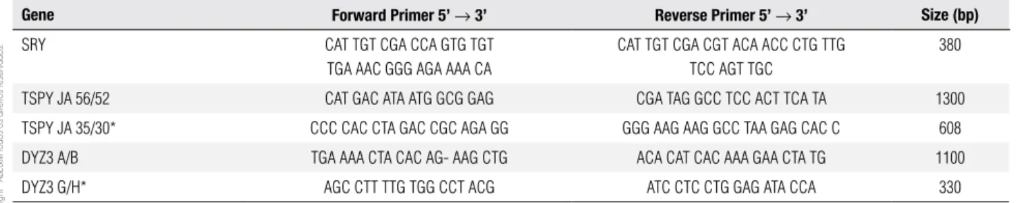
Molecular studies were performed after DNA ex-traction from peripheral blood, that followed standard techniques (21). Y-chromosome sequence analysis was conducted by PCR and nested-PCR. SRY gene was am-pliied with primers depicted in table 1, which also de-scribes those primers used in the irst and in the second rounds of nested-PCR for the TSPY gene and for the Y-centromeric region DYZ3. After PCR ampliication, reaction products (10 µl) were submitted to 2% agarose gel electrophoresis with a molecular weight marker and stained with ethidium bromide. Each PCR reaction con-tained normal female and male controls. Nested-PCR experiments contained also a template-free sample. Hap-loinsuiciency of DMRT1 gene was evaluated with mic-rosatellites mapping within the criptical interval 9p24.3 according to Calvari and cols. (22), Guioli and cols. (23) and Veitia and cols. (24) (Table 2). MLPA assay was performed using SALSA MLPA P185 Intersex probe-mix (MRC-Holland) following provider instructions. 46,XX male: molecular studies
Table 1. Primer sequences for PCR and nested-PCR of the Y chromosome
Gene Forward Primer 5’ → 3’ Reverse Primer 5’ → 3’ Size (bp)
SRY CAT TGT CGA CCA GTG TGT
TGA AAC GGG AGA AAA CA
CAT TGT CGA CGT ACA ACC CTG TTG TCC AGT TGC
380
TSPY JA 56/52 CAT GAC ATA ATG GCG GAG CGA TAG GCC TCC ACT TCA TA 1300
TSPY JA 35/30* CCC CAC CTA GAC CGC AGA GG GGG AAG AAG GCC TAA GAG CAC C 608
DYZ3 A/B TGA AAA CTA CAC AG- AAG CTG ACA CAT CAC AAA GAA CTA TG 1100
DYZ3 G/H* AGC CTT TTG TGG CCT ACG ATC CTC CTG GAG ATA CCA 330
Cop
yright
© ABE&M t
odos os dir
eit
os r
eser
vados
.
Each MLPA reaction was analyzed on an ABI 310 Ge-netic Analyzer (ABI PRISM/PE Biosystems, Foster City, CA, USA) and results were evaluated using Ge-nescan and Genotyper softwares (Applied Biosystems, Foster City, CA, USA). The probemix included in the MLPA kit contains probes for SOX9 gene and its lank-ing region at 17q24.3, NR5A1 gene (SF-1) at 9q33, WNT4 (1p36.12), NR0B1 gene (DAX-1) at Xp21.2 and, also, control probes mapping within other regions on X and Y chromosomes. Data were analyzed using free Coffalyser MLPA data analysis software available in the provider website (www.mrc-holland.com). Data were normalized with results of ive control individuals and with internal controls (reference probes). Normal-ized relative values ranging within a conidence interval of 0.8 to 1.3, which was established by control data variations, corresponded to two gene copies in the gen-otype, whereas values below 0.8 and above 1.3 would correspond to deletion (one gene copy) and duplica-tion (three gene copies), respectively.
This paper was submitted to the Institutional Re-view Board of our Institution, which approved its ethi-cal aspects.
Table 3. Human chorionic gonadotrophin stimulation test at the chronological age of 16 months
Time Hormonal evaluation
LH FSH Total testosterone
Baseline < 0,07 UI/mL (normal < 14 UI/L)
< 0,3 UI/mL (normal < 10 UI/L)
12 ng/dL (normal < 40 ng/dL)
Post- -stimulation
40 ng/dL (normal > 150 ng/dL)
FSH: follicle-stimulating hormone; LH: luteinizing hormone.
RESULTS
The hormonal studies yielded low serum levels of free testosterone before and after the beta-hCG stimulation test suggesting a deicient testicular production (Table 3).
Molecular analyses demonstrated absence of both SRY and TSPY genes and also Y-centromeric DYZ3
sequence indicating absence of Y-chromosome-derived sequences in the genotype. The infant was heterozygous for all 9p microsatellites indicating haplosuficiency for DMRT1 gene. Deletions or duplications of genes involved in sex differentiation processes were investigated by MLPA analysis. Normal copy number for all genes can be observed in igure 1. Absence of Y-chromosome
Figure 1. MLPA analysis. Integrated and normalized MLPA data for genes:SOX9 (17q24.3), NR5A1 (9p33), WNT4 (1p36.12) and NR0B1 (Xp21.2). Genes or gene regions are indicated below each bar in the graphic; e = exon. Chromosome regions are delimitated by black horizontal bars. Absence of signals for both UTY region and SRY gene probes on the Y chromosome is observed. The height of vertical bars corresponds to integrated and normalized peak areas obtained in capillary electrophoresis; values between 0.8 and 1.3 indicate two copies of the fragment.
1.6 1.4 1.2 1 0.8 0.6 0.4 0.2 0
Relative peak area
Chr. 17 Chr. 9 Chr. 6 Chr. 1 Chr. X Chr. Y Reference probes
SOX9-e1 SOX9-e1 SOX9-e2 SOX9-e3
SOX9-area 1 SOX9-area 2 SOX9-area 3 SOX9-area 4 SOX9-area 5 NR5A1-e1 NR5A1-e3 NR5A1-e4 NR5A1-e5 NR5A1-e6 CYP21A2-e1 CYP21A2-e3 CYP21A1P-e1
CYP21A1P-e10
WNT4-e1 WNT4-e2 WNT4-e3 WNT4-e4 WNT4-e5 NROB1-5’e1 NROB1-e1 NROB1-e2
COL4A5
PHEX GJB1
PQBP1
UTY SRY RF-1 RF-2 RF-3 RF-4 RF-5 RF-6 RF-7 RF-8
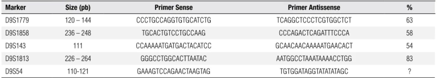
Table 2. Primer sequences utilized for the 9p microsatellite studies
Marker Size (pb) Primer Sense Primer Antissense %
D9S1779 120 – 144 CCCTGCCAGGTGTGCATCTG TCAGGCTCCCTCGTGGCTCT 63
D9S1858 236 – 248 TGCACTGTCCTGCCAAG CCCAGACTCAGATTTCCCA 58
D9S143 111 CCAAAAATGATGACTACATCC GCAACAACAAAAATGAACACT 54
D9S1813 226 – 264 GGGCCTGGCACTTAATAC AATGGCCTAAATAAAACCTGG 83
D9S54 110-121 GAAAGTCCAGAACTAAGTAG TGTGGATAGGTATATATAGC ?
Cop
yright
© ABE&M t
odos os dir
eit
os r
eser
vados
.
46,XX male: molecular studies
probe signals can also be noted conirming PCR assays. Therefore, MLPA did not show any copy number variation for either SOX9, NR5A1 (SF-1), WNT4 or DAX-1, which is coded by NR0B1 gene.
DISCUSSION
About 90% of 46,XX (testicular DSD) men are SRY gene carriers. In most cases this gene is translocated to the short arm-end of the paternal X chromosome (1,3). Therefore the mechanism generating this syndrome in SRY carriers involves mainly a mistake in the cross-over between pseudoautosomal regions of sexual chromo-somes during the paternal meiosis (10,18). However, 10% of the patients are SRY-negative (1,3). Molecular analysis in our patient showed absence of SRY, which is an unusual form of the XX male syndrome.
It has been considered that, in the absence of a functional SRY protein, male phenotype can also be induced in 46,XX individuals by excessive or absent ac-tions of other determinants within the sexual differen-tiation cascade such as SOX9, DAX-1, Ad4BP/SF-1, WT1, GATA4, WNT4, FGF9 and RSPO1 (10,18). In the patient described here, there was no evidence for SOX9, SF-1, WNT4 and DAX-1 duplications or dele-tions that were investigated by MLPA technique.
In general, 46,XX male presents with three pheno-types: normal internal and external genitalia; ambiguous genitalia; and, ovotesticular DSD (1-3). All are charac-terized by infertility caused by absence of the region AZF present in the long arm of the Y chromosome (1-3). In the classical presentation, 46,XX males are found with normal penile length, infertility and presence of Wolff duct structures without Mullerian duct structures (18). There is a higher prevalence of microrchidism and gynecomastia and a trend to female pubic hair distribu-tion, with decreased facial hair (1,10). Short stature can occur due to absence of either pubertal spurt depen-dent on testosterone or Y-chromosome genes related to speciic growth forms.
Most SRY-positive patients have normal external genitalia, whereas SRY-negative patients are found within a large spectrum of masculinization ranging from ambiguous to normal genitalia (1,3). The diag-nosis of the patient reported here was performed dur-ing childhood due to ambiguous genitalia. Ovotesticu-lar DSD, an important differential diagnosis, was ruled out in the patient after gonadal biopsy. During puberty, testosterone levels are normal evolving, in adulthood,
to hypergonadotrophic hypogonadism (6,7,10,15,18). Some authors evidenced a deicient testosterone pro-duction after β-hCG stimulation test; whereas, others reported a normal testosterone production by the Ley-dig cells, although not ruling out the possibility of a testosterone deiciency during adulthood (3,10).
In conclusion, the SRY-negative XX male incom-plete masculinization in this case cannot be attributed to SOX9, SF-1, WNT4 and DAX-1 duplications or de-letions, however, gain of function mutations in one or more genes downstream the SRY gene pathway or a loss of function in some gene associated with the inhi-bition of male development should be considered for further investigation.
Disclosure: no potential conlict of interest relevant to this article was reported.
REFERENCES
1. Vorona E, Zitsmann M, Gromoll J, Schüring N, Nieschlag E. Clini-cal, endocrinologiClini-cal, and epigenetic features of the 46,XX male syndrome, compared with 47,XXY Klinefelter patients. J Clin En-docrinol Metab. 2007;92:3458-65.
2. Rajender S, Vutukuri R, Gupta NJ, Chakravarty B, Singh L, Than-garaj K. SRY-negative 46,XX male with normal genitals, complete masculinization and infertility. Mol Hum Reprod. 2006;5:341-6. 3. Ergun-Longmire B, Vinci G, Alonso L, Matthew S, Tansi S, Lin-Su
K, et al. Clinical, hormonal and cytogenetic evaluation of 46,XX male and review of the literature. J Pediatr Endocrinol Metab. 2005;18(8):739-48.
4. Berta P, Hawkins JR, Sinclair AH, Taylor A, Griffiths BL, Goo-dfellow PN, et al. Genetic evidence equating SRY and the testis-determining factor. Nature. 1990;348(6300):448-50.
5. Koopman P, Münsterberg A, Capel B, Vivian N, Lovell-Badge R. Expression of a candidate sex-determining gene during mouse testis differentiation. Nature. 1990;348(6300):450-2.
6. Kim JW, Bak CW, Chin MU, Cha DH, Yoon TK, Shim SH. SRY-ne-gative 46,XX infertile male with Leydig cell hyperplasia: clinical, cytogenetic, and molecular analysis and review of the literature. Fertil Steril. 2010;94(2):753.e5-9.
7. Wang T, Liu JH, Yang J, Chen J, Ye ZQ. 46, XX male sex reversal syndrome: a case report and review of the genetic basis. Andro-logia. 2009;41(1):59-62.
8. de la Chapelle A. The etiology of maleness in XX men. Hum Ge-net. 1981;58(1):105-16.
9. Fechner PY, Marcantonio SM, Jaswaney V, Stetten G, Goodfellow PN, Migeon CJ, et al. The role of the sex-determining region Y gene in the etiology of 46,XX maleness. J Clin Endocrinol Metab. 1993;76(3):690-5.
10. Dauwerse JG, Hansson KBM, Brouwers AAM, Peters DJM, Breuning MH. An XX male with the sex-determining region Y gene inserted in the long arm of cromossome 16. Fertil Ste-ril.2006:86:463.e2-e5.
Cop
yright
© ABE&M t
odos os dir
eit
os r
eser
vados
.
12. Queralt R, Madrigal I, Vallecillos MA, Morales C, Ballescá JL, Oli-va R, et al. Atypical XX male with the SRY gene located at the long arm of chromosome 1 and a 1qter microdeletion. Am J Med Genet A. 2008;146A(10):1335-40.
13. Parada-Bustamante A, Ríos R, Ebensperger M, Lardone MC, Piot-tante A, Castro A. 46,XX/SRY-negative true hermaphrodite. Fertil Steril. 2010;94(6):2330.13-6.
14. Berger-Zaslav AL, Mehta L, Jacob J, Mercado T, Gadi I, Tepperberg JH, et al. Ovotesticular disorder of sexual development (true her-maphroditism). Urology. 2009;73(2):293-6.
15. Maciel-Guerra AT, de Mello MP, Coeli FB, Ribeiro ML, Miranda ML, Marques-de-Faria AP, et al. XX Maleness and XX true her-maphroditism in SRY-negative monozygotic twins: additio-nal evidence for a common origin. J Clin Endocrinol Metab. 2008;93(2):339-43.
16. Ramos ES, Moreira-Filho CA, Vicente YA, Llorach-Velludo MA, Tucci S, Duarte MH, et al. SRY-negative true hermaphrodites and an XX male in two generations of the same family. Hum Genet. 1996;97:596-8.
17. Zenteno JC, Lopez M, Vera C, Mndez JP, Kofman-Alfaro S. Two SRY-negative XX male brothers without genital ambiguity. Hum Genet. 1997;100:606-10.
18. Kojima Y, Hayashi Y, Mizuno K, Sasaki S, Fukui Y, Koopman P, et al. Up-regulation of SOX9 in human sex-determining region on
the Y chromosome (SRY)-negative XX males. Clin Endocrinol. 2008:68(5):791-9.
19. Refai O, Friedman A, Terry L, Jewett T, Pearlman A, Perle MA, et al. De novo 12;17 translocation upstream of SOX9 resulting in 46,XX testicular disorder of sex development. Am J Med Genet A. 2010;152A(2):422-6.
20. Polanco JC, Wilhelm D, Davidson TL, Knight D, Koopman P. Sox10 gain-of-function causes XX sex reversal in mice: implications for human 22q-linked disorders of sex development. Hum Mol Ge-net. 2010;19(3):506-16.
21. Sambrook J, Fristsch EF, Maniatis TE. Molecular cloning: a labo-ratory manual. Cold Spring Harbour Labolabo-ratory, Press. New York: Cold Spring Harbour, 1989.
22. Calvari V, Bertini V, De Grandi A, Peverali G, Zuffardi O, et al. A new submicroscopic deletion that refines the 9p region for sex reversal. Genomics. 2000:65(3):203-12.
23. Guioli S, Schmitt K, Critcher R, Bouzyk M, Spurr NK, Ogata T, et al. Molecular analysis of 9p deletions associated with XY sex rever-sal: refining the localization of a sex-determining gene to the tip of the chromosome. Am J Hum Genet. 1998:63(3):905-8. 24. Veitia RA, Nunes M, Quintana-Murci L, Rappaport R, Thibaud