Original article
cyclin-dependent kinase inhibitor 1A (CDKN1A) in central
nervous system tumors
Frequência dos polimorismos e da expressão protéica do inibidor de quinase dependente de
ciclina 1A
(CDKN1A)
em tumores do sistema nervoso central
Mev Dominguez Valentin
I, Renata Canalle
II, Rosane de Paula Queiroz
III, Luiz Gonzaga Tone
IVHospital das Clínicas (HC) of Faculdade de Medicina de Ribeirão Preto (FMRP), Universidade de São Paulo (USP), Ribeirão Preto, São Paulo, Brazil
IPhD, Genomic and Molecular Laboratory, Research Center of Hospital A. C. Camargo, São Paulo, Brazil.
IIPhD. Professor, Biomedicine School, Ministro Reis VellosoCampus, Universidade Federal do Piauí (UFPI), Parnaíba, Piauí, Brazil.
IIIPhD. Investigator, Childcare and Pediatric Department, Faculdade de Medicina de Ribeirão Preto (FMRP), Universidade de São Paulo (USP), Ribeirão Preto, São Paulo, Brazil. IVMD, PhD. Professor, Childcare and Pediatric Department. Faculdade de Medicina de Ribeirão Preto (FMRP), Universidade de São Paulo (USP), Ribeirão Preto, São Paulo, Brazil.
ABSTRACT
CONTEXT AND OBJECTIVE: Genetic investigation of central nervous system (CNS) tumors provides valuable information about the genes regulating proliferation, differentiation, angiogenesis, migration and apoptosis in the CNS. The aim of our study was to determine the prevalence of genetic
polymorphisms (codon 31 and 3’ untranslated region, 3’UTR) and protein expression of the cyclin-dependent kinase inhibitor 1A (CDKN1A) gene in patients with and without CNS tumors.
DESIGN AND SETTING: Analytical cross-sectional study with a control group, at the Molecular Biology Laboratory, Pediatric Oncology Department, Hospital das Clínicas de Ribeirão Preto.
METHODS: 41 patients with CNS tumors and a control group of 161 subjects without cancer and paires for sex, age and ethnicity were genotyped using polymerase chain reaction-restriction fragment length polymorphism (PCR-RFLP). Protein analysis was performed on 36 patients with CNS tumors, using
the Western Blotting technique.
RESULTS: The frequencies of the heterozygote (Ser/Arg) and polymorphic homozygote (Arg/Arg) genotypes of codon 31 in the control subjects were 28.0% and 1.2%, respectively. However, the 3’UTR site presented frequencies of 24.2% (C/T) and 0.6% (T/T). These frequencies were not statistically different (P > 0.05) from those seen in the patients with CNS tumors (19.4% and 0.0%, codon 31; 15.8% and 2.6%, 3’UTR site). Regarding the protein
expression in ependymomas, 66.67% did not express the protein CDKN1A. The results for medulloblastomas and astrocytomas were similar: neither of them expressed the protein (57.14% and 61.54%, respectively).
CONCLUSION: No signiicant differences in protein expression patterns or polymorphisms of CDKN1A in relation to the three types of CNS tumors were observed among Brazilian subjects.
RESUMO
CONTEXTO E OBJETIVO: A investigação genética dos tumores do sistema nervoso central (SNC) provê valiosa informação sobre os genes que regulam a proliferação, diferenciação, angiogênese, migração e apoptose. O objetivo deste estudo é determinar a prevalência entre os polimorismos genéticos
(códon 31 e da região 3’ não traduzida, 3’UTR) e a expressão protéica do gene inibidor de quinase dependente de ciclina 1A (CDKN1A) em pacientes com e sem tumor do SNC.
TIPO DE ESTUDO E LOCAL: Estudo transversal analítico com grupo controle, desenvolvido no Laboratório de Biologia Molecular do Departamento de Oncologia Pediátrica do Hospital das Clínicas de Ribeirão Preto.
MÉTODOS: 41 pacientes com tumor do SNC e um grupo controle de 161 indivíduos sem câncer pareados por idade, sexo e etnia foram genotipados mediante uma reação de polimorismo no comprimento de fragmentos de restrição (RFLP). A análise das proteínas foi realizada em 36 pacientes com
tumor de SNC mediante Western Blotting.
RESULTADOS: A frequência do genótipo heterozigoto (Ser/Arg) e do homozigoto polimórico (Arg/Arg) do códon 31 nos controles foi 28,0% e 1,2%, respectivamente. Entretanto, o sítio 3’UTR apresentou uma frequência de 24,2% (C/T) e 0,6% (T/T). Estas frequências não são signiicativamente diferentes (P > 0,05) daquelas observadas no grupo dos pacientes com tumor de SNC (19,4% e 0,0%, códon 31; 15,8% e 2,6%, sítio 3’UTR).
Com respeito à expressão protéica, nos ependimomas, 66,67% não expressaram a proteína CDKN1A. Estes resultados foram similares entre os meduloblastomas e os astrocitomas, os quais não expressaram a proteína com 57,14% e 61,54%, respectivamente.
CONCLUSÃO: Não foram encontradas diferenças signiicativas entre o padrão de expressão protéica, polimorismos de CDKN1A e os três tipos de tumores de SNC em indivíduos brasileiros.
Key words:
Cyclin-dependent kinase inhibitor
p21. Brain neoplasms.
Polymorphism, genetic. Polymorphism, restriction fragment
length. Blotting, western.
PALAVRAS-CHAVES:
Inibidor de quinase dependente de ciclina p21.
Neoplasias encefálicas. Polimorismo genético.
Polimorismo de fragmento de restrição.
INTRODUCTION
Central nervous system (CNS) tumors are the second most com-mon type of pediatric cancer, exceeded only by leukemia, and they are the most common solid tumor during childhood. Every year, approxi-mately 2,200 subjects younger than 20 years old are diagnosed with CNS tumors in the United States.1 he etiology of these tumors is
mul-tifactorial and probably varies according to the kind of tumor. Although the etiology of brain and CNS tumors is largely unknown, genetic investigation of CNS tumors provides valuable information about the genes regulating proliferation, diferentiation, angiogenesis, migration and apoptosis in the CNS. Studying the gene expression of this type of neoplasm may identify genes that are candidates for direct-ed gene therapy, in an attempt to increase the chances of cure for CNS tumor subgroups that have an unfavorable prognosis.2 One of these
genes, cyclin-dependent kinase inhibitor 1A (CDKN1A), is a universal inhibitor of cyclin-dependent kinases (CDKs) and plays critical roles in G1/S and G2/M transition regulation. CDKN1A may lead to diferen-tiation of normal and transformed cells and suppression of malignant cell growth in vitro and in vivo.2 Moreover, it may lead to apoptosis
in-volving the tumor protein 53 (TP53) and retinoblastoma protein (RB)
signaling pathways, in the same way as in senescence.3 Changes in the
CDKN1A gene and its expression may have an important role in can-cer pathogenesis, since its normal function comprises suspension of the cell cycle, terminal diferentiation and apoptosis. Diferent types of neo-plasms correlate with CDKN1A abnormalities, including cervical neo-plasms, colorectal carcinoma, ovary carcinoma, bladder cancer, prostate cancer, hepatomas and others.4-8
Two polymorphisms have been identiied and characterized in the CDKN1A gene. One of these, the codon 31 transversion (C→A), changes serine into arginine in the protein. he other one is located 20 nucleotides downstream of the termination codon, in the 3’ untranslat-ed region (3’UTR) of exon 3, with a transition of C→T.9,10 hese two
variants have recently been seen in a signiicant number of patients with lung, esophageal and breast cancer and sarcomas.
Some studies have analyzed CDKN1A gene expression as a possi-ble tool for diagnosis or as a prognosis indicator. Nadal et al.11 found
that six out of seven laryngeal tumors of grade IV presented low lev-els of CDKN1A,compared with 41 out of 42 tumors of grade I to III, which presented intermediate levels of CDKN1A. here is controver-sy regarding the prognostic value of CDKN1A expression in breast tis-sue. Two groups have associated increased expression with a good prog-nosis in osteosarcoma,3,12 while other groups have correlated this with
a worse prognosis.13,14 he relationships between CDKN1A expression
levels and the prognostic factors in other types of tumor are similarly contradictory.
Because of the high prevalence of CNS tumors in Brazil (18.3%), and continuing the previous work15 that detected the presence of
CD-KN1A gene polymorphisms through sequencing, the goal of this study was to determine the prevalence of CDKN1A gene polymorphisms in CNS tumor patients and control individuals. Furthermore, this study sought to analyze associations between these polymorphisms and the risk of developing the disease and its expression, along with the
relation-ship with tumor malignancy grade. As far as we know, this is the irst study in Brazil on these polymorphisms among a general population and among CNS tumor patients.
OBJECTIVE
he aim of this study was to determine the prevalence of genetic polymorphisms (codon 31 and 3’UTR) and protein expression of the cyclin-dependent kinase inhibitor 1A (CDKN1A) gene, and to deter-mine the risk of developing CNS-like medulloblastoma (16 cases), as-trocytoma (15 cases) and ependymoma (10 cases).
MATERIAL AND METHODS
Patients and controls
Tumor tissue stored in the Pediatrics Laboratory tumor bank was used. his tissue came from patients younger than 18 years of age who were treated for medulloblastoma (n = 16), ependymoma (n = 10) and astrocytoma (n = 15) in the Pediatric Oncology and Hematology De-partment and the Neurosurgery DeDe-partment of Hospital das Clínicas da Faculdade de Medicina de Ribeirão Preto(HC-FMRP-USP). he use of human tissue was approved by the institution’s Ethics Committee (pro-cedural number 9375/2003).
he criterion used for sample inclusion was the anatomopatho-logical diagnosis, in addition to the availability of samples containing deoxyribonucleic acid (DNA) and proteins in amounts and of qual-ity appropriate for the analysis. he classiication of the tumors ana-lyzed was based on the World Health Organization (WHO) nomen-clature, 2000.
Genomic DNA samples extracted from peripheral blood lympho-cytes were used as controls. hese came from the Pediatrics Laborato-ry controls bank and their use was approved by the Ethics Committee of the participating institution (HC-FMRP-USP) (procedural number 9374/2003). he control group was paired with the patients according to sex, age and ethnicity, and was formed by 97 female and 64 male sub-jects, with ages ranging from ive months to 20 years, without any his-tory of neoplastic or genetic disease. Epidemiological data on the study population were obtained by means of a standard interviewer-adminis-tered questionnaire that gathered data on social habits, health problems, family history of cancer and ancestry.
he human subject protocol was approved by the Ethics Commit-tee of HC-FMRP-USP (procedural number 7709/2004), and written informed consent was obtained from all subjects or their parents.
Polymerase chain reaction
he primers were designed for the 273 base pairs (bp) of exon 2 of the CDKN1A gene (codon 31), with 5’-GGATGTCCGTCAGAAC-CCAT–3’ (upstream) and 5’- GGTGCCAGGCCGCCTGCCTC – 3’ (downstream). he cycling conditions were: 94 °C for 5 minutes, 34 cy-cles of 94 °C for 40 seconds, 69 °C for 1 minute and 72 °C for 40
sec-onds, followed by 10 minutes at 72 °C for the inal extension. he prim-ers designed for the ampliication of the 300 bp of exon 3 of the CDK-N1A gene (3’UTR) were 5’-GGGCGGCCAGGGTATGTAC–3’ (up-stream) and 5’-CCCAGGGAAGGGTGTCCT G–3’ (down(up-stream). he cycling conditions were 95 °C for 5 minutes, 30 cycles of 95 °C for 30 seconds, 63 °C for 30 seconds and 72 °C for 30 seconds, followed by 10 minutes at 72 °C for the inal extension.
Analysis of restriction fragment length polymorphism
he 273 bp ampliied fragment of exon 2 of the CDKN1A gene was producedand digested by the restriction enzyme BsmAI (New Eng-land Biolabs, Beverey, Massachusetts, United States) using the modiied protocol of Mousses et al.16 he digestion of the wild type allele (Ser/
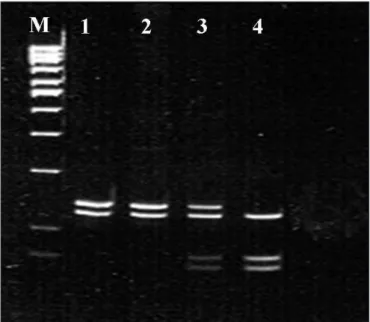
Ser) presented a constant site for restriction enzyme recognition and yielded two bands: one of 142 bp and the other of 131 bp. he ho-mozygote genotype for the polymorphic (Arg/Arg) allele was character-ized by the presence of a second site for the BsmAI enzyme, yielding three bands: 131, 75 and 67 bp; and the heterozygote genotype (Ser/ Arg) showed four bands: 142, 131, 75 and 67 bp. After digestion, the reaction was analyzed by means of electrophoresis on 10% polyacrylam-ide gel at 120V (Figure 1). he 300 bp fragment of exon 3 of the CDK-N1A gene was producedand digested by the PstI enzyme (New England Biolabs) using the modiied protocol of Law et al.17 he polymorphism
C→T gave rise to the loss of the PstI site. PstI digestion of the wild al-lele, with the PstI site intact, presented two bands: one of 126 bp and the other of 174 bp. After digestion, the reaction was analyzed by means of electrophoresis on 2.0% agarose gel (Invitrogen, São Paulo, Brazil) at 50V (Figure 2).
Western Blotting
he total protein fraction was obtained by Trizol reagent (Invitro-gen, São Paulo, Brazil). he protein proile was analyzed by means of SDS-PAGE, using the discontinuous system proposed by Laemmli et al.18 Mini-gels of dimensions 8 x 7 x 0.01 cm, with acrylamide
concen-tration of 12% in the separation gel and 4% in the stacking gel, were used. he samples were prepared with 50 µg of protein for each 20 µl of sample bufer. he molecular weight marker used was Kaleidoscope Prestained (Bio-Rad, Hercules, California, United States). he proteins were transferred from the polyacrylamide gel into the nitrocellulose membranes through the Bio-Rad system, after their components had previously been immersed in the transfer solution. After running the gel, it was left for 15 minutes in transfer bufer and the transfer was per-formed at 100V for one hour at 4-6 °C. he primary antibody used was anti-p21 mouse monoclonal (Santa Cruz, Biotechnology, Inc), diluted in 1% Tris-bufered saline Tween (TBST) at a concentration of 1:500, and the secondary antibody was anti-mouse IgG (Amersham Pharmacia Biotech, United States) bound to peroxidase, diluted in 1% TBST at a concentration of 1:2500. he intensity of the bands seen on the X-ray ilms was quantiied using a computerized densitometer (GS 800 Cali-brated Densitometer, Bio-Rad). he results were digitized and analyzed (Figure 3) using the reader software (Quantity One quantitation soft-ware, Bio-Rad) and their intensity values were expressed in optical den-sity units/square millimeter (OD/mm2) and recorded on a chart.
Figure 1. Polymerase chain reaction-restriction fragment length
polymorphism (PCR-RFLP) proile showing the polymorphisms in the cyclin-dependent kinase inhibitor 1A (CDKN1A) gene. Analysis of codon 31 of the CDKN1A-BsmAI gene, in which M = molecular weight marker (100 bp); B = blank; lines 1 and 2 = homozygote for wild allele (Ser/Ser); line 3 = heterozygote (Ser/Arg); and line 4 = homozygote for polymorphic allele (Arg/Arg).
Figure 2. Polymerase chain reaction-restriction fragment length
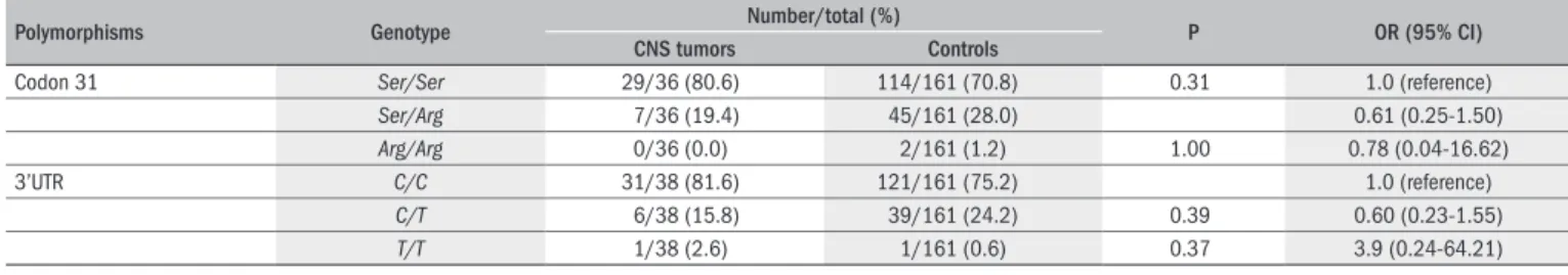
Table 1. Frequency of cyclin-dependent kinase inhibitor 1A (CDKN1A) gene polymorphisms in central nervous system (CNS) tumors and controls
Polymorphisms Genotype Number/total (%) P OR (95% CI)
CNS tumors Controls
Codon 31 Ser/Ser 29/36 (80.6) 114/161 (70.8) 0.31 1.0 (reference)
Ser/Arg 7/36 (19.4) 45/161 (28.0) 0.61 (0.25-1.50)
Arg/Arg 0/36 (0.0) 2/161 (1.2) 1.00 0.78 (0.04-16.62) 3’UTR C/C 31/38 (81.6) 121/161 (75.2) 1.0 (reference)
C/T 6/38 (15.8) 39/161 (24.2) 0.39 0.60 (0.23-1.55)
T/T 1/38 (2.6) 1/161 (0.6) 0.37 3.9 (0.24-64.21)
Ser/Ser,C/C = homozygote for wild allele; Ser/Arg, C/T = heterozygote; Arg/Arg,T/T = homozygote for polymorphic allele; P = values were calculated using Fisher’s exact probability test; OR = odds ratio; CI = conidence interval.
Table 2. Frequencies of the cyclin-dependent kinase inhibitor 1A (CDKN1A) genotypes in the different types of central nervous system (CNS) tumor and
controls
Tumor
Genotype
Codon 31 3’UTR
Ser/Ser Ser/Arg Arg/Arg C/C C/T T/T
Ependymomas 6/9 (66.7) 3/9 (33.3) 0/9 (0.0) 6/10 (60.0) 4/10 (40.0) 0/10 (0.0) Controls 114/161 (70.8) 45/161 (28.0) 2/161 (1.2) 121/161 (75.2) 39/161 (24.2) 1/161 (0.6)
P 0.72 1.00 0.28 1.00
OR (95% CI) 1.0 (reference) 1.27 (0.30-5.29) 3.5 (0.15-81.26) 1.0 (reference) 2.07 (0.55-7.71) 6.23 (0.23-168.47) Medulloblastomas 11/14 (78.6) 3/14 (21.4) 0/14 (0.0) 12/14 (85.7) 1/14 (7.15) 1/14 (7.15)
Controls 114/161 (70.8) 45/161 (28.0) 2/161 (1.2) 121/161 (75.2) 39/161 (24.2) 1/161 (0.6)
P 0.76 1.00 0.28 1.00
OR (95% CI) 1.0 (reference) 0.69 (0.18-2.59) 1.99 (0.09-44.08) 1.0 (reference) 2.07 (0.55-7.71) 6.23 (0.23-168.47) Astrocytomas 12/13 (92.3) 1/13 (7.7) 0/13 (0.0) 13/14 (92.9) 1/14 (7.1) 0/14 (0.0)
Controls 114/161 (70.8) 45/161 (28.0) 2/161 (1.2) 121/161 (75.2) 39/161 (24.2) 1/161 (0.6)
P 0.19 1.00 0.28 1.00
OR (95% CI) 1.0 (reference) 0.21 (0.03-1.67) 1.83 (0.08-40.37) 1.0 (reference) 0.24 (0.03-1.88) 3.00 (0.12-77.40)
Ser/Ser,C/C = homozygote for wild allele; Ser/Arg, C/T = heterozygote; Arg/Arg,T/T = homozygote for polymorphic allele; P = values were calculated using Fisher’s exact probability test; OR = odds ratio; CI = conidence interval. patient (2.6%) out of the 38 analyzed showed the C→T transition, and one patient in the control group also showed this (0.6%). here were no statistically signiicant diferences between the patients and the controls (P = 0.37), with an odds ratio of 3.9 (CI = 0.24-64.21) (Table 1).
Analysis of the frequencies of codon 31 genotypes for each CNS tu-mor type that was evaluated, in comparison with the controls, showed that there were no statistically signiicant diferences between ependymo-mas and controls (P = 1.00), medulloblastoependymo-mas and controls (P = 1.00) or astrocytomas and controls (P = 1.00). he same was found regarding polymorphism at the 3’UTR site, and its frequencies were not statisti-cally signiicant (Table 2).
Statistical analysis
Fisher’s exact probability test (Agresti)19 was used to analyze the
fre-quency of each genotype studied, in comparison with the total sample of patients and controls. It was also used for comparisons with the dif-ferent types of tumor, with the aim of possibly correlating the genotypes found with the presence or absence of increased risk of development of CNS tumors. To analyze protein expression, the chi-square test, Fisher’s exact test and the nonparametric Kruskal-Wallis test were used. he sig-niicance criterion used was a probability (P) level of less than or equal to 0.05. Odds ratios (OR) and 95% conidence intervals (CI)20 were
cal-culated as estimates of risk and degree of association.
RESULTS
Genetic analyses of
CDKN1A
in CNS tumors and controls
he frequencies of the C→A polymorphism in codon 31 and C→T polymorphism at the 3’UTR site of the CDKN1A gene in patients with CNS tumors and control subjects are shown in Table 1. Among the 36 CNS tumor patients studied for the polymorphism in codon 31, none of them (0.0%) presented the transversion C→A in codon 31. How-ever, out of the 161 controls studied, two subjects (1.2%) showed the transversion leading to the replacement of serine by arginine. he dif-ferences in frequency seen between the patients and control group were not statistically signiicant (P = 1.00), with an odds ratio of 0.78 (CI = 0.04-16.62) (Table 1). For the polymorphism at the 3’UTR site, one
Figure 3. Results from Western Blot experiment showing the
Analysis of CDKN1A protein expression in CNS tumors
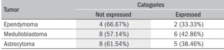
To determine the clinical relevance of the polymorphisms in codon 31 and at the 3’UTR site, we determined their correlation with CDK-N1A protein expression in the CNS tumor samples (Table 3). Out of the 33 tumors evaluated, 39.39% expressed this protein and 60.61% did not (Figure3). he diferences between these two categories were not statistically signiicant (P = 0.22). he CNS tumors were analyzed in pairs (ependymomas-medulloblastomas, ependymomas-astrocytomas and medulloblastomas-astrocytomas) in order to evaluate their protein expression patterns. It was observed that in ependymomas, 66.67% did not express the CDKN1Aprotein, whereas the others did so. he re-sults were similar for medulloblastomas and astrocytomas, with 42.86% and 38.46%, respectively. On the other hand, 57.14% and 61.54% did not express this protein. By means of Fisher’s test, it was seen that there was no signiicant diference (P = 1.00) in expression pattern percent-age between the diferent types of tumor (Table 3). Correlation between the protein expression analyses and polymorphism showed that the two genotypes (Ser/Ser and Ser/Arg) for polymorphism in codon 31 had sim-ilar frequencies for the categories “not expressed” (64.0% and 50.0%) and “expressed” (36.0% and 50.0%). his inding was not statistically signiicant (P = 0.62), nor was the correlation for polymorphism at the 3’UTR site (P = 0.99) (Table 4).
he polymorphism and protein expression analysis was compared to the degree of malignancy of each tumor. For this, pilocytic astrocy-tomas (WHO grade I) and ependymomas (WHO grade II) were taken to be low-grade tumors, and anaplastic astrocytomas and ependymomas
(WHO grade III) and medulloblastomas (WHO grade IV) were taken to be high-grade tumors. his comparison did not show any signiicant diferences in codon 31 polymorphism (P = 0.99), 3’UTR site polymor-phism (p=0.99) or protein pattern (P = 0.86), in relation to the degree of tumor malignancy (Table 5).
DISCUSSION
CDKN1A plays a central role in suspension of the cell cycle. How-ever, it is possible that gene abnormalities could be responsible for the progression of a number of types of neoplasm.
his was the irst Brazilian study to analyze the most frequent CD-KN1A gene polymorphisms (codon 31 and 3’UTR site) and protein ex-pression relating to the second most frequent category of pediatric can-cer (CNS tumors), in comparison with a control population.
Based on these data, we determined the prevalence of genetic poly-morphisms of CDKN1A in a sample of 10 ependymomas, 16 medullo-blastomas and 15 astrocytomas from the northwestern part of the State of São Paulo, compared with 161 control subjects. Our aim was to estab-lish a correlation between these genetic polymorphisms and the risk of developing CNS tumors among children and adolescents. In this study, analyzing the total sample, no diference in the homozygote frequency for the codon 31 polymorphic allele (Arg/Arg) was found in CNS tu-mors (0.0%), in comparison with the controls (1.2%). Regarding the normal homozygote (Ser/Ser) and heterozygote (Ser/Arg) genotypes, no signiicant diference was observed between the CNS tumors (80.6% and 19.4%, respectively) and the controls (70.8% and 28.0%, respec-tively). Our allele frequency for the Arg variant (10.0%) was also similar to that observed by Koopmann et al.21 in brain tumors (8.5%).
he frequency of the Arg allele found in the control population of our study (0.15) was similar to that found by Chedid et al.22 in the United
States (0.14). It has been suggested that the American and Brazilian pop-ulations consist of miscegenation of whites and blacks, since these allele frequencies were found among the values observed by Birgander et al.23
in African blacks (0.291) and in European Caucasians (0.039-0.0461). In Brazil, the African inluence exerted through the slave population for three centuries, along with the Native Indian and colonizing European popula-tions, could explain these results.24 Correlations between each
polymor-phism and one speciic type of tumor did not show any signiicant dif-ferences between ependymomas and controls (P = 0.73), medulloblasto-mas and controls (P = 0.76) or astrocytomedulloblasto-mas and controls (P = 0.19), for codon 31. hese data did not demonstrate any association between these polymorphisms and the risk of developing cancer. Moreover, similarly to our result, other studies also did not ind any association between codon 31 polymorphism and the risk of developing tumors.5,9
he other polymorphism of interest was the 3’UTR site, which plays an important role in mRNA stabilization, cell proliferation and tumor diferentiation and suppression.25,26 he prevalence of the homozygote
genotype (C/C) was found to be 81.6% in the CNS tumors and 75.2% in the controls. he heterozygote genotype (C/T) was seen in 15.8% of the CNS tumors and in 24.2% of the controls. he homozygote geno-type showed similar percentages for the polymorphic allele (T/T) in the CNS tumors (2.6%) and the controls (0.6%).
Table 3. Correlation between protein expression patterns among central
nervous system tumors
Tumor Categories
Not expressed Expressed
Ependymoma 4 (66.67%) 2 (33.33%) Medulloblastoma 8 (57.14%) 6 (42.86%) Astrocytoma 8 (61.54%) 5 (38.46%)
Table 4. Correlation between polymorphism frequencies and protein
expression in central nervous system tumors
Categories
Polymorphisms
Codon 31 3’UTR
Ser/Ser Ser/Arg C/C C/T
Not expressed 16/35 (64.0) 2/4 (50.0) 17/28 (60.7) 1/2 (50.0) Expressed 9/35 (36.0) 2/4 (50.0) 11/28 (39.3) 1/2 (50.0)
P 0.62 0.99
Ser/Ser,C/C = homozygote for wild allele; Ser/Arg, C/T = heterozygote; Arg/Arg,T/T = homozygote for poly-morphic allele; P = values were calculated using Fisher’s exact probability test.
Table 5. Correlation of the polymorphism and protein expression analysis
with the clinical behavior of central nervous system tumors
Tumor Codon 31 3’UTR WB
Ser/Ser Ser/Arg C/C C/T Not expressed Expressed
LD 14/29 (48.3)
3/7 (42.9)
17/32 (53.1)
3/5 (60.0)
11/16 (68.8)
5/16 (31.3) HD 15/29
(51.7) 4/7 (57.1)
15/32 (46.9)
2/5 (40.0)
9/17 (52.9) 8/17 (47.1)
P 0.99 0.99 0.86*
he frequencies of the polymorphic allele (T) were 0.11 in the CNS tumors and 0.13 in the controls. hese were higher than the frequency (8%) observed by Shiohara et al.,27 and this diference may be related to
the ethnic mix of the Brazilian population.24
It was observed that all codon 31 and 3’UTR polymorphisms pre-sented linkage disequilibrium. hus, if a tumor was heterozygotic for codon 31, it was also heterozygotic for the 3’UTR site. Likewise, if it was homozygotic for the wild allele in codon 31, it was also homozy-gotic for 3’UTR. Facher et al.10 found cosegregation of these two
poly-morphisms in a high percentage of cancer patients, thus suggesting that there was some functional diference in these allele variants that allowed subjects to be more susceptible to certain types of cancer. Polymorphism of the 3’UTR site may be located in a position that is required for rapid degradation of messenger CDKN1A and, for this reason, it would pre-vent transient loss of activity. CDKN1A mRNA containing the codon 31 variant could become more stable through association with 3’UTR polymorphism.
Konishi et al.26 also suggested that codon 31 polymorphism seemed
to be only an innocent ligand and that the 3’UTR site that is tightly bound to codon 31 could be involved in the association between this polymorphism and the risk of tumors. Future experiments, taking this information into account, are required in order to evaluate the possi-ble functional diferences that contribute towards carcinogenesis, along with studies involving larger numbers of patients and controls.
CDKN1A expression in vivo, in human tumors, and its possible role in tumor progression and cell diferentiation have been examined by investigators, with conlicting results.13 One of the purposes of the
present study was to evaluate CDKN1A protein expression in CNS tumors, in order to correlate the expression levels with the develop-ment and degree of malignancy of these tumors. hirty-three CNS tumor samples were analyzed, 6 ependymomas, 13 astrocytomas, and 14 medulloblastomas. We irst compared CDKN1A protein expres-sion among the three types of tumor, considering the intensity of the bands obtained through the Western Blotting technique. Analysis of the expression patterns using only two categories (“not expressed” and “expressed”) showed that the diferences were not signiicant, thus in-dicating that these tumors do not present a uniform pattern of CDK-N1A expression.It is likely that this non-uniformity of expression pat-tern occurs because of heterogeneity of the samples and, therefore, we analyzed these two categories in comparison with the diferent types of tumor. Even though the CDKN1A gene has a tumor suppressive func-tion, it has been correlated with increased protein expression in some types of tumor, such as lung carcinoma, hepatocellular carcinoma and head and neck cancer.13,14 In relation to CNS tumors, Jung et al.28
found that CDKN1A was frequently expressed in greater amounts in astrocytomas, anaplastic astrocytomas and glioblastomas. In addition to investigating the association between polymorphisms and protein expression, we analyzed our results through comparison of these poly-morphisms and their expression with the degree of tumor malignancy. he statistical analysis showed that there was no signiicant diference between the relative clinical behavior of the polymorphisms (P = 0.99 for codon 31 and 3’UTR) and the relative clinical behavior of the pro-tein expression (P = 0.86). hese results conirmed the previous
analy-sis, thus showing that there was no correlation between the presence of polymorphisms and the protein expression.
One of the possible explanations for these results is the fact that
CDKN1A alone is insuicient to stop the cell cycle and inhibit tu-mor cell growth, considering that mutations or loss of expression of other cell cycle/apoptosis genes such as PTEN, CDKN2A, MDM2, TP53 and RB is also associated with brain tumor etiology. More gen-erally, these pathways are of importance in other cancer types, includ-ing breast carcinoma, melanoma and colon carcinoma.29 Despite the
potential importance of the cell cycle and apoptosis pathways in brain tumor etiology, very little has been published regarding the risk of brain tumors that is associated with the more common gene variants in these pathways, with the exception of the TP53 gene.29 On the
oth-er hand, investigation of protein expression levels alone indicates that this is not the active form. It is important to emphasize that the cell cycle regulation pathways are extremely complex and not completely clariied yet.
CONCLUSIONS
No signiicant diference was observed between expression pat-terns and polymorphisms of CDKN1A and three kinds of CNS tumors among Brazilian subjects. herefore, the CDKN1A gene could have po-tential application in gene therapy, because of its role in regulating cell cycle progression or in inducing interruption of the cycle cell.
REFERENCES
1. Baldwin RT, Preston-Martin S. Epidemiology of brain tumors in childhood--a review. Toxicol Appl Pharmacol. 2004;199(2):118-31.
2. Wechsler-Reya R, Scott MP. The developmental biology of brain tumors. Annu Rev Neurosci. 2001;24:385-428.
3. Nakatani F, Tanaka K, Sakimura R, et al. Identiication of p21WAF1/CIP1 as a direct target of EWS-Fli1 oncogenic fusion protein. J Biol Chem. 2003;278(17):15105-15. 4. Kokunai T, Tamaki N. Relationship between expression of p21WAF1/CIP1 and
radioresistan-ce in human gliomas. Jpn J Canradioresistan-cer Res. 1999;90(6):638-46.
5. Milner BJ, Hosking L, Sun S, Haites NE, Foulkes WD. Polymorphisms in P21CIP1/ WAF1 are not correlated with TP53 status in sporadic ovarian tumours. Eur J Cancer. 1996;32A(13):2360-3.
6. Viale G, Pellegrini C, Mazzarol G, Maisonneuve P, Silverman ML, Bosari S. p21WAF1/CIP1 expression in colorectal carcinoma correlates with advanced disease stage and p53 muta-tions. J Pathol. 1999;187(3):302-7.
7. Cheng L, Lloyd RV, Weaver AL, et al. The cell cycle inhibitors p21WAF1 and p27KIP1 are associated with survival in patients treated by salvage prostatectomy after radiation therapy. Clin Cancer Res. 2000;6(5):1896-9.
8. Jahnson S, Karlsson MG. Tumor mapping of regional immunostaining for p21, p53, and mdm2 in locally advanced bladder carcinoma. Cancer. 2000;89(3): 619-29.
9. Shih CM, Lin PT, Wang HC, Huang WC, Wang YC. Lack of evidence of association of p21WAF1/ CIP1 polymorphism with lung cancer susceptibility and prognosis in Taiwan. Jpn J Cancer Res. 2000;91(1):9-15.
10. Facher EA, Becich, MJ, Deka A, Law JC. Association between human cancer and two poly-morphisms occurring together in the p21Waf1/Cip1 cyclin-dependent kinase inhibitor gene. Cancer. 1997;79(12):2424-9.
11. Nadal A, Jares P, Cazorla M, et al. p21WAF1/Cip1 expression is associated with cell diffe-rentiation but not with p53 mutations in squamous cell carcinomas of the larynx. J Pathol. 1997;183(2):156-63.
12. Liao WM, Zhang CL, Li FB, Zeng BF, Zeng YX. p21WAF1/CIP1 gene DNA sequencing and its expression in human osteosarcoma. Chin Med J (Engl). 2004;117(6):936-40. 13. Harada K, Ogden GR. An overview of the cell cycle arrest protein, p21(WAF1). Oral Oncol.
14. Marchetti A, Doglioni C, Barbareschi M, et al. p21 RNA and protein expression in non-small cell lung carcinomas: evidence of p53-independent expression and association with tumo-ral differentiation. Oncogene. 1996;12(6):1319-24.
15. Souza RJSP. Estudo das alterações genéticas envolvendo o gene p21 (WAF1/CIP1/CDNK1) em meduloblastomas, ependimomas e ependimoblastomas, em menores de 18 anos. [dis- dis-sertation]. Ribeirão Preto;. Faculdade de Medicina de Ribeirão Preto da Universidade de São Paulo;– 2003.
16. Mousses S, Ozçelik H, Lee PD, Malkin D, Bull SB, Andrulis IL. Two variants of the CIP1/WAF1 gene occur together and are associated with human cancer. Hum Mol Genet. 1995;4(6):1089-92. 17. Law JC, Deka A. Identiication of a PstI polymorphism in the p21Cip1/Waf1
cyclin-depen-dent kinase inhibitor gene. Hum Genet. 1995;95(4):459-60.
18. Laemmli UK. Cleavage of structural proteins during the assembly of the head of bacterio-phage T4. Nature. 1970;227(5259):680-5.
19. Agresti A. Modelling patterns of agreement and disagreement. Stat Methods Med Res. 1992;1(2):201-18.
20. Kleinbaum J, Shamoon H. Selective counterregulatory hormone responses after oral glucose in man. J Clin Endocrinol Metab. 1982;55(4):787-90.
21. Koopmann J, Maintz D, Schild S, et al. Multiple polymorphisms, but no mutations, in the WAF1/CIP1 gene in human brain tumours. Br J Cancer. 1995;72(5):1230-3.
22. Chedid M, Michieli P, Lengel C, Huppi K, Givol D. A single nucleotide substitution at codon 31 (Ser/Arg) deines a polymorphism in a highly conserved region of the p53-inducible gene WAF1/CIP1. Oncogene. 1994;9(10):3021-4.
23. Birgander R, Själander A, Saha N, Spitsyn V, Beckman L, Beckman G. The codon 31 poly-morphism of the p53-inducible gene p21 shows distinct differences between major ethnic groups. Hum Hered. 1996;46(3):148-54.
24. Rodrigues FC, Kawasaki-Oyama RS, Fo JF, et al. Analysis of CDKN1A polymorphisms: ma-rkers of cancer susceptibility? Cancer Genet Cytogenet. 2003;142(2):92-8.
25. Rastinejad F, Blau HM. Genetic complementation reveals a novel regulatory role for 3’ un-translated regions in growth and differentiation. Cell. 1993;72(6):903-17.
26. Konishi R, Sakatani S, Kiyokane K, Suzuki K. Polymorphisms of p21 cyclin-dependent kinase inhibitor and malignant skin tumors. J Dermatol Sci. 2000;24(3):177-83.
27. Shiohara M, el-Deiry WS, Wada M, et al. Absence of WAF1 mutations in a variety of human malignancies. Blood. 1994:84(11):3781-4.
28. Jung JM, Bruner JM, Ruan S, et al. Increased levels of p21WAF1/Cip1 in human brain tumors. Oncogene. 1995;11(10):2021-8.
29. Rajaraman P, Wang SS, Rothman N, et al. Polymorphisms in apoptosis and cell cycle control genes and risk of brain tumors in adults. Cancer Epidemiol Biomarkers Prev. 2007;16(8):1655-61.
Acknowledgements: We thank Geraldo Cassio dos Reis for the statistical analyses of this work, and the clinicians who referred the families with cases of CNS tumors
Sources of funding: This work was supported by the Brazilian Public Financial Agencies Coordenação de Aperfeiçoamento de Pessoal de Nível Superior (Capes) [2003] and Fundação de Apoio ao Ensino, Pesquisa e Assistência (Faepa) [2004]
Conlict of interest: None
Date of irst submission: December 9, 2008
Last received: October 28, 2009
Accepted: October 29, 2009
Address for correspondence:
Mev Dominguez Valentin
Laboratório de Biologia Genômica e Molecular Centro de Pesquisa do Hospital A C Camargo Rua Professor Antônio Prudente, 211 São Paulo (SP) — Brasil
CEP 01525-001