INTRODUCTION
Address to: Dr. Carlos Eduardo Blanco Linares. Depto Ciências da Saúde/URI. Av. Assis Brasil, 98400-000 Frederico Westphalen, RS, Brasil.
Fax: 55 55 3744-9265 e-mail: [email protected] Received 19 September 2013 Accepted 26 November 2013
Fluconazole and amphotericin-B resistance are associated
with increased catalase and superoxide dismutase activity
in
Candida albicans
and
Candida dubliniensis
Carlos Eduardo Blanco Linares
[1], Sandro Rogério Giacomelli
[2], Delsi Altenhofen
[2],
Sydney Hartz Alves
[3], Vera Maria Morsch
[4]and Maria Rosa Chitolina Schetinger
[4][1].Departamento de Bioquímica, Instituto de Ciências Básicas da Saúde, Universidade Federal do Rio Grande do Sul, Porto Alegre, RS. [2]. Curso de Farmácia, Centro de Ciências da Saúde, Universidade Regional do Alto Uruguai e das Missões, Frederico Westphalen, RS. [3]. Departamento de Microbiologia, Centro de Ciências da Saúde, Universidade Federal de Santa Maria, Santa Maria, RS. [4]. Departamento de Química, Centro de Ciências Naturais e Exatas, Universidade Federal de Santa Maria, Santa Maria, RS.
ABSTRACT
Introduction: Candida dubliniensis, a new species of Candida that has been recovered from several sites in healthy people, has been associated with recurrent episodes of oral candidiasis in AIDS and HIV-positive patients. This species is closely related to
C. albicans. The enzymatic activity of C. dubliniensis in response to oxidative stress is of interest for the development of drugs to combat C. dubliniensis. Methods: Fluconazole- and amphotericin B-resistant strains were generated as described by Fekete-Forgács et al. (2000). Superoxide dismutase (SOD) and catalase assays were performed as described by McCord and Fridovich (1969) and Aebi (1984), respectively. Results: We demonstrated that superoxide dismutase (SOD) and cata lase activities were
signifi cantly higher (p<0.05) in the fl uconazole- and amphotericin B-resistant strains of C. dubliniensis and C. albicans than
in the sensitive strains. The catalase and SOD activities were also signifi cantly (p<0.01) higher in the sensitive and resistant
C. albicans strains than in the respective C. dubliniensis strains. Conclusions: These data suggest that C. albicans is better protected from oxidative stress than C. dubliniensis and that fl uconazole, like amphotericin B, can induce oxidative stress in Candida; oxidative stress induces an adaptive response that results in a coordinated increase in catalase and SOD activities.
Keywords: Catalase. Superoxide dismutase. Candida albicans. Candida dubliniensis.
Candida dubliniensis, a new emerging opportunistic species,
was fi rst described in 1995 in Dublin, Ireland. The cultures were obtained from human immunodefi ciency virus
(HIV)-positive patients with recurrent oral candidiasis. Although
C. dubliniensis was initially recovered from the oral cavity, this species has also been reported at other body sites, including the respiratory tract, central nervous system, vagina, and skin, as well as in the blood, urine, and feces of both HIV-positive and HIV-negative patients1-4. Nunn et al.5 also described the isolation of C. dubliniensis from nonhuman environmental sources. Thus,
this species is not confi ned to humans5.
Candida dubliniensis is phenotypically and genotypically similar to C. albicans and C. stellatoidea. Like these Candida
species, C. dubliniensis forms chlamydospores and is germ
tube-positive in phenotypic identifi cation tests. The detection
of unusual carbohydrate assimilation profi les, which can be identifi ed using commercially available systems such as the
API® ID 32C and API® 20C AUX kits, were one of the earliest indications that C. dubliniensis represented a new species within the Candida genus4. Studies have since documented a high prevalence of this organism in patients suffering from
diabetes (3.6 to 18.2%), cystic fi brosis (11.1%), cancer (2 to
4.6%), vulvovaginal candidiasis (0.17 to 2.4%), and candidemia (0.5 to 7.9%)6.
Candida dubliniensis is more susceptible to polyenes and azoles than C. albicans; however, a remarkable feature of C. dubliniensis is the rapid development of secondary
resistance to fl uconazole7. Azoles, such as fl uconazole, act on the ergosterol biosynthesis pathway by inhibiting the enzyme
C14-α-demethylase, a cytochrome P450-dependent enzyme8. Amphotericin B, a polyene antifungal agent, binds to sterols such as ergosterol and disrupts the osmotic integrity of the fungal membrane, resulting in the leakage of intracellular ions and metabolites9. Despite intense study of C. dubliniensis, the enzymatic activities of superoxide dismutase (SOD) and
catalase in fl uconazole-resistant and amphotericin B-resistant
METHODS
hydrogen peroxide (H2O2), which can be converted into water by catalase11,12. Catalase has been implicated in amphotericin B resistance by inhibiting the oxidative damage produced by this polyene13. Ergosterol, a sterol present in the fungal plasma membrane, is the target of amphotericin B. The binding of amphotericin B to ergosterol produces channels that leak potassium ions, resulting in the disruption of the proton gradient14,15. The antioxidant enzymes SOD and catalase may act synergistically to protect cells from oxidative damage16. Some authors have suggested that the coordinated inactivation of ROS by the sequential action of superoxide- and hydrogen peroxide-inactivating enzymes is essential for the effective protection of cells against ROS toxicity17.
To further explore the antioxidant enzymatic activities and the coordinated inactivation of ROS by SOD and catalase in
C. dubliniensis and C. albicans, we investigated the SOD and
catalase activities in fl uconazole- and amphotericin B-sensitive
and resistant strains of C. dubliniensis and C. albicans.
Strains
The Candida albicans and Candida dubliniensis strains used in this study [Ca 10, Ca 81, Ca 119, Ca 170, Ca ATCC 44373, Cd 1, Cd 2, Cd 3, Cd 4, Cd 5, Cd 7, Cd 9, Cd 11, and Cd 13] were isolated from patients with oral candidiasis at the University Hospital of
Universidade Federal de Santa Maria (Brazil). Species identifi cation
was based on phenotypic and genotypic characteristics. The
C. dubliniensis genotype was confi rmed by random amplifi cation of
polymorphic deoxyribonucleic acid (DNA) using the primers CDU
(5′ GCG ATC CCC A 3′) and B-14 (5′ GAT CAA GTC C 3′)18,4. The C. albicans strain ATCC 44373 from the American Type Culture Collection was used as a control strain.
Generation of fl uconazole-resistant strains
The minimal inhibitory concentration (MIC) of fl uconazole
was first determined for C. albicans and C. dubliniensis
according to the Clinical and Laboratory Standards Institute (CLSI) document M27-A319. After the MIC determinations, the cultures were grown overnight in Sabouraud glucose broth
(SDB), and the cells were added to fl asks containing 20ml of media to achieve a fi nal absorbance of 0.1 at 640nm. The cultures were incubated at 30°C for 10h, and fl uconazole was then added to a fi nal concentration of 8μg ml−1. After 14h of
further incubation, the cells of the fl uconazole-containing culture
were consecutively subcultured 3 times in fresh SDB medium
containing 8μg ml−1 fl uconazole; in each case, the cultures were incubated at 30°C with shaking for 24h. After the third
incubation, the cells were added to fl asks containing 20ml of SDB medium containing 8μg ml−1 fl uconazole, and the turbidity
of the cultures was adjusted to achieve a fi nal absorbance of 0.1 at 640nm. After a 10h incubation, fl uconazole was added at a fi nal concentration of 16μg ml−1, and after 14h, the cells from this culture were subcultured 3 times in fresh SDB medium
containing 16μg ml−1 fl uconazole; each subculture was incubated
at 30°C with shaking for 24h. The fl uconazole concentration
was always duplicated under this procedure, and the fi nal concentration was 64μg ml−1. Cells from this culture were plated on Sabouraud glucose agar (SDA), and the colonies were designated C. albicans fl uconazole resistant and C. dubliniensis
fl uconazole resistant20.
Generation of amphotericin B-resistant strains
Induction of amphotericin B resistance was performed as
described for the generation of the fl uconazole-resistant strains, with some modifi cations. The time needed for cell growth between exposures to different amphotericin B concentrations was 48h. Resistant cells obtained at 2µg ml-1 were plated on SDA, and the colonies were designated C. albicans amphotericin B-resistant and C. dubliniensis amphotericin B-resistant.
Susceptibility tests
Antifungal agents: standard fluconazole (Pfizer Central Research) and amphotericin B (Bristol-Myers, Squibb Australia Pty Ltd) powders were obtained from their respective manufacturers. Stock solutions were prepared in water
(fl uconazole) and dimethyl sulfoxide (amphotericin B). Serial
two-fold dilutions were prepared exactly as outlined in the CLSI document M27-A319. Final dilutions were made in Roswell Park Memorial Institute (RPMI) 1640 medium buffered to pH 7.0 with 0.165mmol l-1 morpholinopropanesulfonic acid (MOPS). Aliquots
(100µl) of each antifungal agent at a 2X fi nal concentration were
dispensed into the wells of plastic microdilution trays, which were
sealed and frozen at -70ºC until use. The fl uconazole dilutions ranged from 0.12 to 128μg ml-1, and the amphotericin B dilutions
ranged from 0.0313 to 16μg ml-1.
Inoculum standardization -inoculum suspensions of the yeast strains were prepared in sterile saline (0.85% NaCl) from 24h cultures grown on SDA at 35°C. The turbidity of the suspensions
was adjusted to achieve a fi nal absorbance of 0.1 at 530nm8,19.
CLSI broth microdilution method (document M27-A3):
microplates containing 100μl of two-fold serial dilutions of
the antifungal drugs in standard RPMI 1640 medium (0.2%
glucose) were inoculated with 100μl of inoculum containing
1 × 10³ to 5 × 10³ colony-forming units per milliliter (CFU/ml). Following inoculation, the reference microdilution plates were incubated at 35°C, and the Minimum Inhibitory Concentration
MICs were determined after 24 and 48h. The fl uconazole and
amphotericin B reference MICs corresponded to the lowest drug
dilutions that resulted in prominent growth inhibition (≥ 80%)
or the absence of growth, respectively19. Enzyme assays
Candida albicans and Candida dubliniensis strains: strains
were cultured for 48h in SDA containing 64μg ml−1 fl uconazole
or 2μg ml−1 amphotericin B. The yeast cells were added to sterile
saline (0.85%) to achieve a fi nal absorbance of 0.1 at a wavelength
of 625nm. Aliquots (1ml) of this suspension were added to 50ml of SDB containing 4% glucose and 1% mycological peptone, and these cultures were incubated at 30ºC with shaking for 24h
for the fl uconazole experiments and for 48h for the amphotericin
RESULTS
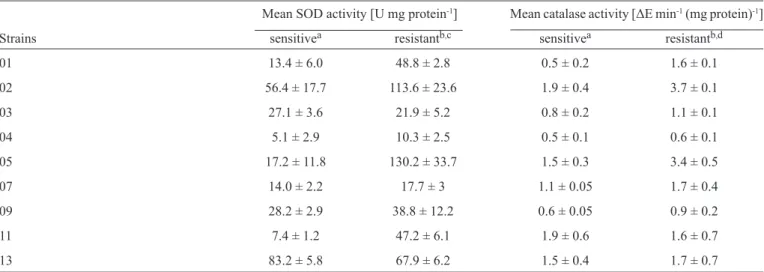
TABLE 1 - Superoxide dismutase and catalase activities of the fl uconazole-sensitive and fl uconazole-resistant Candida dubliniensis strains. Mean SOD activity [U mg protein-1] Mean catalase activity [ΔE min-1 (mg protein)-1]
Strains sensitivea resistantb,c sensitivea resistantb,d
01 13.4 ± 6.0 48.8 ± 2.8 0.5 ± 0.2 1.6 ± 0.1
02 56.4 ± 17.7 113.6 ± 23.6 1.9 ± 0.4 3.7 ± 0.1
03 27.1 ± 3.6 21.9 ± 5.2 0.8 ± 0.2 1.1 ± 0.1
04 5.1 ± 2.9 10.3 ± 2.5 0.5 ± 0.1 0.6 ± 0.1
05 17.2 ± 11.8 130.2 ± 33.7 1.5 ± 0.3 3.4 ± 0.5
07 14.0 ± 2.2 17.7 ± 3 1.1 ± 0.05 1.7 ± 0.4
09 28.2 ± 2.9 38.8 ± 12.2 0.6 ± 0.05 0.9 ± 0.2
11 7.4 ± 1.2 47.2 ± 6.1 1.9 ± 0.6 1.6 ± 0.7
13 83.2 ± 5.8 67.9 ± 6.2 1.5 ± 0.4 1.7 ± 0.7
The data are the mean ± SD of triplicate experiments. U: unit of SOD; ΔE: absorbance change at 240nm over 240s. aFluconazole-sensitive
C. dubliniensis; bFluconazole-resistant C. dubliniensis (64µg/ml-1); cStatistical analyses revealed signifi cant differences between the SOD activities of the fl uconazole-resistant and fl uconazole-sensitive C. dubliniensis groups (p < 0.01); dStatistical analyses revealed signifi cant differences between the catalase activities of the fl uconazole-resistant and fl uconazole-sensitive C. dubliniensis groups (p < 0.01).
SOD: superoxide dismutase; SD: standard deviation; C.: Candida.
Preparation of cell-free extracts: the crude extracts were prepared using the glass bead lysis method. Cells that had been cultured in SDB supplemented with fluconazole or amphotericin B were washed 3 times with MilliQ water and resuspended in lysis buffer (50mmol l-1 potassium phosphate pH 7.0) containing 0.5g of 500-µm-diameter glass beads. The
mixture was homogenized in a Tefl on-glass homogenizer for 6
cycles of alternating homogenization for 20s and cooling on ice. The mixture was then centrifuged for 30 min in a refrigerated centrifuge to remove the cell debris and glass beads. The supernatant was used for enzyme assays.
Superoxide dismutase activity
Superoxide dismutase activity was assayed according to McCord and Fridovich21. The assay was performed in a total volume of 1ml containing 50mmol l-1 glycine buffer (pH 10), 60mmol l-1 epinephrine, and the enzyme. Epinephrine was added, and adrenochrome formation was recorded at 480nm with a ultraviolet-visible (UV-Vis) spectrophotometer for 4 min. One unit of SOD activity was equivalent to the amount of enzyme required to inhibit epinephrine oxidation by 50% under the experimental conditions. The assays were performed in triplicate.
Catalase activity
Catalase activity was determined in the cell-free extracts using the method of Aebi et al.22. H
2O2 solution (10mM), enzyme extract, and 50mM phosphate buffer (pH 7) were pipetted into a cuvette. The reduction of H2O2 was followed at a wavelength of 240nm for 4 min against a blank containing 50mM phosphate buffer and enzyme extract. Catalase activity was expressed in
ΔE min-1 (mg protein)-1. The assays were performed in triplicate.
Protein analysis
Protein concentrations were measured using the Bradford method and Coomassie Blue; serum albumin was used as the standard23.
Statistics
Paired t tests were used to compare the sensitive and resistant cells. Comparisons among the C. albicans and C. dubliniensis
groups were made using unpaired t tests. The calculations were performed using the GraphPad InStat statistical program.
Ethical considerations
This protocol was approved by the Bioethics Committee of the Universidade Regional Integrada do Alto Uruguai e das Missões from Frederico Westphalen - RS, Brazil, under registration n° 061-2/PIH/04.
Fluconazole- and amphotericin B-resistant C. dubliniensis and
C. albicans strains were obtained by exposing sensitive isolates to increasing concentrations of these antifungal agents. For the sensitive C. dubliniensis cells, the MICs ranged from 0.06 to
0.5μg ml-1 for fl uconazole and from 0.0312 to 0.125μg ml-1 for amphotericin B; for the sensitive C. albicans cells, the MICs
ranged from 0.5 to 4.0μg ml-1 for fl uconazole and from 0.0625 to
0.25μg ml-1 for amphotericin B. After the induction of resistance,
tests of the susceptibility to fl uconazole and amphotericin B
demonstrated that resistance developed in all strains assayed. Table 1 shows the SOD and catalase activities in the cell-free
C. dubliniensis strains. In general, the mean SOD activity was
1.97-fold greater for fl uconazole-resistant C. dubliniensis than for the sensitive cells. The difference between the 2 groups was
signifi cant (p < 0.01). The catalase activity of the fl resistant cells was 1.58-fold greater than that of the fl uconazole-sensitive cells; this difference was signifi cant (p < 0.01).
Table 2 shows the SOD and catalase activities of the
C. dubliniensis strains resistant to 2µg ml-1 amphotericin B. The mean SOD activity of the C. dubliniensis strain resistant to 2µg ml-1 amphotericin B was 1.97-fold greater than that of the sensitive cells;
this difference was statistically signifi cant (p < 0.01). The mean
catalase activity of the group resistant to 2µg ml-1 amphotericin B was 2-fold greater than that of the sensitive group. Statistical
TABLE 2 - Superoxide dismutase and catalase activities of the amphotericin B-sensitive and amphotericin B-resistant Candida dubliniensis
strains.
Mean SOD activity [U mg protein-1] Mean catalase activity [ΔE min-1 (mg protein)-1]
Strains sensitivea resistantb,c sensitivea resistantb,d
01 15.5 ± 0.4 69.7 ± 20.7 0.6 ± 0.1 2.4 ± 0.5
02 15 ± 1.5 22.6 ± 3.0 0.8 ± 0.1 0.9 ± 0.1
03 13.5. ± 0.4 37.8 ± 6.8 0.9 ± 0.01 2.1 ± 0.1
04 27.2 ± 1.8 50.7 ± 5.0 1.1 ± 0.1 1.8 ± 0.01
05 22.1 ± 3.2 37.3 ± 3.5 1.0 ± 0.1 1.8 ± 0.2
07 15.7 ± 1.9 44.1 ± 13.3 1.1 ± 0.01 3.1 ± 0.01
09 47.2 ± 12.6 101.1 ± 24.1 1.6 ± 0.2 2.2 ± 0.3
11 57.9 ± 33.2 69.3 ± 4.7 1.7 ± 0.2 3.6 ± 0.6
13 35.5 ± 10.2 57.8 ± 8.1 0.8 ± 0.1 1.0 ± 0.1
The data are the mean ± SD of triplicate experiments; U: unit of SOD; ΔE: absorbance change at 240nm over 240s. aAmphotericin B-sensitive
C. dubliniensis; bC. dubliniensis resistant to 2µg/ml-1 amphotericin B; cStatistical analyses indicated signifi cant differences between the SOD
activities of the C. dubliniensis cells resistant to 2µg/ml-1 amphotericin B and the sensitive C. dubliniensis cells (p < 0.01). dStatistical analyses indicated signifi cant differences between the catalase activities of the C. dubliniensis cells resistant to 2µg/ml-1 amphotericin B and the sensitive
C. dubliniensis cells (p < 0.01). SOD: superoxide dismutase; SD: standard deviation; C.: Candida.
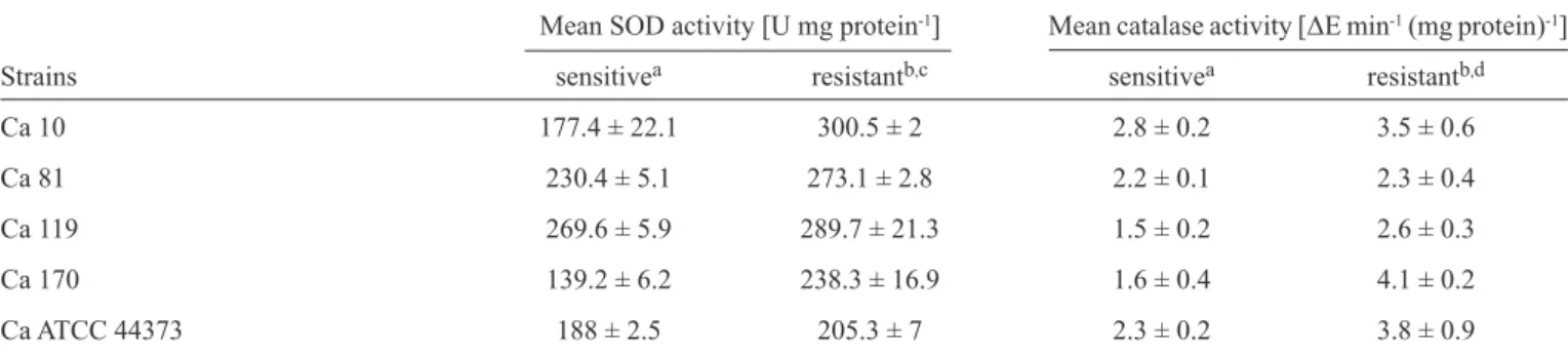
TABLE 3 - Superoxide dismutase and catalase activities of the fl uconazole-sensitive and fl uconazole-resistant Candida albicans strains. Mean SOD activity [U mg protein-1] Mean catalase activity [ΔE min-1 (mg protein)-1]
Strains sensitivea resistantb,c sensitivea resistantb,d
Ca 10 177.4 ± 22.1 300.5 ± 2 2.8 ± 0.2 3.5 ± 0.6
Ca 81 230.4 ± 5.1 273.1 ± 2.8 2.2 ± 0.1 2.3 ± 0.4
Ca 119 269.6 ± 5.9 289.7 ± 21.3 1.5 ± 0.2 2.6 ± 0.3
Ca 170 139.2 ± 6.2 238.3 ± 16.9 1.6 ± 0.4 4.1 ± 0.2
Ca ATCC 44373 188 ± 2.5 205.3 ± 7 2.3 ± 0.2 3.8 ± 0.9
The data are the mean ± SD of triplicate experiments. U: unit of SOD; ΔE: absorbance change at 240nm over 240s. aFluconazole-sensitive
C. albicans; bFluconazole-resistant C. albicans (64µg/ml-1); cStatistical analyses revealed signifi cant differences between the SOD activities of the fl uconazole-resistant and fl uconazole-sensitive C. albicans groups (p < 0.01); dStatistical analyses revealed signifi cant differences between the catalase activities of the fl uconazole-resistant and fl uconazole-sensitive C. albicans groups (p < 0.01). SOD: superoxide dismutase;
SD: standard deviation; C.: Candida.
analyses showed signifi cant differences (p < 0.01) between the
C. dubliniensis catalase activities of the cells resistant to 2µg ml-1 amphotericin B and the sensitive cells.
The SOD and catalase activities of the fl uconazole-sensitive
and fluconazole-resistant C. albicans cells are shown in
Table 3. The mean SOD activity of the fl uconazole-resistant
C. albicans was 1.3-fold greater than that of the sensitive cells;
this difference was statistically signifi cant (p < 0.01). The mean catalase activity of the fl uconazole-resistant group was 1.57-fold greater than that of the fl uconazole-sensitive group; this difference was statistically signifi cant (p < 0.01).
Table 4 shows the SOD and catalase activities of the
TABLE 4 - Superoxide dismutase and catalase activities of the amphotericin B-sensitive and resistant Candida albicans strains.
Mean SOD activity [U mg protein-1] Mean catalase activity [ΔE min-1 (mg protein)-1]
Strains sensitivea resistantb,c sensitivea resistantb,d
Ca 10 22.1 ± 8.5 178.4 ± 15 3.1 ± 0.3 4.4 ± 0.3
Ca 81 116.2 ± 6.4 93 ± 4.8 2 ± 0.2 2.9 ± 0.3
Ca 119 106.6 ± 11.7 298.2 ± 21.3 1.5 ± 0.2 2.9 ± 0.8
Ca 170 186.1 ± 3.2 195 ± 6.9 1.3 ± 0.4 3.9 ± 0.1
Ca ATCC 44373 82.4 ± 4 269.3 ± 10.1 2.8 ± 0.1 4.8 ± 1.1
The data are the mean ± SD of triplicate experiments. U: unit of SOD. ΔE: absorbance change at 240nm over 240s. aAmphotericin B-sensitive
C. albicans; bC. albicans resistant to 2 µg ml-1 amphotericin B; cStatistical analyses revealed signifi cant differences between the SOD activities
of the C. albicans cells resistant to 2µg/ml-1 amphotericin B and the sensitive C. albicans cells (p < 0.01); dStatistical analyses revealed
differences between the catalase activities of the C. albicans cells resistant to 2µg/ml-1 amphotericin B and the sensitive C. albicans cells
(p < 0.05). SOD: superoxide dismutase; SD: standard deviation; C.: Candida.
amphotericin B. The mean SOD activity of the C. albicans
strain resistant to 2µg ml-1 amphotericin B was 1.97-fold greater than that of the sensitive group; this difference was statistically
signifi cant (p < 0.01). The mean catalase activity of the cells
resistant to 2µg ml-1 amphotericin B was 1.77-fold greater than that of the sensitive group; this difference was statistically
signifi cant (p < 0.05).
DISCUSSION
Since the recognition of C. dubliniensis as a new species in 1995, many studies have investigated the differences between this species and C. albicans. Studies of the responses of these two species under identical conditions have uncovered phenotypic as well as genotypic differences between these two species. Enzymes involved in virulence, such as aspartyl proteinases, phospholipases, chondroitin sulfatase, and hyaluronidase, have been thoroughly studied. De Bernardis et al.24 and Linares et al.25 demonstrated that the level of aspartyl
proteinase was signifi cantly higher in C. albicans than in
C. dubliniensis, while no differences in phospholipase, chondroitin sulfatase, and hyaluronidase activities were observed24-26. In this study, we investigated the effect of
fl uconazole and amphotericin B resistance on the SOD and
catalase activities of C. albicans and C. dubliniensis. These enzymes are not related to virulence factors but play important roles in the oxidative stress response in both species27. SOD
activity is involved in the detoxifi cation of free superoxide
radicals, such as the radical produced by phagocyte nicotinamide adenine dinucleotide phosphate (NADPH) oxidase29. Phagocytic cells have 2 antimicrobial systems that are responsible for the generation of reactive oxygen species (ROS) and reactive nitrogen species (RNS). To counter these antimicrobial systems, microorganisms such as Candida have multiple defense responses that can be adjusted according to the nature and amount of ROS produced by the host or environment30. Our
results revealed that SOD activity was signifi cantly increased
in C. dubliniensis and C. albicans strains that were resistant to
amphotericin B and fl uconazole compared to sensitive strains.
The increase in SOD activity in response to amphotericin B may be due to the production of oxidative damage in Candida
through lipid peroxidation15. In our study, yeast cultures were exposed to increasing concentrations of amphotericin B, up to a maximum of 2 µg ml-1. In general, at this concentration, amphotericin B kills Candida by inducing oxidative damage through lipid peroxidation; however, we determined that
Candida SOD activity was activated in response to amphotericin B exposure. The mean SOD activity of the C. dubliniensis and
C. albicans strains resistant to 2µg ml-1 amphotericin B was 1.97-fold greater than that of the sensitive strains. This SOD activation likely occurred due to the induction of an adaptive response upon exposure of Candida to sublethal concentrations of peroxide or superoxide. This adaptive response results in a several-fold increase in catalase, SOD, and glucose 6-phosphate dehydrogenase (G6PDH) activity29,31,32. In addition, the elevation of SOD activity in the yeast strains resistant to amphotericin B may contribute to an increased resistance to phagocytic cell attack, making the yeast cells more resistant to host defenses. We also observed that the SOD activities of the C. albicans
strains that were sensitive to fl uconazole and amphotericin B were signifi cantly (p < 0.01) higher than the SOD activities of
the sensitive C. dubliniensis strains. When SOD activity was compared among the resistant groups, C. albicans also expressed
signifi cantly (p < 0.01) higher levels of SOD than C. dubliniensis. As previously demonstrated for other enzymes (e.g., proteinases), SOD activity was higher in C. albicans than in
C. dubliniensis. This study is the fi rst to evaluate the effect
of fl uconazole resistance on SOD activity in C. albicans and
C. dubliniensis. Our results suggest that fl uconazole has an
oxidant effect on Candida, resulting in the activation of its antioxidant system, including SOD. The mean SOD activities
of the fl uconazole-resistant C. albicans and C. dubliniensis
strains were 1.93- and 1.97-fold higher, respectively, than those
of the sensitive strains. If fl uconazole induces SOD activity, like amphotericin B, then this reaction to fl uconazole would contribute
Similar to SOD activity, catalase activity was elevated in
the fl uconazole- and amphotericin B-resistant C. albicans and
C. dubliniensis cells compared to the sensitive groups. The enzyme catalase converts hydrogen peroxide (H2O2) to oxygen (O2) and water (H2O)16. In Candida, catalase has been suggested to be involved in the resistance to amphotericin B and is involved in hypha formation15,28. Catalase activity has not been previously
studied in fl uconazole-resistant C. albicans and C. dubliniensis
strains. Here, we demonstrated that the catalase activities of the amphotericin B-resistant C. dubliniensis and C. albicans
strains were 2- and 1.77-fold higher, respectively, than those of
the sensitive groups. The catalase activities of the fl
uconazole-resistant C. albicans and C. dubliniensis groups were 1.58- and 1.57-fold higher, respectively, than those of the sensitive groups.
These fi ndings demonstrate that similar changes in catalase and SOD activities occur in response to exposure to fl uconazole and
amphotericin B under the same conditions and corroborate the hypothesis that an adaptive response occurs when Candida is exposed to oxidative stress32. Based on the literature, we expected that these enzymes would have similar responses because the product of SOD is a substrate for catalase11. Here, we confi rmed
this enzymatic profi le in C. albicans and C. dubliniensis. Similar
to SOD, catalase activity was signifi cantly (p < 0.01) higher
in the sensitive and resistant C. albicans strains compared to the respective C. dubliniensis groups. In combination with the increased susceptibility of C. dubliniensis to hydrogen peroxide and macrophage killing, this result suggests that C. albicans is better protected from oxidative stress than C. dubliniensis, in accordance with the higher virulence of C. albicans33,34,35.
The results obtained in this study also demonstrate a synergism between SOD and catalase in the C. dubliniensis and C. albicans
strains that were resistant to fl uconazole and amphotericin B,
in which protection by SOD generates hydrogen peroxide but not other ROS, such as lipid hydroperoxides or the reactive nitric intermediate (RNI) peroxynitrite, which would be more detrimental to the cells due to the absence of defenses against these types of compounds. Because the hydrogen peroxide generated by SOD activity is still toxic to the cell, it must be eliminated by catalase, which converts this toxic compound into water14.
In conclusion, this study is the fi rst to determine the effect of fl uconazole and amphotericin B resistance on catalase and SOD
activities in C. albicans and C. dubliniensis. The stress-inducing
effect of fl uconazole on Candida has not been previously described, and we have demonstrated that catalase and SOD activities are
increased in fl uconazole-resistant cells. We also confi rmed that
C. albicans has more active antioxidant pathways than C. dubliniensis.
REFERENCES
The authors declare that there is no confl ict of interest.
CONFLICT OF INTEREST
1. Fotedar R, Al-Hedaithy SS. Candida dubliniensis at a University in Saudi Arabia. J Clin Microbiol 2003; 41:1907-1911.
2. Odds FC, Nuffel LV, Dams G. Prevalence of Candida dubliniensis
isolates in a yeast stock collection. J Clin Microbiol 1998; 36:2869-2973.
3. Polacheck I, Strahilevitz J, Sullivan D, Donnelly S, Salkin IF, Coleman DC. Recovery of Candida dublinensis from non-human immunodefi ciency
virus-infected patients in Israel. J Clin Microbiol 2000; 38:170-174.
4. Sullivan DJ, Westerneng TJ, Haynes KA, Bennett DE, Coleman DC.
Candida dubliniensis sp. nov.: phenotypic and molecular characterization
of a novel species associated with oral candidosis in HIV-infected individuals. Microbiology 1995; 141:1507-1521.
5. Nunn MA, Schäfer SM, Petrou MA, Brown RM. Environmental source of
Candida dubliniensis. Emerg Infect Dis 2007; 13:747-750.
6. Loreto ES, Scheid LA, Nogueira CW, Zeni G, Santurio JM, Alves SH.
Candida dubliniensis: epidemiology and phenotypic methods for
identifi cation. Mycopathologia 2010; 169:431-443.
7. Moran GP, Sullivan DJ, Henman MC, McCreary CE, Harrington BJ, Shanley DB, et al. Antifungal drug susceptibilities of oral Candida
dubliniensis isolates from human immunodefi ciency virus (HIV)-infected
and non-HIV-infected subjects and generation of stable fl
uconazole-resistant derivatives in vitro. Antimicrob Agents Chemother 1997; 41: 617-623.
8. Bodey GP. Azole antifungal agents. Clin Infect Dis 1992; 14: 161-169.
9. Thomas AH. Suggested mechanisms for the antimycotic activity for the polyene antibiotics and the N-substituted imidazoles. J Antimicrob Chemother 1986; 17:269-279.
10. Vázquez N, Walsh TJ, Friedman D, Chanock SJ, Lyman CAInterleukin-15 augments superoxide production and microbicidal activity of human monocytes against Candida albicans. Infect Immun 1998; 66:145-150.
11. Johnson F, Giulivi C. Superoxide dismutases and their impact upon human health. Mol Aspects Med 2005; 26:340-352.
12. Martchenko M, Alarco A, Harcus D, Whiteway M. Superoxide Dismutases
in Candida albicans: Transcriptional Regulation and Functional
Characterization of the Hyphal-induced SOD5 Gene. Mol Biol Cell 2004; 15:456-467.
13. Sokol-Anderson M, Sligh JE, Jr Elberg S, Brajtburg J, Kobayashi GS, Medoff G. Role of cell defense against oxidative damage in the resistance
of Candida albicans to the killing effect of amphotericin B. Antimicrob
Agents Chemother 1998; 32:702-705.
14. Nakagawa Y, Kanbe T, Mizuguchi I. Disruption of the human pathogenic yeast Candida albicans catalase gene decreased survival in mouse-model infection and elevated susceptibility to higher temperature and to detergents. Microbiol Immunol 2003; 47:395-403.
15. Sokol-Anderson ML, Brajtburg J, Medoff G. Amphotericin B-induced oxidative damage and killing of Candida albicans. J Infect Dis 1986; 154:76-83.
16. Michiels C, Raes M, Toussaint O, Remacle J. Importance of Se-glutathione peroxidase, catalase, and Cu/Zn-SOD for cell survival against oxidative stress. Free Radic Biol Med 1994; 17:235-248.
17. Lortz S, Tiedge M. Sequential inactivation of reactive oxygen species by combined overexpression of SOD isoforms and catalase in insulin-producing cells. Free Radic Biol Med 2003; 34:683-688.
18. Bauer D, Muller H, Reich J, Riedel H, Ahrenkeil V, Warthoe P, et al.
Identifi cation of differentially expressed mRNA species by an improved
display technique (DDRT-PCR). Nuc Acids Res 1993; 21:4272-4280.
19. Clinical and Laboratory Standards Institute (CLSI). (2008) Reference Method for Broth Dilution Antifungal Susceptibility Testing of Yeasts. Approved Standard, M27-A3. Wayne, PA: Clinical and Laboratory Standards Institute.
20. Fekete-Forgács K, Gyüre L, Lenkey B. Changes of virulence factors
accompanying the phenomenon of induced fl uconazole resistance in
Candida albicans. Mycoses 2000; 43:273-279.
21. McCord JM, Fridovich I. Superoxide dismutase: an enzymic function for erythrocuprein (hemocuprein). J Biol Chem 1969; 244:6049-6055.
23. Bradford MM. A rapid and sensitive method for the quantifi cation of microgram quantities of protein utilizing the principle of protein-dye binding.Anal Biochem 1976; 72:248-254.
24. De Bernardis F, Sullivan PA, Cassone A. Aspartyl proteinases of Candida
albicans and their role in pathogenicity. Med Mycol 2001; 39:303-313.
25. Linares CEB, Loreto ES, Silveira CP, Pozzatti P, Scheid LA, Santurio JM, et al. Enzymatic and hemolytic activities of Candida dubliniensis strains. Rev Inst Med Trop S Paulo 2007; 49:203-206.
26. Kothavade RJ, Panthaki MH. Evaluation of phospholipase activity of
Candida albicans and its correlation with pathogenicity in mice. J Med
Microbiol 1998; 47:99-102.
27. Lee J, Godon, C, Lagniel G, Specto D, Garini J, Labarre J, et al.Yap1 and Skn7 Control Two Specialized Oxidative Stress Response Regulons in Yeast. J Biol Chem 1999; 274:16040-16046.
28. Nakagawa Y. Catalase gene disruptant of the human pathogenic yeast
Candida albicans is defective in hyphal growth, and a catalase-specifi c
inhibitor can suppress hyphal growth of wild-type cells. Microbiol Immunol 2008; 52:16-24.
29. Giro M, Carrillo N, Krapp AR. Glucose- 6- phosphate dehydrogenase and ferredoxin- NADP(H) reductase contribute to damage repair during the soxRS response of Escherichia coli. Microbiol 2006; 152:1119-1128.
30. Ikner A, Shizaki K. Yeast signalling pathways in the oxidative stress response. Mut Res 2005; 569:13-17.
31. Jamieson DJ. Saccharomyces cerevisiae has distinct adaptive responses to both hydrogen peroxide and menadione. J Bacteriol 1992; 174: 6678-6681.
32. Turton HE, Dawes IW, Grant CM. Saccharomyces cerevisiae exhibits a yAP-1-mediated adaptive response to malondialdehyde. J Bacteriol 1997; 179:1096-1101.
33. Enjalbert B, Moran GP, Vaughan C, Yeomans T, Maccallum DM, Quinn J,
et al. Genome-wide gene expression profi ling and a forward genetic
screen show that differential expression of the sodium ion transporter Ena21 contributes to the differential tolerance of Candida albicans and
Candida dubliniensis to osmotic stress. Mol Microbiol 2009; 72:216-228.
34. Moran GP, MacCallum DM, Spiering MJ, Coleman DC, Sullivan DJ. Differential regulation of the transcriptional repressor NRG1 accounts for altered host-cell interactions in Candida albicans and Candida
dubliniensis. Mol Microbiol 2007; 66:915-929.
35. O'Connor L, Caplice N, Coleman DC, Sullivan DJ, Moran GP. Differential