An interview with
How to cite this interview: Capelli Júnior J. Interview. Dental Press J Orthod. 2013 July-Aug;18(4):10-28.
Submitted: February 14, 2013 - Revised and accepted: March 7, 2013
» Patients displayed in this interview previously approved the use of their facial and intraoral photographs.
Jonas Capelli
Júnior
All young people have an idol to follow, which can be a rock star or a great professor. Many people, including the greatest professors, do not realize their positive influence in their students’ education. When I started the specialization course in Orthodontics at the Rio de Janeiro State University (UERJ), I knew nothing about this specialty, dentist’s offices or teaching, and I needed orientation. Having good-hearted and altruist professors determined the path I should follow. Among such professors, I would like to highlight Prof. Jonas Capelli Jr. — the expert I present in this interview. Being from the first class of the course, he has always been a model to us. Dr. Capelli Jr. is the Director of the Brazilian Board of Orthodontics and Dentofacial Orthopedics (BBO) and has received many awards, such as the CDABO Case Report of the Year, for the best case report published during 2007 in the AJO-DO. In addition, he has had more than 60 articles published in scientific journals. Therefore, coordinating this interview could not be more rewarding and satisfactory to me. I proudly claim that it will pro-vide us with a wide range of expertise concerning our clinical and scientific practice within our beloved field. In addition, this interview will allow us to know more about the professor who has been considered a positive and inspiring milestone in his students’ lives as well as in the history of Brazilian Orthodontics.
Sissy Maria Mendes Machado
» PhD and Full Professor in Orthodontics, Rio de Janeiro State University (UERJ). » Associate Professor of Orthodontics, UERJ.
» Director of the Brazilian Board of Orthodontics and Dentofacial Orthopedics (BBO). » Peer-reviewer of the following journals: Archives of Oral Biology, Dental Press Journal of
Is there any clinical diference in the quality of smile esthetics diagnosis performed by photo-graphs and ilming? Carlos Alexandre Câmara
In Orthodontics, three methods are commonly sug-gested when studying the smile, namely: Qualitative, semi-quantitative and quantitative methods. The quali-tative method is strictly visual; the orthodontist looks at the patient’s smile and assesses, for instance, the smile line height. In the semi-quantitative method, analysis of the smile is performed by means of photographs, while in the quantitative method, the smile line height is determined with the aid of measurement instruments and may vary from the simplest to the most sophisticated approaches.1
Capturing the smile image through photography presents many drawbacks. Photograph standardization is diicult due to diferences in camera positioning, dis-tance control between the patient and the focal point, head angle and the impossibility of reproducing the same smile in diferent photography sessions.2
Capturing an image through ilming, creating a video for subsequent analysis with computer sotware seems to be a very eicient method for the dynamic analysis of speech and smile.3
Clinically speaking, I believe that the quality of es-thetic diagnosis may be well established through pho-tography. Moreover, the orthodontist is in contact with the patient during the irst appointment, which gives the specialist the opportunity to observe all variables in-volved in the process of diagnosis. On the other hand,
with regard to researches involving smile analysis at dif-ferent time intervals, in which didif-ferent measures are carried out, recording video images seems to be a valu-able tool. Our studies with videos in which patients are encouraged to say a sentence and then smile seem to be highly eicient for analyzing the exposure of teeth and sot tissues at diferent time intervals.4,5
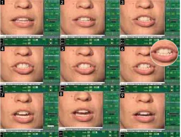
These videos last for about 12 seconds, comprising a total of 360 frames per video. They are analyzed and split up – at rest, during speech and smile — in order to produce four static frames (corresponding to a pho-tograph) for analysis of the least and the greatest expo-sure of the incisor, for example. Figure 1 demonstrates an example of this method.
What are the main esthetic parameters that guide your orthodontic treatment plan?
Carlos Alexandre Câmara
Our main parameters are listed below and illustrated in Figures 2 to 7:
1) Incisor exposure (Fig 2): Taking into account the length and elevation of the lip as well as the vertical height of the maxilla.
2) Negative lateral space or buccal corridor (Fig 3): There are many publications, comprising studies developed with digital image manipulation, which present different options of buccal corridor exposure. It seems to be common sense that minimal corridors are more attractive.2,3,5
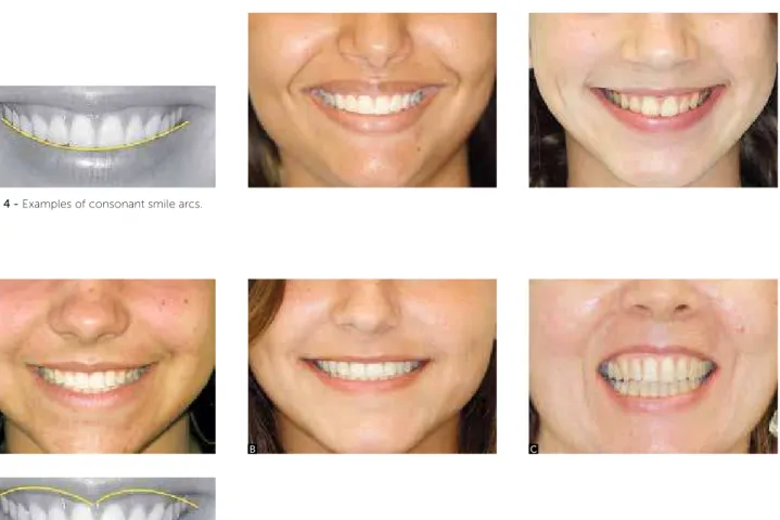
3) Smile arc (Fig 4): Taking into account that, in an ideal smile arc, the curvature of the incisal edge of the maxillary incisors coincides with the mar-gin of the lower lip when smiling (“consonant”). The lower lip may touch, not touch or slightly cover the incisal edge of the maxillary incisors.
4) Upper lip curvature (Fig 5) — Three options: up-ward, when the commissure is higher than the center of the lip; straight, when the commissure and the center of the lip are leveled; downward, when the commissure is lower than the center of the lip. Upward and straight curvatures are considered more esthetic than the down-ward curvature. The upper lip curvature is guided by the facial muscles; therefore, it cannot be changed through orthodontic treatment.
5) Smile symmetry (Fig 6): Commissures move upward and laterally when smiling, but there may be some diference in amount and direction of movement
Figure 1 - Video split up into static frames. Frame number 6 (ampliied)
shows the maximum incisor exposure when patients pronounced the sound “Czech”, as in Czechoslovakia.
1
4
7 8 9
5 6
between the right and the let sides. Should there be any signiicant diference, it may be due to deiciency of muscular tonus in one of the sides of the face.
6) Frontal occlusal plane (Fig 7): Represented by a line that goes from the incisal edge of the upper right canine to the incisal edge of the upper left canine. Transversal inclination may be caused by a differ-ence in eruption time or due to skeletal asymmetry.
Figure 4 - Examples of consonant smile arcs.
Figure 5 - Upper lip curvature (D).Examples of the three types of curvature: Upward (A), straight (B)
and downward (C).
A
D
B C
Figure 2 - Upper incisor exposure directly associated with both length and
elevation of the upper lip.
Figure 3 - Examples of negative lateral space or buccal corridor.
Clinical examinations are essential to perform a differential diagnosis to distinguish between smile asymmetry, occlusal plane inclination and facial asymmetry. The influence of the lower teeth on the smile cannot be seen in dental cast models.
Figure 6 - In a symmetric smile (A), commissures move upward harmoniously. In B, asymmetry is remarkable, due to right commissure elevation being higher than the left one — such conditions cannot be changed through orthodontic therapy.
Figure 7 - Frontal occlusal plane represented by
a line that goes from the right canine to the left canine (A).Transversal inclination may be caused by a diference between the time of eruption or skeletal asymmetry. In B, example of frontal oc-clusal plane inclination.
A
A
B
B
Patient’s age is another esthetic parameter which I consider essential for treatment plan. Over time, men and women present less exposure of maxillary incisors and greater exposure of mandibular incisors, due to the fact that the length of the upper lip increases, while the length of the lower lip decreases with age. It has been proven that the amount of exposure of mandibular inci-sors ater 60 years of age is equivalent to the amount of exposure of maxillary incisors before 30 years of age.10
With aging, the lips go through many changes which afect the exposure of teeth. For example, muscle atrophy that implies in decreased lip volume, changes in lip shape and increased length of the lips. Such changes highlight the importance of normative values according to age, al-lowing older patients to have their treatment plan objec-tively and precisely carried out.11
Ater the age of 40, there is a 2 mm mean loss of muscle capacity to lit up the upper lip. This contributes to age one’s appearance, since upper anterior teeth are hidden by the upper lip at smile.11
When a patient of such age comes to me complain-ing about dissatisfaction concerncomplain-ing crowdcomplain-ing of man-dibular incisors, claiming that he/she is dissatisied with his/her appearance in photographs and that he/she has recently noticed this situation, I suppose that such
crowding had already been there for a long time, but has been recently noticed.
Deepening of the nasolabial angle is another conse-quence of aging, causing great esthetic impact. As time goes by, there is loss of cheek prominence due to atrophy, making the skin loose. As the cheek becomes laccid, the elevator muscles of the upper lip pull the nasolabial folds upward, resulting in a deep nasolabial angle.
Our researches carried out at the Rio de Janeiro State University/Brazil (UERJ) also suggest that men are more likely to sufer the efects of aging than wom-en. A comparative example regarding these diferences is demonstrated in photographs of young and older men, as shown in Figure 8. Many factors may turn a young skin into a precociously aged skin, namely: sun exposure, smoking, stress, hormonal changes, medi-cines and unhealthy eating habits. The main villain has always been excessive sun exposure. Therefore, exces-sive sun exposure, due to sports practice or outdoor ac-tivities, without proper protection from UV radiation, may be one of the factors that inluence the fact that men are more afected by the efects of aging. How-ever, it is important to consider that smoking has been the main cause of early aging, as it directly acts upon the epithelium elastic ibers and collagen matrix.5
In the last few years, some authors have ques-tioned how valid is to use a simple photograph to assess esthetics and determine the patient’s treatment plan. They have suggested the use of dynamic images recorded in video. In your opin-ion, capturing dynamic images through ilming, in order to use them for the diagnosis and
treat-ment plan in Orthodontics, is a valid method? Sissy Mendes Machado
Smile analysis performed through standardized photographs may be used as a subsidiary method for diagnosing and planning orthodontic and surgical treatments, allowing assessments before and ater the treatment. Additionally, it may be considered a reli-able instrument for comparing pre and posttreatment phases. However, it is important to highlight that this technique may be considered dubious, as it does not represent a smile apex. Video recording may provide larger information for facial esthetic assessment.4,5
Dynamic analysis of speech and smile has been made possible because of technology. Current low prices and popularization of digital cameras have fa-cilitated access to this type of equipment. Photo edit-ing programs are available in most computers. This facilitated the identification of both positive and neg-ative aspects of facial esthetics, enabling, for exam-ple, observation of the effects of aging in the perioral soft tissues. Videography may be used in orthodontic practice with many purposes, such as diagnosis and treatment plan, communication between specialist and patient during appointments and communication between specialists.4
Figure 8 - Exposure of upper incisor is greater in younger individuals (A, B, C) while exposure of lower incisor is greater in elder individuals (D, E, F).
A
D
B
E
C
There has been increasing awareness among professors, researchers and clinicians about the importance of biology for orthodontic practice and this has been relected in the curriculum of the main Orthodontics programs in the world. Why is it important for the orthodontist’s formal education to be familiar with the biology of tooth movement? Ildeu Andrade Jr.
The postgraduate programs in Orthodontics have always favored formal education and training based on orthodontic mechanics, as all credit hours are spent with studies performed with typodont simulator. However, Orthodontic clinical practice is based in both mechani-cal and biologimechani-cal parameters. The mechanimechani-cal approach is enough for treatments performed on the typodont, how-ever, in clinical practice, knowledge of biology involved in the process is necessary. I am a die-hard fan of the mechanotransduction deinition: Mechanical forces ap-plied to teeth do not result in mechanical movement only, they also produce biological stimuli capable of promot-ing desirable tissue reactions in order to achieve a stable and long-lasting change in tooth position.13 The clinical practice of this deinition lead us to question the claims concerning the types of brackets or techniques ofering drastic reduction in treatment duration. Whenever I see an advertisement or a presentation stating this type of of-fer, I begin to think: Is this capable of promoting a dif-ferentiated biological stimulus? If not, what explains the hypothesis of reducing treatment duration?
The main orthodontic journals in the world have provided more opportunities for the publication of researches into biology of tooth movement. In your opinion, what are the beneits — for us, or-thodontists and for our patients — of the increas-ing interaction between biological and clinical researches in Orthodontics? Ildeu Andrade Jr.
We have conducted a survey at PubMed, in March 2013, which reveals an increase in the number of pub-lications available in the literature,when the keywords “orthodontic tooth movement” were used (data is shown in Fig 9).
There are many reasons for this increasing number of publications. First, the total amount of scientiic journals has signiicantly increased, providing the opportunity for the publication of scientiic indings. We are currently facing the dilemma of what we should read due to the
great amount of information that is available. However, I believe that the main reason for this increasing number of publications is the development of researches that have as inal outcome the publication of their results. Researches carried out with biomarkers aim at studying not only orthodontic tooth movement in many diferent situa-tions, but also root resorption as well as accelerated and decelerated tooth movement. Such researches, which fre-quently involve molecular biology analysis, have contrib-uted for a signiicant increase in the number of publica-tions in the 2000s, especially in the last ive years.14,15 Our studies involving the quantiication of diferent markers present in the gingival crevicular luid while moving teeth aim at understanding what happens when we apply forces to induce tooth movement.16 As a researcher, I aim at un-derstanding the cascade of biological events involved in tooth movement. The main beneit for the orthodontist is to understand why his patients react diferently to the same mechanics, same appliances and same activations. Why do patients react diferently towards the gains and sequelae of orthodontic treatment? Force modulation be-ing the only variable used to answer that seems to be a simplistic approach towards a more complex issue.
In your opinion, what are the future prospects and the potential clinical impacts of researches on the cellular and molecular mechanisms in-volved in biology of tooth movement?
Ildeu Andrade Jr.
Our initial studies on gingival crevicular fluid (GCF) were based on Periodontology researches which claim that the GCF flow may increase by 30 times in periodontitis, when compared to healthy sulcus.17 As orthodontic tooth movement has
inflam-Figure 9 - Total number of publications available in the literature using
“orthodontic tooth movement” as keywords. Source: www.ncbi.nlm.nih.gov/pubmed. 1400 1200 1000 800 600 400 200
1950 – 1959 1960 – 1969 1970 – 1977 1978 – 1983 1984 – 1989 1990 – 1995 1996 – 2001 2002 – 2007 2008 – 2013
mation-dependent characteristics, we believe that the GCF volume would increase during this movement. However, we have observed that such data are not consistently expressed. The GCF volume does not seem to be a reliable biomarker used for the analysis of tissue remodeling during orthodontic treatment.18-21
With regard to periodontal patients, we based our studies upon previous evidence which claim that clinically healthy sites of patients with periodontitis have greater levels of GCF and periodontal pathogen biomarkers than clinically healthy sites of patients without periodontitis.22 In other words, periodontal patients would be considered as high-risk patients for orthodontic treatment, as pro-inflammatory mark-ers originated from periodontal diseases have been superposed onto those markers activated by tooth movement. It is a chaotic scenario! However, a study carried out with periodontal patients undergoing tooth movement reveals that orthodontic movement of periodontally compromised teeth does not result in significant changes of matrix metalloproteinase in the GCF.23 Clinically speaking, this is a great finding as it assures us that moving periodontally compro-mised teeth, yet healthy, is possible.
With regard to future prospects, it is necessary to have a better understanding concerning the molecular constituents of the gingival crevicular luid which are more sensitive to orthodontic tooth movement, the meaning of GCF as well as its biological function. Stud-ies on GCF have enabled non-invasive monitoring of tissue remodeling and may serve as a good tool for re-searches on tooth movement, root resorption or den-tal ankylosis. The more we know about the biological mechanisms of orthodontic tooth movement, the earlier we shall develop local administration methods of medi-cation. Local application of bioactive agents associated with conventional biomechanics may be used to facili-tate or hinder tooth movement.
In clinical practice, orthodontic treatment formed in adults is diferent from treatment per-formed in adolescents, as adults are more likely to have compromised dentitions. Based on your ex-perience with biological studies on tooth move-ment, what would be the most appropriate treat-ment plan for adult patients with bone loss and clinical attachment loss caused by jeopardizing
the biological space or by previous periodontal diseases, taking into account biomechanical and functional properties? Sissy Mendes Machado
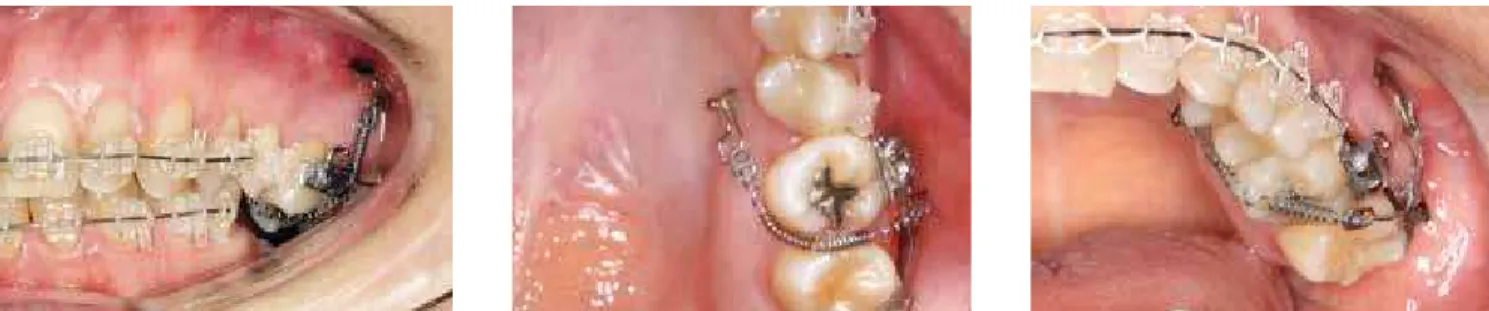
Orthodontic treatment of patients with periodon-tal disease may not be faced as a common treatment. All orthodontic cases require efective diagnosis and planning. However, this practice is more diicult in periodontal patients. We must bear in mind that all types of treatment must beneit the patient. When the periodontium is compromised and the orthodontic plan is defective, the chances of harming the patient, due to an improperly performed treatment, are high. Therefore, such cases require integration between spe-cialists. The patient should not start orthodontic tooth movement without his periodontist’s approval, and the periodontist must follow-up the periodontal situation during orthodontic treatment.25,26 Figures 10, 11 and 12 show a good example of tooth movement in a pa-tient with little periodontal attachment.
Tooth movement should not be performed in pa-tients with periodontal diseases for many reasons, namely: 1) It may cause periodontal abscess, especially in deep periodontal pockets; 2) Ater periodontal ther-apy, edema is eliminated and gingival collagen ibers consolidate, leading to tooth accommodation which may be favorable to tooth alignment and, thus, reduce the duration of a subsequent orthodontic treatment; 3) At the beginning of orthodontic treatment, it is im-portant to know the height as well as the density of the bone surrounding the teeth. Such information may be observed by the periodontist during surgery and pro-vided later on to the orthodontist.25,26
Figure 10 - Intraoral photographs and periapi-cal radiograph taken at the beginning of the orthodontic treatment in patient with controlled periodontitis. Tooth #22, with little attachment, must be moved with low intensity forces.
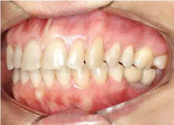
Figure 11 - Tooth #22 position improves after
For adult patients, the orthodontic plan with ide-al objectives must be reconsidered when the peri-odontium is compromised, when there is great tooth loss or other limiting conditions. Such patients need an objective and directed treatment in order to have their problem quickly and efficiently solved. Three factors must be taken into account at this moment, namely: 1) Simplification — seek mechanics to fa-vor patient’s oral hygiene. 2) Goals — ponder which would be the best result taking esthetics, stability and functional occlusion into account. Patients usu-ally seek improvements in esthetics, which does not always provide stable results. Therefore, the special-ist must discuss with the patient in order to reach a
Figure 12 - Intraoral photographs and periapical
ra-diograph taken at the beginning of the orthodontic treatment in patient with controlled periodontitis.
Figure 13 - Example of an objective and directed treatment. Need for
intru-sion of right mandibular molars to place the crowns over the implants in the antagonist area.
mutual agreement on the final outcome. 3) Risk-cost/benefit — talk to the patient about the benefits of the treatment. The costs involve not only the fi-nancial value, but also pain, discomfort and tooth mobility. Risks are seen as the possibility of creating problems which did not exist in the first place, such as decalcification, root resorption, mucogingival problems and even tooth loss. At this moment, the orthodontist must also notify the patient about the risks of not taking the treatment.25 Figures 13 to 16 illustrate a case with a realistic objective: the need for intrusion of left mandibular molars for prosthetic
restoration of antagonist teeth. Figure 15 - After intrusion and placement of crowns over the implants.
Figure 16 - Study models demonstrating the performed treatment: Initial (A); after intrusion of teeth #26 and #27 (B); crowns placed over implants in the
mandibular arch (C).
A B C
What is your opinion about early orthosurgical treatment with esthetic and psychosocial pur-poses? Carlos Alexandre Câmara
When treating Class III, we have good results in patients from 7 to 10 years old with mild to moderate malocclusion and in patients from 17 to 19 years old undergoing the renowned orthosurgical treatment, or in cases with little maxillomandibular discrepancies in which we have the option of orthodontic camoulage.6 In clinical practice, I have always noticed a gap con-cerning the treatment of Class III patients from 12 to 14 years old with dental and facial deformities: it is too late for an interceptive approach and too early for surgery. Awaiting growth to end prior to starting or-thosurgical treatment is sometimes extremely diicult
Figure 19 - Facial and intraoral photographs taken at the end of treatment (surgery performed by Dr. Henrique Martins).
Figure 20 - Cephalometric radiographs at initial (A), presurgical (B) and postsurgical (C) phases.
Figure 21 - Facial and intraoral photographs of patient aged 18. Five years after the surgery, we notice good facial esthetics and, again, dental compensation in the anteroinferior segment. Potential need for a new surgery shall be assessed at the age of 21.
Is there a minimum age required to indicate or-thognathic surgery for growing patients? If so, what would it be concerning the diferent types of deformities? Carlos Jorge Vogel
The minimum age required to indicate orthogna-thic surgery for growing patients is directly associated with the development of dentition. Therefore, the dental development stage is decisive for surgical proce-dures involving all kinds of facial deformities in which early surgery is recommended — mandibular hyper-plasia or hypohyper-plasia, maxillary hypohyper-plasia or maxil-lary vertical hyperplasia. The development degree of maxillary canines is important to assess the viability of maxillary surgery with no harm to the dental structure.
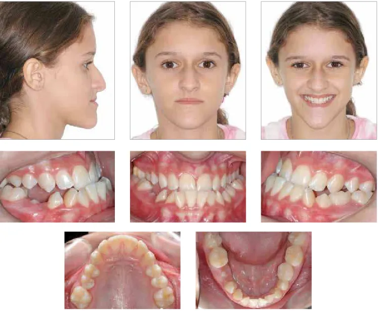
Figure 22 - Facial and intraoral photographs taken at the beginning of the orthodontic treatment in patient aged 12, with Class III dental and skeletal relationship.
Is surgical treatment indicated for mandibular asymmetry cases during growth?
Carlos Jorge Vogel
The mandibular asymmetry cases in which surgery is indicated during growth are, mainly, the condylar hyperplasia ones. In this asymmetric growth process, accentuated by the age of 13, the hypothesis of high condylectomy procedure must be taken into account. Some surgical maneuvers that section the upper part of the mandibular condyle, which grows increasingly, have presented good results for a subsequent balanced mandibular growth process.
Which surgery cases are more likely to succeed: Correction of hypoplasia or hyperplasia?
Carlos Jorge Vogel
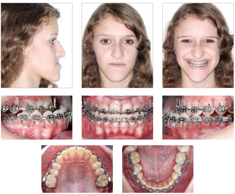
Figure 23 - Facial and intraoral photographs after orthodontic preparation for orthognathic surgery.
alone was considered satisfactory, currently many Class III cases that are treated with an early surgical approach un-dergo more maxillary advancement surgeries than man-dibular setback surgeries.7,24
The Anticipated Beneit orthognathic surgery has presented many advantages, such as the reduc-tion of about 40 to 50% in convenreduc-tional treat-ment estimated total time. Based on your experi-ence, do you also see such advantage? If so, what are the causes of this reduction in time?
Sissy Maria Mendes Machado
The expression “Anticipated Beneit” was created by my colleague Dr. Jorge Faber.27 It refers to an ap-proach towards orthosurgical treatment in which there
is an inversion of stages: From Orthodontics prior to Surgery to Surgery prior to Orthodontics.
The “surgery-irst” approach refers to a process in which orthognathic surgery is performed prior to orthodontic treatment, while the “Orthodontics-irst” approach refers to a process in which orthodontic treat-ment is impletreat-mented prior to orthognathic surgery. Presurgical orthodontic treatment aims at aligning both upper and lower dental arches in order to coordi-nate their respective skeletal bases, eliminating dental compensations. Orthodontic treatment performed at the presurgical phase is proved very important to yield the best results with the least relapse.6,28
occlu-Figure 24 - Facial and intraoral photographs taken at the end of treatment. Combined surgery was performed,with 5 mm of maxillary advancement and 2 mm of mandibular setback (surgery performed by Dr. Henrique Martins).
Figure 25 - Cephalometric radiographs at initial (A) and inal (B) phases.
sion prior to facial esthetics. The surgery-irst approach solves, at the same time, skeletal problems despite occa-sional dental compensations, and a transitional occlusion is corrected ater surgery. The surgery-irst approach is recommended for cases with no need of signiicant de-compensation or presurgical orthodontic alignment. Such cases include: 1) properly aligned or slightly crowd-ed teeth; 2) mild curve of Spee and 3) incisor inclination ranging from normal to slightly buccal or retroclined.28
The main reported advantages are: 1) improvement in patient’s chief complaint (dental function) and facial es-thetics right at the beginning of treatment; 2) reduction in treatment total time, depending on the complexity of the orthodontic treatment; 3) reduction in complexity and duration of orthodontic treatment due to accelerated orth-odontic tooth movement at the presurgical phase.28
Answering your question, we presume that reduc-tion in total treatment time can be associated with recom-mending the surgery-irst approach for less complex cases. When comparing the average total time of the surgery-irst approach with the average total time of the traditional ap-proach, which includes all types of orthosurgical treatments, we found that the average time for the former is shorter.
However, we should also pay attention to the fact that tooth movement was faster immediately ater surgery. An increase in osteoclastic activity and metabolic chang-es in the dentoalveolar region, with higher exprchang-ession of bone activity markers — such as alkaline phosphatase (ALP) and C-terminal telopeptide (CTX-1) — are val-id reasons for such accelerated bone remodeling.29 It is as if orthognathic surgery worked as the injury caused by the selective alveolar corticotomy. This is an inter-esting inding which requires further research.
During the ABOR (Brazilian Association of Ortho-dontics and Facial Orthopedics) congress, held in the city of Belo Horizonte/MG, the Brazilian Board of Orthodontics and Dentofacial Ortho-pedics (BBO) promoted the pilot project for the certiication process of recent graduates and students in the last year of the post-graduation course in Orthodontics. What are your expecta-tions towards those participating in this activity? What are the plans for the future?
Carlos Jorge Vogel
The participants in this activity should follow cri-teria such as: 1) minimum 12-month graduate from a post-graduation course in Orthodontics with a min-imum of 2,000 credit hours; 2) student attending the last year of a post-graduation course in Orthodontics with a minimum of 2,000 credit hours; 3) member of a state-owned entity affiliated with the ABOR.
The chairman of the ABOR invited the coordi-nators of the courses with a minimum of 2,000 credit hours to take part in this pilot project.
The 47 candidates who applied passed the pilot project examination (equivalent to Phase I). Once they have passed the examination, they have up to 10 years to sit in for Phase II of the certification exam. Should they pass it, they will become a BBO Diplo-mate, being approved in the “Clinical Excellence” examination.
It is highly likely that a similar exam will be held in Natal/RN (Brazil), during the next ABOR Congress.
As for the BBO, our expectations obligatorily lay on our young colleagues: They shall decide the future of our specialty.
Carlos Alexandre Câmara
» Specialist in Orthodontics, UERJ.
» Director of the College of Diplomates of the Brazilian Board of Orthodontics and Dentofacial Orthopedics (CDBBO).
» Peer reviewer, Revista Dental Press de Estética.
Ildeu Andrade Jr.
» MSc and Specialist in Orthodontics, Marquette University, USA.
» PhD in Cell Biology, UFMG.
» Professor of the Masters course in Orthodontics, PUC-MG.
Carlos Jorge Vogel
» PhD in Orthodontics, USP. MSc in Orthodontics, University of Illinois, USA.
» Member of the Angle Society, USA.
» Former Director of the Brazilian Board of Orthodontics and Dentofacial Orthopedics (BBO).
Sissy Maria Mendes Machado
» Specialist in Orthodontics, UERJ.
» Specialist in Occupational Dentistry, Faculdade São Leopoldo Mandic.
» MSc in Dentistry, Dental Materials, UFPA.
» Professor at the Specialization course in Orthodontics at ABO-PA.
1. Van der Geld PAAM, Oosterveld P, Van Waas MAJ, Kuijpers-Jagtman AM. Digital videographic measurement of tooth display and lip position in smiling and speech: Reliability and clinical application. Am J Orthod Dentofacial Orthop. 2007;131(3):301.e1-8.
2. Ackerman MB, Ackerman JL. Smile analysis and design in the digital era. J Clin Orthod. 2002;36(4):221-36.
3. Sarver DM, Ackerman MB. Dynamic smile visualization and quantiication: Part 1. Evolution of the concept and dynamic records for smile capture. Am J Orthod Dentofacial Orthop. 2003;124(1):4-12.
4. Cosendey VL, Drummond S, Capelli Jr J. Capture, analysis and measurement of images of speech and smile dynamics. Dental Press J Orthod. 2012;17(5):151-6.
5. Drummond S. Exposição dos incisivos durante a fala e o sorriso: correlação com a idade e o gênero [dissertação]. Rio de Janeiro (RJ): Universidade do Estado do Rio de Janeiro; 2011.
6. Franco A, Cosendey V, Almeida MA, Capelli Jr J. Tratamento da Classe III: cirurgia ou camulagem? Orthod Sci Pract. 2012;5(19):333-45.
7. Capelli Jr J, Almeida RC. Orthosurgical treatment of patients in the growth period: at what cost? Dental Press J Orthod. 2012;17(1):159-77.
8. Wolford LM, Karras SC, Mehra P. Considerations for orthognathic surgery during growth, Part I: Mandibular deformities. Am J Orthod Dentofacial Orthop. 2001;119(2):95-101.
9. Câmara CA. Aesthetics in orthodontics: six horizontal smile lines. Dental Press J Orthod. 2010;15(1):118-31.
10. Vig G, Brundo C. The kinetics of anterior tooth display. J Prosthet Dent. 1978;39(5):502-4.
11. Desai S, Upadhyay M, Nanda R. Dynamic smile analysis: changes with age. Am J Orthod Dentofacial Orthop. 2009 ;136(3):310.e1-10; discussion 310-1. 12. Escossia N. Utilização de toxina botulínica do tipo A para minimizar o sorriso
gengival. [monograia]. Rio de Janeiro (RJ): Universidade do Estado do Rio de Janeiro; 2012.
13. Consolaro A. Movimentação dentária induzida: biologia aplicada à prática clinica. In: Consolaro A. Reabsorções dentárias nas especialidades clínicas. Maringá: Dental Press; 2005. cap. 11, p. 303-52.
14. Krishnan V, Nair S, Ranjith A, Davidovitch Z. Research in tooth movement biology: the current status. Semin Orthod. 2012;18:308-16.
15. Andrade I, Taddei S, Souza P. Inlammation and tooth movement: the role of cytokines, chemokines, and growth factors. Seminars in Orthod. 2012;18(4):257-69.
16. Capelli J Jr, Kantarci A, Hafajee A, Teles RP, Fidel R Jr, Figueredo CM. Matrix metalloproteinases and chemokines in the gingival crevicular luid during orthodontic tooth movement. Eur J Orthod. 2011;33(6):705-11.
17. Uitto VJ. Gingival crevice luid: An introduction. Periodontol 2000. 2003;31:9-11. 18. Capelli Jr J, Fidel Jr Rivail, Figueredo CM, Teles RP. Change in the gingival
luid volume during maxillary canine retraction. Dental Press J Orthod. 2010;15(2):52-7.
19. Drummond S, Canavarro C, Perinetti G, Teles R, Capelli Jr J. The monitoring of gingival crevicular luid volume during orthodontic treatment: a longitudinal randomized split-mouth study. Eur J Orthod. 2012;34(1):109-13. 20. Almeida RC, Santos D, Teles R, Capelli Jr J. Gingival crevicular luid volume
evaluation in patients with controlled periodontal disease submitted to orthodontic treatment. J World Fed Orthod. 2012;22:e 9-12. REFERENCES
21. Perinetti G, Primozic J, Castaldo A, Di Lenarda R, Contardo L. Is gingival crevicular luid volume sensitive to orthodontic tooth movement? A systematic review of split-mouth longitudinal studies. Orthod Craniofac Res. 2013;16(1):1-19.
22. Teles R, Sakellari D, Teles F, Konstantinidis A, Kent R, Socransky S, et al. Relationships among gingival crevicular luid biomarkers, clinical parameters of periodontal disease, and the subgingival microbiota. J Periodontol. 2010;81(1):89-98.
23. Almeida R C. Efeito do movimento dentário nos níveis de metaloproteinases da matriz no luido gengival de pacientes com periodontite controlada [tese]. Rio de Janeiro (RJ): Universidade do Estado do Rio de Janeiro; 2012. 24. Teixeira A, Medeiros P, Capelli Jr J. Intervenção ortocirúrgica em paciente
adolescente com acentuada displasia esquelética de Classe III. Rev Dental Press Ortod Ortop Facial. 2007;12(54):55-62.
25. Almeida RC, Martins CC, Capelli Jr J. Inter-relação Periodontia-Ortodontia. In: Ortodontia em um contexto multidisciplinar. Maringá: Dental Press; 2013. cap. 4, p. 85-120.
26. Heasman PA, Millett D, Chapple I L. The periodontium and orthodontics in health and disease. Oxford: Oxford University; 1996. p. 137-59.
27. Faber J. Benefício antecipado: uma nova abordagem para o tratamento com cirurgia ortognática que elimina o preparo ortodôntico convencional. Dental Press J Orthod. 2010;15(1):144-57.
28. Liou EJ, Chen PH, Wang YC, Yu CC, Huang CS, Chen YR. Surgery-irst accelerated orthognathic surgery: orthodontic guidelines and setup for model surgery. J Oral Maxillofac Surg. 2011;69(3):771-80.
29. Liou EJ, Chen PH, Wang YC, Yu CC, Huang CS, Chen YR. Surgery-irst accelerated orthognathic surgery: postoperative rapid orthodontic tooth movement. J Oral Maxillofac Surg. 2011;69(3):781-5.
Acknowledgments