Angle Class II malocclusion treated with
extraction of permanent teeth
» The author reports no commercial, proprietary or financial interest in the products or companies described in this article.
» The patient displayed in this article previously approved the use of her facial and intraoral photographs.
Submitted: May 10, 2013 - Revised and accepted: May 29, 2013
Contact address: Gustavo Mattos Barreto
Rua Campo do Brito, 47 – Apt. 1002 – Edf. Mansão Cristal – Bairro 13 de Julho CEP: 49.020-380 – Aracaju/SE — Brazil
E-mail: [email protected] INTRODUCTION
A 17.11-year-old female patient came to the clinic reporting diiculty in gripping food with her front teeth and dissatisfaction regarding dental esthetics, particularly concerning her smile. In the anamnesis, the patient presented history of mouth breathing, with adenoids treated during childhood, allergic rhinitis, good oral health with few restorations, lingual inter-position and absence of the third molars which had al-ready been extracted.
DIAGNOSIS
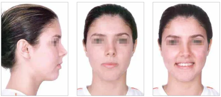
On assessing the patient’s face, a dolichofacial pat-tern was observed in addition to absence of passive lip seal, increased lower facial third, signiicative exposure of the lower lip vermilion, wide buccal corridor when smiling, straight proile, normal upper and lower lips and increased nasolabial angle (Fig 1).
Clinically, the dental arches presented Angle Class II, division 1 malocclusion with greater magnitude on the right side (full), being cusp-to-cusp on the let side.
Gustavo Mattos Barreto1
1Specialist in Radiology, UNICAMP. MSc and PhD in Orthodontics and Facial
Orthopedics, State University of São Paulo — Araraquara. Certified by the Brazilian Board of Orthodontics and Dentofacial Orthopedics (BBO). Visiting professor at the PostGraduate Course in Orthodontics, UNIT.
* Clinical case, category 2, accepted by the Brazilian Board of Orthodontics and Dentofacial Orthopedics, BBO.
How to cite this article: Barreto GM. Angle Class II malocclusion treated with extraction of permanent teeth. Dental Press J Orthod. 2013 July-Aug;18(4):126-33.
Angle Class II malocclusion associated with anterior open bite in adult patients demands a carefully elaborated orthodon-tic planning, aiming at restoring not only harmonious dental and facial estheorthodon-tics, but also a balanced masorthodon-ticatory function. Orthognathic surgery or permanent teeth extraction are oten the choice of treatment, therefore, treatment decision is related to all dental, skeletal and functional aspects. The present report discusses orthodontic compensation carried out by means of upper premolar extraction performed to correct the Class II canine relationship and, consequently, the an-terior open bite, accepting that the upper incisors be retroclined. This clinical case was presented to the Brazilian Board of Orthodontics and Dentofacial Orthopedics (BBO) as part of the requirements for obtaining the BBO Certiication.
Keywords:Class II malocclusion. Anterior open bite. Orthodontic compensation.
A má oclusão Classe II de Angle associada à mordida aberta anterior, em pacientes adultos, envolve um planejamento ortodôntico bem elaborado e cuidadoso, tendo como objetivo principal devolver a estética facial e dentária harmonio-sa, e uma função mastigatória equilibrada. O tratamento, muitas vezes, está no limite para cirurgia ortognática ou para extrações de dentes permanentes, fazendo com que a decisão esteja ligada a todos os aspectos dentários, esqueléticos e funcionais. O presente relato mostra a compensação ortodôntica por meio de extrações de pré-molares superiores, a im de corrigir a relação de Classe II dos caninos e, consequentemente, a mordida aberta anterior, aceitando que os incisivos superiores estejam retroinclinados. Esse caso clínico foi apresentado à diretoria do Board Brasileiro de Orto-dontia e Ortopedia Facial (BBO) como parte dos requisitos para a obtenção do título de Diplomado pelo BBO.
BBO Case Report Barreto GM
Figure 1 - Initial facial photographs.
With regard to the latter, parents were aware that it would take longer, with the possibility of slight upper lip retraction. Moreover, in this planning, the upper irst premolars would remain in Class II relationship, but with the canines in key of occlusion.1,2 For the correction of
the anterior open bite, the space let ater the extractions would allow retroclination to be planned and consequent extrusion of the upper incisors, favoring bite closure.3,4
As for arch atresia, expansion performed with a Haas ap-pliance was recommended. Treatment prognosis would be favorable, but careful, in order to slightly maintain or change the upper lip position due to retraction. Addi-tionally, speech monitoring would be necessary due to lingual interposition. With regard to stability, it is im-portant to consider the removal of the etiologic factor of the anterior open bite (lingual interposition ater mouth breathing during childhood).5 Occlusal adjustments were
also planned ater treatment inishing.
PERFORMED TREATMENT
Initially, the Haas expander appliance was installed for upper expansion. It was activated at ¼ of a turn ev-ery 12 hours, during 12 days, with a retention period lasting for ive months. Aterwards, a 0.022 x 0.028-in Edgewise standard system conventional ixed appli-ance was installed, however, not including teeth #14 and #24, which would be removed. The extraction of teeth #14 and #24 was requested and a 0.9 mm palatal Therefore, severe anterior open bite, restricted from
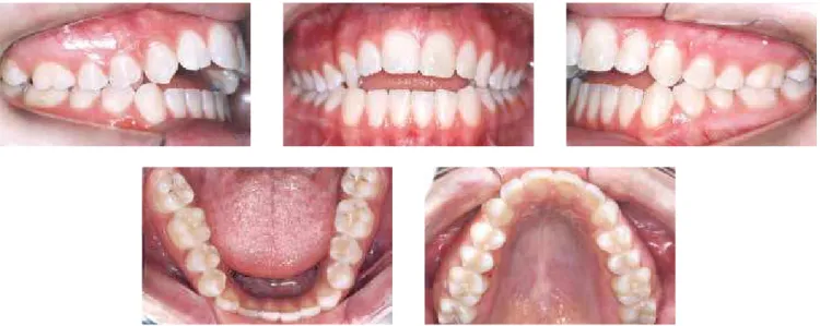
canine to canine, was also observed in addition to a 2 mm deviation of the lower midline to the right, overjet of 8 mm and lingual inclination of the posterior teeth. With regard to interdental distances, the upper intercanine dis-tance was of 30.5 mm while the lower one was of 27 mm. Functional guides were absent and there was diiculty in gripping food by the anterior region (Figs 2 and 3).
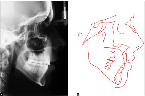
The maxillomandibular relationship was within the range of normality (ANB = 3°), however, with both the maxilla and the mandible retruded in relation to the cra-nial base. SNA= 75° and SNB= 72°, with a vertical growth pattern (SN.GoGn = 45°, FMA = 36° and Y axis = 66°). Moreover, the patient presented buccal inclination and protrusion of the upper incisors (1.NA = 35.5° and 1-NA = 9 mm). Additionally, she presented retroclination of lower incisors (1.NB = 16° and IMPA = 76°) (Fig 4 and Table 1). On assessing the panoramic radiograph, absence of the third molars which had already been removed, and slight root shortening of teeth #11, #12, #21, #22 and api-cal rounding of teeth #33, #32, #31, #41, #42 and #43 (Fig 5) were observed.
TREATMENT PLAN
Figure 2 - Initial intraoral photographs.
Figure 3 - Initial casts.
Figure 4- Initial proile cephalometric radiograph (A) and initial cephalometric tracing (B).
BBO Case Report Barreto GM
bar was installed for anchorage control. On the upper arch, alignment and leveling were performed on three segments: On teeth #13 to #17, #12 to #22 and #23 to #27. All segments began with 0.014-in NiTi wire, followed by 0.016, 0.018 and 0.020-in stainless steel wires, and, at last, 0.018 x 0.025-in stainless steel wire. At this phase, an extrusion arch was placed for the up-per incisors, with a 0.017 x 0.025-in TMA wire — coming out from the auxiliary tubes of the upper irst molars, bypassing, being superimposed on the region of the upper incisors and being activated for extrusion. Ater leveling, the upper occlusal plan was realigned with a continuous arch prior to mass space closure conventional mechanics, using stainless steel 0.018 x 0.025-in and 0.019 x 0.025-in archwires, with Bull loops between teeth #13 and #14, and #23 and #24.
At treatment inishing, stainless steel 0.019 x 0.025-in rectangular archwires were used with irst, second and third order bends individualized as necessary. In the lower arch, alignment and leveling were initiated with NiTi archwire 0.014-in, followed by 0.014 to 0.020-in SS archwires with irst and second order bends. Stainless steel 0.019 x 0.025-in rectangular inishing archwires, coordinated, with irst, second and third order bends individualized as necessary, were used next.
As retention protocol, a ixed bar was installed be-tween teeth #11 and # 21 (Bond a Braid) and a full remov-able upper plate was installed with stainless steel 0.032-in archwire. In the lower arch, an intercanine ixed bar was installed with 0.032-in Twist Flex archwire. The patient needed speech monitoring for six months, starting one
month before the removal of the ixed appliances. Occlu-sal adjustments were performed in three moments: at the inishing phase, six months and one year ater removal of the appliance. Furthermore, anatomic recontouring of the upper incisors was performed in addition to gingival plasty carried out on the posterior teeth for better dental exposure and, consequently, better esthetics.
OBTAINED RESULTS
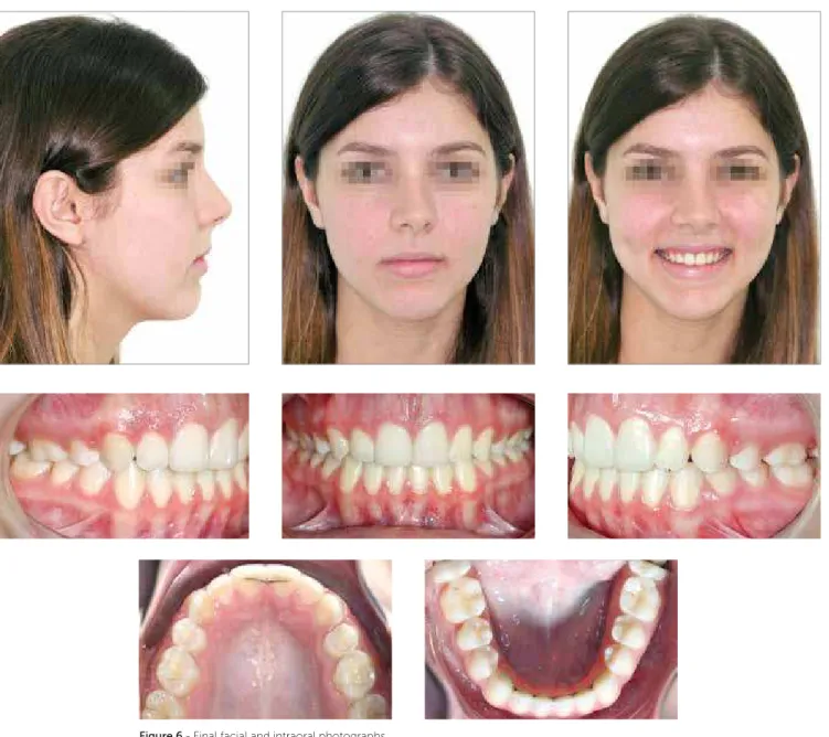
Ater orthodontic inishing, exams were performed in two moments: At inal phase (Figs 6 and 7) and af-ter two years of control (Figs 8 and 9). Improvement in lip seal, smile harmony with good visualization of the posterior teeth and good exposition of the incisors (gingival plasty was extremely relevant for the beauty of the smile) and mild alteration in the upper lip (UL-Line S = -1 mm ater treatment and -1.5 mm ater two years of control) were observed.
The molars inished in Class II relationship, as ex-pected, but with canines in key of occlusion. The ante-rior open bite was corrected and the results were main-tained ater the control period. Improvement in mid-line deviation ( with slight deviation), correction of the overjet (1 mm inal and ater two years) and in the upper arch shape (Figs 6 to 9) were observed.
Vertically, the patient presented the same charac-teristics, as expected, since an exclusively orthodontic treatment was chosen. On the anteroposterior direction, there was slight improvement in the ANB angle (3° ini-tial, 2° inal and ater two years of stability). Cephalo-metrically, the upper incisors were retroclined and re-tracted (1.NA = 13° and 1-NA = 3.5 mm, maintained ater two years of control) (Figs 10 and 11, Table 1).
On assessing the inal panoramic radiograph, good root parallelism ater space closure was observed. Such paral-lelism was preserved ater two years of control and root lengths underwent minor alterations, only (Figs 12 and 13).
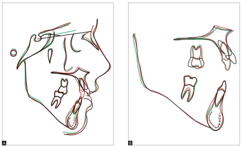
Interpretation of the cephalometric superimposi-tions demonstrated improvement in facial proile and lip position, which promoted not only a better seal, but also mild alteration of the upper lip and correction of the overjet and overbite. Partial assessment of the max-illa demonstrated signiicant retroclination, retraction and extrusion of the upper incisors, in addition to me-sialization of the upper molars due to anchorage loss. Partial assessment of the mandible did not demonstrate any signiicant alteration (Fig 14).
Figure 6 - Final facial and intraoral photographs.
BBO Case Report Barreto GM
Figure 8 - Facial and intraoral photographs of control after two years.
Figure 10- Final proile cephalometric radiograph (A) and cephalometric tracing (B).
Figure 11- Cephalometric radiograph (A) and proile cephalometric tracing (B) of control after two years.
Figure 12- Final panoramic radiograph. Figure 13- Panoramic radiograph of control after two years.
A B
BBO Case Report Barreto GM
1. Proit WR, Phillips C, Douvartzidis N. A comparison of outcomes of
orthodontic and surgical-orthodontic treatment of Class II malocclusion in adults. Am J Orthod Dentofacial Orthop. 1992;101(6):556-65.
2. Proit WR. Ortodontia contemporânea. 3a ed. Rio de Janeiro: Guanabara
Koogan; 2002.
3. Mihalik CA, Proit WR, Phillips C. Long-term follow-up of Class II adults treated with orthodontic camoulage: a comparison with orthognathic surgery outcomes. Am J Orthod Dentofacial Orthop. 2003;123(3):266-78.
4. Cal-Neto JP, Quintão CC, de Menezes LM, Almeida MA. Severe anterior
open-bite malocclusion. orthognathic surgery or several years of orthodontics? Angle Orthod. 2006;76(4):728-33.
5. Janson G, Valarelli FP, Beltrão RTS, Freitas MR, Henriques JFC. Stability of anterior open-bite extraction and nonextraction treatment in the permanent dentition. Am J Orthod Dentofacial Orthop. 2006;129(6):768-74.
REFERENCES
Measures Normal A B A/B dif. Control
Skeletal pattern
SNA (Steiner) 82° 75° 73.5° 1.5° 73°
SNB (Steiner) 80° 72° 71.5° 0.5° 71°
ANB (Steiner) 2° 3° 2° 1° 2°
Facial angle (Downs) 0° 2° 2° 0° 2°
Y axis (Downs) 59° 66° 67° 1° 67°
Facial angle (Downs) 87° 83° 82.5° 0.5° 82°
SN-GoGn (Steiner) 32° 45° 45° 0° 45°
FMA (Tweed) 25° 36° 36.5° 0.5° 37°
Dental pattern
IMPA (Tweed) 90° 76° 78° 2° 77.5°
1.NA (degrees) (Steiner) 22° 35.5° 13° 22.5° 13°
1-NA (mm) (Steiner) 4 mm 9 mm 3.5 mm 5.5 mm 4 mm
1.NB (degrees) (Steiner) 25° 16° 16° 0° 16°
1-NB (mm) (Steiner) 4 mm 3 mm 3.5 mm 0.5 mm 4
1
1- Interincisal angle (Downs) 130° 127° 150° 23° 150°
1-APo (mm) (Ricketts) 1 mm -1 mm 0 mm 1 mm 0 mm
Profile Upper lip – S line (Steiner) 0 mm -0.5 mm -1 mm 0.5 mm -1.5 mm
Lower lip – S line (Steiner) 0 mm 0 mm 0.5 mm 0.5 mm 0 mm
Table 1 - Cephalometric measurement summary.
FINAL CONSIDERATIONS
The results were satisfactory and the objectives were achieved. It should be highlighted that in this treatment, orthognathic surgery was considered, but discarded by the patient as well as by her parents. Additionally, the inal smile was pleasant and expressive, with good dental ex-posure, correction of anterior open bite and overjet, with good stability ater a two-year follow-up. The patient was very glad, since treatment exceeded her expectations as well as her family’s. It is worth emphasizing the importance of a multidisciplinary approach involving Cosmetic Dentistry and Periodontics, for the success of the treatment.
Figure 14- Total (A) and partial (B) superimpositions of initial tracing (black), inal tracing (red), and tracing of control after two years (green).