Risk factoRs of heteRotopic ossification
in tRaumatic spinal coRd injuRy
Cláudia Virgínia C. Coelho
1, Paulo Sergio S. Beraldo
2abstract – Objective: Heterotopic ossification (HO) is a complication of the spinal cord injury (SCI). It can result in anchylosis, harming the rehabilitation and quality of life. Previous publications had not elucidated the relation between possible independent variables, the aim of this study. Method: From 230 patients with SCI, admitted in 1998 at Hospital SARAH Brasilia, 33 with HO (14.3%; CI95% 10.1–19.6) were compared with 33 controls. The risk factors had been tested in bivariate analysis and in a model of logistic regression. Results:
Spasticity (odds ratio=3.8; CI95% 1.15–12.30), number of pressure ulcers (2.1; CI95% 1.08–3.89) and time lapsed since the injury (1.1; CI95% 1.02–1.24) were independently associated with HO. There was a confounder effect among these variables, without interaction. Conclusion: Spasticity, pressure ulcer and time of injury are associated with HO in spinal cord injury. The first two factors can be prevented and treatable.
Key wORdS: heterotopic ossification, spinal cord injuries, pressure ulcer, muscle spasticity, risk factors, multivariate analysis.
fatores de risco da ossificação heterotópica na lesão medular traumática
Resumo – Objetivo: A ossificação heterotópica (OH) é uma complicação da lesão medular traumática (LMT). Pode resultar em anquilose, prejudicando a reabilitação e a qualidade de vida. estudos prévios não elucidaram a relação entre as potenciais variáveis independentes, propósito desse estudo. Método: de 230 pacientes com LMT, admitidos em 1998 no Hospital SARAH Brasília, 33 tiveram o diagnóstico de OH (14,4%; IC95% 10,1–19,6), que foram comparados a 33 controles. Os fatores de risco foram testados de forma bivariada e num modelo de regressão logística. Resultados: espasticidade (razão de chances 3,8; IC95% 1,15–12,30), número de escaras (2,1; 1,08–3,89) e tempo de lesão (1,1; 1,02–1,24) encontraram-se associadas, de forma independente, à presença de OH. Havia um efeito confundidor entre essas variáveis, porém sem interação. Conclusão: espasticidade, escaras e tempo de lesão estão associados à OH na lesão medular traumática. Os dois primeiros são passíveis de prevenção e tratamento.
PALAvRAS-CHAve: ossificação heterotópica, traumatismos da medula espinal, úlcera de pressão, espasticidade muscular, fatores de risco, análise multivariada.
1Médica Radiologista, Mestre em Ciências da Reabilitação, Hospital SARAH Centro, Brasília dF, Brazil; 2Médico, doutor em Clínica Médica, Centro SARAH de Formação e Pesquisa, Programa de Pós-Graduação em Ciências da Reabilitação, Hospital SARAH Centro, Brasília dF, Brazil.
Received 15 december 2008. Accepted 23 March 2009.
Dr. Paulo Sergio S. Beraldo – SMPW Q18 Conj 5 Lote 3 casa H - 71741-805 Brasília DF - Brasil. E-mail: [email protected] Heterotopic ossiication is a common complication of
traumatic spinal cord injuries with a prevalence ranging from 5 to 50%, depending upon the studies design and the diagnosis method1-4. The progression of heterotopic ossiication may lead to decreases in the range of motion and anchylosis and has a negative impact on the rehabil-itation processes3. The most affected joints are the hips (60%–70%) and knees (20%–30%)1.
The etiology and pathogenesis of heterotopic ossii-cation are still unknown, but some factors appear to fa-vor its onset in patients with traumatic spinal cord
inju-ries: gender, age, completeness of the injury, presence of spasticity and decubitus ulcers5-8. However, various con-founding and interactive factors may be present. despite the importance of heterotopic ossiication, no studies were found which employed multivariate analyses to de-termine independent risk factors.
method
A total of 331 adult patients with traumatic spinal cord inju-ries were admitted to the Sarah Brasilia Hospital during the year of 1998. From this sample, patients younger than 15 years of age and with time since injury over two years were excluded. Thus, out of the 230 (70%) selected patients, 33 (14.4%, conidence in-tervals, CI95% 10.1 to 19.6) had periarticular heterotopic ossiica-tion in the hip and/or knee joints, as conirmed by X-rays. dur-ing the same period, 33 other patients were also investigated by images to exclude heterotopic ossiication and this group served as controls, coniguring a case-control design with a 1:1 ratio. All individuals’ data included in the analysis were collected retro-spectively based on medical records of patients hospitalized at Hospital SARAH Brasília, Spinal Cord Injury Program. The hospi-tal’s ethics committee approved the study.
The following variables were investigated: gender, age, and time since injury, both at hospital admission; length of stay; type of accident; other associated traumatic injuries; motor level and completeness of the injury9; reports of previous rehabilitation;
degrees of spasticity; urinary tract complications; number of pressure ulcers; presence of deep venous thrombosis; and cur-rent or previous histories of smoking.
For characterization purposes, a classiication of the severity of heterotopic ossiication was developed as follows: mild (ten-uous neo-bone formation in soft tissues adjacent to the joints),
moderate (ossiication in tissues adjacent to various anatomical bone prominences with well deined contours and without ar-ticular anchylosis), and severe (wide extensions of the hetero-topic ossiication and the presence of anchylosis).
Spasticity of the limbs was assessed by the Ashworth scale10,
which quantiies the resistance of the movement during passive extension movements. These scores range from zero (laccid, F or zero) to 4 (the affected joint is rigid in lexion or extension). In the analyses, the degrees of spasticity were reclassiied combin-ing the scores: absent/mild (scores F, 1 and 2), moderate (score 3) and severe (score 4); absent/mild (scores F, 1 and 2) and mod-erate/severe (scores 3 and 4).
All data were processed using the ePI-INFO software (CdC version 6.04b). Chi-square, chi-square for tendency and Mann-whitney were employed to investigate the differences be-tween groups according to the type of data. The criteria val-ue to consider the variables for the multivariate analyses was p<0.10. The binary logistical regression analysis using the step-wise method (forward selection) was performed with SPSS 13.0. The logistical model made it possible to estimate the occur-rence of heterotopic ossiication, based upon the risk factors included in the analysis. The evaluation of model adjustment and adequacy was obtained by the method of Hosmer-Leme-show11. Statistical signiicance for these analyses was
estab-lished at p<0.05.
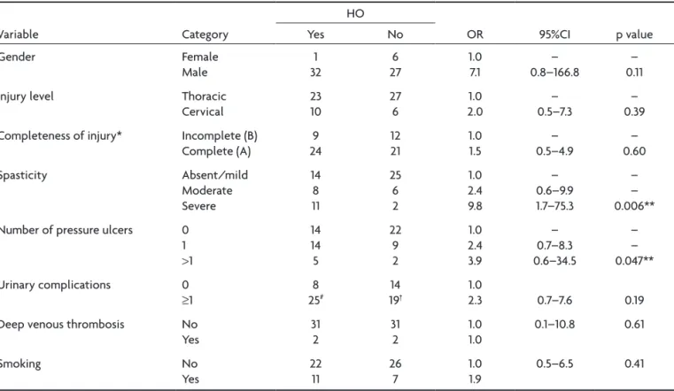
Table 1. Categorical variables for the heterotopic ossiication (HO) and control groups with the odds ratios (OR), conidence intervals (95%CI) and signiicance levels (p values).
variable Category
HO
OR 95%CI p value yes No
Gender Female
Male 321 276 1.07.1 0.8–166.8– 0.11– Injury level Thoracic
Cervical 2310 276 2.01.0 0.5–7.3– 0.39– Completeness of injury* Incomplete (B)
Complete (A) 249 1221 1.01.5 0.5–4.9– 0.60– Spasticity Absent/mild Moderate Severe 14 8 11 25 6 2 1.0 2.4 9.8 – 0.6–9.9 1.7–75.3 – – 0.006** Number of pressure ulcers 0
1 >1 14 14 5 22 9 2 1.0 2.4 3.9 – 0.7–8.3 0.6–34.5 – – 0.047** Urinary complications 0
≥1
8
25# 1914† 2.31.0 0.7–7.6 0.19
deep venous thrombosis No yes 31 2 31 2 1.0 1.0 0.1–10.8 0.61 Smoking No yes 22 11 26 7 1.0 1.9 0.5–6.5 0.41
Results
Among the 33 patients with heterotopic ossiication, the hip joints were most affected with 21 cases, 14 of which were bilateral. The knees were also affected in six patients and, of those, just one was bilaterally. The se-verity of the heterotopic ossiication was equally distrib-uted with 11 patients for each classiication levels (mild, moderate, and severe). In all cases and controls the auto-mobile accidents prevailed (53%), following by irearm le-sions (26%) and height fall (11%). However, there was a larg-er proportion of lesions from irearm among cases in rela-tion to controls (36% vs. 15%; c2=3.88, p=0.048). There was no difference between the groups about associated le-sions, with 14 cases and 18 controls (44% vs. 56%; c2=0.971; p=0.325). Fractures and dislocations (57%) predominate, equally distributed among the groups. In both groups pre-dominated the fractures of the bones from the trunk in cases (58%) and the controls (46%). Previous physiothera-py was informed for more than two thirds of the individ-uals, without distinction between the groups (28 cases vs.
23 controls, c2=2.16; p=0.142).
There were no differences between medians of cas-es and controls for age (30.0 vs. 27.0 years) and hospi-tal length of stay (1.5 vs. 1.8 months). However, the me-dian time since injury was 3 times larger in cases, com-pared to controls (8.6 vs. 2.8 months; p=0.001). The num-ber of pressure ulcers was categorized according to its median, i.e., none, one, or more than one. differences be-tween the groups were found only for the variables relat-ed to the spasticity and number of pressure ulcers, both in the positive sense, in other words, as larger its magni-tude, larger the association with the heterotopic ossii-cation (Table 1).
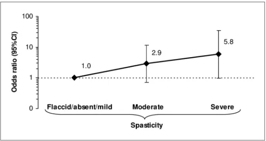
After adjusting for the confounding variables, the num-ber of pressure ulcers and the time since the injury con-tinued to show associations with the presence of hetero-topic ossiication. The association of spasticity and het-erotopic ossiication followed a dose-response pattern, although no signiicant (Figure). Considering that the fre-quency of severe spasticity was low, with 20% of the total number of cases and controls, this variable was dichoto-mized: laccid/absent/mild and moderate/severe. After Figure. Odds ratio of spasticity and heterotopic ossiication in patients with spinal cord injuries,
ad-justed to the number of pressure ulcers and time since injury. Although not signiicant, at initial mod-el, there is a clear tendency of a positive relationship between spasticity and heterotopic ossiica-tion, which became evident when spasticity was dichotomized to laccid/absent/mild and moder-ate/severe (inal model, Table 2).
Table 2. Independent variables associated with the presence of heterotopic ossification in patients with traumatic spinal cord.
variable Coeficient** Odds Ratio CI (95%) p value Spasticity* 1.32 3.76 1.15 -12.30 0.029 Number of pressure ulcers 0.72 2.05 1.08 - 3.89 0.028 Time since injury (months)† 0.12 1.12 1.02 - 1.24 0.023
these adjustments, the inal logistical model was obtained (Table 2). Based on the adjusted odds ratio, it was possible to estimate that the presence of spasticity into three and four grades compared to those related to the absence or mild (F, 1, and 2 grades), lead to increases of 276% in the probability of being associated with heterotopic ossiica-tion. In addition, for each pressure ulcer, there was a prob-ability of it being 105% higher in the patients who devel-oped heterotopic ossiication. Finally, every year of lesion increases in 322% this association (Table 2).
discussion
The present study evaluated patients with traumat-ic spinal cord injuries with heterotoptraumat-ic ossitraumat-ication com-pared to controls. The bivariate analyses detected signif-icant associations between the time of injury, number of pressure ulcers, degree of spasticity and irearm lesions with the development of heterotopic ossiication. Based upon the inal logistical model, the irst three variables were independently associated, without interactions. Be-fore considering these results, it is important to discuss methodological aspects involved in the investigation, as well as their implications.
The case-control design starts from the effects to identify the possible causes, coniguring a typical retro-spective study12,13. In this type of study, the relative risks are estimated by the odds ratio. The knowledge of the odds ratio permits the prediction of the occurrence of the heterotopic ossiication, but this does not mean that its presence necessarily implied the development of het-erotopic ossiication. The odds ratios were only indirect measures of this probability, since there may also exist associations with other determinants with various con-ditions of interest12. The case-control design has various limitations, such as the bias of prevalence, which can only be avoided when the most recent cases are included12,13. The ideal situation would be to consider the initial stag-es of heterotopic ossiication; however, this is a chronic condition and it is dificult to be identiied early, unless employing a cohort design. To minimize this bias, it was decided not to include patients who had more than two years of spinal cord injury.
Another problem regarding case-control design is the selection of the control group and the lack of compa-rability between groups’ characteristics. It is possible to restrict potential discrepancies by applying procedures, such as category limitations, matching, or making adjust-ments during the data analyses, like multivariate analy-sis12. Retrospective data may not be appropriate due to the lack of information in the medical records or because they were only based upon the subjects’ recall. Because of this, it is important that the assessments should be con-ducted by an independent investigator, who is blind to the
group assignments12. In this study the heterotopic ossii-cation cases were not blind to the investigator. However, this fact should not be considered to be relevant, since the variables were objective and easily identiied in the patients’ medical records. On the other hand, among the advantages of the case-control design, it is a fact that it is adequate for the investigation of rare conditions and a rapid, practical, and low-cost method to test the interac-tion effects of a great number of factors, which could po-tentially be related to the research in question12,13.
There were several dificulties to carry out the study protocol in the present research. First, the establishment of the heterotopic ossiication diagnosis must be consid-ered. The option to employ the imagery methods was due to their reported high sensitivity and speciicity for diag-nosing late heterotopic ossiication14. This method does not supply the precocious diagnosis, because it becomes positive only two to four weeks after the beginning of the clinical signs, when there is mineral increase in the osteoid matrix. Our criterion had purpose of correctly discrimi-nate the patients with and without heterotopic ossiica-tion. Because this criteria we were limited about the num-ber of patients to compose the control group. even so, considering a prevalence of the main risk factors for het-erotopic ossiication around 40%5, an alpha of 5% and a minimum odds ratio of 4.0, the calculated post hoc power of the study is of 76%, which is considered acceptable.
In the literature, various studies were found related to heterotopic ossiication in patients with spinal cord inju-ries, which focused on the clinical, diagnostic, and treat-ment aspects. However, considering causality, particular-ly using case-control design, onparticular-ly six were found, all us-ing only bivariate analyses. Scher7 found association with complete lesions. weiss et al.15, and Hunter et al.16, stud-ied patients with spinal cord and cerebral injuries and did not ind any associations between heterotopic ossiica-tion and the histocompatibility antigens. However, Larson et al.17 demonstrated an increased frequency of HLA-B27 in patients with spinal cord injuries and heterotopic ossi-ication. The indings of Lai et al.8 and Bravo-Payno et al.5 deserve some attention because of their methodologi-cal similarities with our study. They also made diagno-ses of heterotopic ossiication based upon radiographic indings. From 14 variables studied by Lai et al.8, age, com-plete injuries, pressure ulcers, and spasticity showed as-sociation with heterotopic ossiication. Out of the nine variables investigated by Bravo-Payno et al.5, three were associated with the development of heterotopic ossii-cation: complete injury, pressure ulcers, and spasticity. The indings of the present study were somewhat sim-ilar and only differed regarding the age and the extent of the injuries.
iden-tiied here should also be appraised according to causal-ity criteria18: strength, biological gradient (dose-response curve), consistency, analogy, plausibility (coherence) and temporality (temporal sequence). Thus, it should be point-ed out that the associations between the risk factors and heterotopic ossiication have been shown to be strong (odds ratio>2, Table 2). The study also evidenced that as larger the number of pressure sore, degree of spasticity and the time since injury, more susceptible was the pa-tient to developing heterotopic ossiication. Those results are consistent with previous studies5,8 and other non-pro-gressive neurological conditions, as in the cerebral lesion, which also complicated with heterotopic ossiication19,20. The three risk factors identiied, as we will see ahead, are closely interrelated and, fundamentally, point to a cumula-tive effect of micro traumas. Finally, the chronological se-quence among exposition to the risk factors and the out-come is a criterion little assisted in a case-control study12. It is known that an inlammatory process due to local-ized pressure, spasticity, micro traumas and hemorrhag-es, may be responsible for the development of pressure ulcers and ossiication5,21. In the same way, pressure ulcers could unchain spasticity and heterotopic ossiication8. As we can observe, it is a triangle of interrelated condi-tions, where we do not have as to deine precisely which is the trigger event. Among them, the time since injury also associates, demonstrating to be a slow process, pro-gressive, cumulative and, possibly associated to a genet-ic susceptibility17,22. Our results provide further evidence that spasticity, pressure ulcers and time since injury are independent risk factors, with a confounder effect among them, without interaction. It is believed that after prema-ture mobilization, the traumatized and scar connective muscular tissues become the site of ibroblastic prolifer-ation. Local metabolic alterations would be related with neovascular formation and, probably, they would inlu-ence in the development of heterotopic ossiication, as they act in the cellular differentiation23. Furthermore, va-somotor abnormalities, as arteriovenous istulas and vas-cular hyperplasia of the surround tissues, can be also rel-evant in this processes23.
Thus, isolated or in combination, several theories have been proposed to determine the etiology of het-erotopic ossiication, which considered the mechanisms such as inductors of osteal matrices24, chemical factors25, tissue hypersensitivity, auto-immune responses, and ge-netic factors16,22,24,26. Until now, no scientiic support has been suggested for these hypotheses16,27,28. More recently, another interesting suggestion was added, including the contributions of a proprioception dysfunctions29. In sum-mary, it appears that central and local mechanisms are in-volved in the process of heterotopic ossiication. The cen-tral mechanisms may be genetic, hormonal, or
metabol-ic, whereas the local mechanisms may also include micro-traumas, immobilization, infections, pressure ulcers, and vasomotor disturbances.
Heterotopic ossiication is a clinical complication of great impact since, depending on its extent, it may lead to various degrees of limitations in the range of motion and even anchylosis. In its more advanced stages, it can become an extremely disabling condition, limiting the ob-jectives of the rehabilitation. Often, the patient is unable to neither assume the orthostatic and sitting positions nor adopt adequate postures due to anchylosis of one or more joints. while the exact mechanisms involved in its genesis are still unknown, it is important to identify the risk factors to be able to prevent and even detect this condition. In conclusion, it seems that spasticity, pressure ulcer, and time since injury are risk factors of heterotopic ossiication in adult spinal cord injury patients with less than two years of injury. These results conirm previous abroad studies and call attention to the potential of pre-vention of this serious complication.
RefeRences
1. Hernandez AM, Forner JV, de la Fuente T, Gonzalez C, Miro
R. The para-articular ossiications in our paraplegics and tetra
-plegics: a survey of 704 patients. Paraplegia 1978;16:272-275. 2. Blane CE, Perkash I. True heterotopic bone in the paralyzed
patient. Skeletal Radiol 1981;7:21-25.
3. Freehafer AA, Yurick R, Mast W. Para-articular ossiication in spinal cord injury. Med Services J 1966;22:471-478.
4. Wharton GW, Morgan TH. Ankylosis in the paralyzed patient. J Bone Joint Surg [Am] 1970;52:105-112.
5. Bravo-Payno P, Esclarin A, Arzoz T, Arroyo O, Labarta C. In
-cidence and risk factors in the appearance of heterotopic ossi
-ication in spinal cord injury. Paraplegia 1992;30:740-745. 6. Wittenberg RH, Peschke U, Botel U. Heterotopic ossiication
after spinal cord injury. Epidemiology and risk factors. J Bone Joint Surg [Br] 1992;74:215-218.
7. Scher AT. The incidence of ectopic bone formation in post-trau
-matic paraplegic patients of different racial groups. Paraple
-gia 1976;14:202-206.
8. Lal S, Hamilton BB, Heinemann A, Betts HB. Risk factors for heterotopic ossiication in spinal cord injury. Arch Phys Med Rehabil 1989;70:387-390.
9. Maynard FMJ, Bracken MB, Creasey G et al. International Stan
-dards for Neurological and Functional Classiication of Spinal Cord Injury. American Spinal Injury Association. Spinal Cord 1997;35:266-274.
10. Ashworth B. Preliminary trial of carisoprodol in multiple scle
-rosis. Practioner 1964;192:540-542.
11. Lemeshow S, Hosmer DW Jr. A review of goodness of it sta
-tistics for use in the development of logistic regression mod
-els. Am J Epidemiol 1982;115:92-106.
in the application of case-control methodology. Epidemiolog
-ic Review 1994;16:65-76.
13. Pereira MG. Métodos empregados em epidemiologia. In Perei
-ra MG (Ed). Epidemiologia: teoria e prática. Rio de Janeiro: Guanabara Koogan, 1995:269-288.
14. Oliveira GS, Ares M. Calciicação heterotópica em lesão medu
-lar. Acta Fisiátrica 1998;5:128-134.
15. Weiss S, Grosswasser Z, Ohri A, et al. Histocompatibility (HLA) antigens in heterotopic ossification associated with neurological injury. J Rheumatol 1979;6:88-91.
16. Hunter T, Dubo HI, Hildahl CR, Smith NJ, Schroeder ML. His
-tocompatibility antigens in patients with spinal cord injury or cerebral damage complicated by heterotopic ossiication. Rheumatol Rehabil 1980;19:97-99.
17. Larson JM, Michalski JP, Collacott EA, et al. Increased prevalence of HLA-B27 in patients with ectopic ossiication following trau
-matic spinal cord injury. Rheumatol Rehabil 1981; 20:193-197. 18. Hill AB. The environment and disease: association or causa
-tion? Proc R Soc Med 1965;58:295-300.
19. Garland DE, Hanscom DA, Keenan MA, Smith C, Moore T. Re
-section of heterotopic ossiication in the adult with head trau
-ma. J Bone Joint Surg [Am] 1985;67:1261-1269.
20. Garland DE. Clinical observations on fractures and heterotopic ossiication in the spinal cord and traumatic brain injured pop
-ulations. Clin Orthop 1988;687:86-101.
21. Vanden BL, Vanderstraeten G. Heterotopic ossiication: a re
-view. J Rehabil Med 2005;37:129-136.
22. Ritter MA, Biegel AA, Bray RA, Bintz M. HLA antigens and ectopic ossiication following total hip arthroplasty. Contemp Orth 1984;8:45-48.
23. Chantraine A, Heynen G, Franchimont P. Bone metabolism, parathyroid hormone, and calcitonin in paraplegia. Calcif Tis
-sue Int 1979;27:199-204.
24. Urist MR, Strates BS. Bone formation in implants of partial
-ly and whol-ly demineralized bone matrix: including observa
-tions on acetone-ixed intra and extracellular proteins. Clin Or
-thop 1970;71:271-278.
25. Bridges JB, Pritchard JJ. Bone and cartilage induction in rab
-bit. J Anat 1958;92:28-38.
26. Minaire P, Betuel H, Girard R, Pilonchery G. Neurologic inju
-ries, paraosteoarthropathies, and human leukocyte antigens. Arch Phys Med Rehabil 1980;61:214-215.
27. Garland DE, Alday B, Venos KG. Heterotopic ossiication and HLA antigens. Arch Phys Med Rehabil 1984;65:531-532. 28. Ohry A. Paraosteoarthropaties and HLA. Arch Phys Med Re
-habil 1980;61:427.
29. Paz AC, Carod Artal FJ, Kalil RK. The function of propriocep
-tors in bone organization: a possible explanation for neurogen
-ic heterotop-ic ossi-ication in patients with neurolog-ical dam