Arq Neuropsiquiatr 2007;65(1):157-160
Neurology and Neuropediatrics Services, Hospital de Clínicas, Federal University of Paraná (UFPR), Curitiba PR, Brazil. Received 19 June 2006, received in final form 8 August 2006. Accepted 9 October 2006.
Dra. Ana C.S. Crippa Serviço de Neurologia / Hospital de Clínicas da UFPR Rua General Carneiro 181 80060900 Curitiba PR -Brasil. E-mail: [email protected]
MENKES´ DISEASE
Case report
Fabio Agertt, Ana C.S. Crippa, Paulo J. Lorenzoni, Rosana H. Scola,
Isac Bruck, Luciano de Paola, Carlos E. Silvado, Lineu C. Werneck
ABSTRACT - Menkes’ disease is a rare neurodegenerative disorder due to an intracellular defect of a cop-per transport protein. We describe a 7 months male patient who presented with seizures, hypoactivity and absence of visual contact. The investigation disclosed pilli torti and thrycorrexis nodosa in the hair, low serum levels of both copper and ceruloplasmin, brain magnetic resonance study showed atrophy and white matter hypointensities on T1-weighted images, electroencephalogram reveals moderate background activ-ity disorganization and epileptiform activactiv-ity, and muscle biopsy with type 2 fiber atrophy. The clinical, lab-oratorial, genetic, muscle biopsy and neurophysiological findings in Menkes’ disease are discussed.
KEY WORDS: Menkes’ disease, copper, ceruloplasmin.
Doença de Menkes: relato de caso
RESUMO - A doença de Menkes é uma rara desordem neurodegenerativa causada por defeito intracelu-lar na proteína transportadora do cobre. Descrevemos um paciente de 7 meses, masculino, com crises con-vulsivas, hipoatividade e ausência de contato visual. A investigação demonstrou pilli torti e thrycorrexis nodosa; níveis séricos baixos de ceruloplasmina e cobre; RNM de crânio com atrofia e redução de sinal da substância branca (imagens em T1); eletroencefalograma com moderada desorganização da atividade de base e atividade irritativa; e biópsia muscular com atrofia de fibras do tipo 2. As características clínicas, laboratoriais, genéticas, biópsia muscular e estudo neurofisiológico na doença de Menkes são discutidas.
PALAVRAS-CHAVE: doença de Menkes, cobre, ceruloplasmina.
Menkes’ disease (MD) is a degenerative disease, with an X-linked recessive inheritance, characterized by involvement of the nervous system due to an intra-cellular defect of the copper transport protein1-5. Clinical diagnosis can be confirmed by quantifying serum and urinary levels of copper, serum ceruloplas-min level and genetic study5. Nevertheless, neuro-physiological studies and muscle biopsy can be used to helping in the diagnosis6.
We describe the characteristics on patient with MD, because only few cases have been described since the first report of the disease.
CASE
A 7-months-old, white, male patient presented with a history of clonic seizures compromising only the left upper-limb, along with hypoactivity and absent visual contact since 2 months-old. At three months seizures evolved to a more wide-spread compromise, with clonic movements of
right upper-limb and lower-limb, along with blinking move-ments and he was started on phenobarbital 4 mg/kg qid and sodium valproate 30 mg/kg qid, obtaining a partial control of seizures. The patient had been born at term, vaginal delivery, without any sort of complication; his par-ents were not related and prior to his admission he had had 5 episodes of pneumonia.
158 Arq Neuropsiquiatr 2007;65(1)
The investigation showed cerebrospinal fluid, red and white blood cell count, platelet count, sodium, potassium, creatinine, alkaline phosphatase, gamma-GT, alanine and aspartate aminotransferases, creatine kinase, bilirubins, lactate, albumin, thyroid hormones and panel for inborn metabolic errors all normal. The serum ceruloplasmin lev-el (82 mg/L; normal value >200 mg/L) and serum copper level (<0.1 µg/L; normal value: 0.7 to 1.3 µg/L) were de-creased. The X-ray of long bones presented epiphyseal frag-mentation in the distal extremities of humerus and femurs. Optic microscopy of a hair sample disclosed pilli torti with nodular thickening at the fracture points (thrycorrexis nodosa) (Fig 1B).
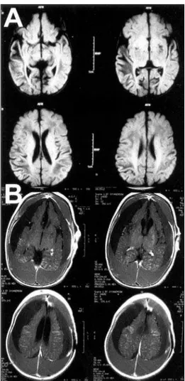
Magnetic resonance imaging (MRI) showed at two months of age abnormalities of signal intensity in the white matter of both cerebral hemispheres, more evident over the right temporal region (Fig 2A). After 5 moths a new MRI study disclosed diffuse brain atrophy, dural thickening and large subdural and epidural collections, suggestive of chronic blood clots in different stages (Fig 2B).
On the electroencephalogram (EEG) there was an abnor-mally disorganized background activity without any pre-dominance of side, intermingled with highly active epilep-tic activity, mostly over the parieto-rolandic areas bilater-ally, which was immediately followed by diffuse voltage depression. Sensitive nerve conduction of median and ulnar nerves and motor nerve conduction were normal. Needle electromyography (EMG) disclosed low amplitude, short motor unit potentials, a great density of short polyphasic potentials and increased muscle recruitment (biceps, tib-ialis anterior and quadriceps muscles). Muscle biopsy
analy-sis, according to standard procedures (hematoxylin-eosin, modified Gomori-trichrome, oil red O, PAS, cresyl violet, siryus red, ATPases, NADH, nonspecific esterase, myophos-phorylase, acid phosphatase, alkaline phosphatase, succi-nate dehydrogenase, cytochrome-C oxidase and adenylate deaminase)6, showed only marked atrophy of type 2
mus-cle fibers.
All studies were done following informed consent of parents.
DISCUSSION
Menkes’ disease main clinical features make a tri-ad of developmental delay, neurological degenera-tion and hair abnormalities found only in this dis-ease1. The incidence is of one case for every 100,000 to 250,000 births; its unique features can be found early in the first few months after birth and include Fig 1. (A) Typical facial features of MD patients include
epican-thus, thin and breakable metallic gray hair strands. (B) Micro-scopic examination of hair showed abnormal torsion of hair strands along their own axis (arrow: pilli torti) when compared to normal hair strands.
Arq Neuropsiquiatr 2007;65(1) 159
skin, hair and connective tissue abnormalities, early onset seizures and decreased muscle tone with pro-gressive deterioration1-3. MD was first described in 1962 by Menkes et al. who reported the affected sub-jects, all of them male and from the same family4. The genetic mutation responsible for the disease was first identified in 1993, leading to a defect in the pro-duction of an intracellular protein involved in cop-per transport7,8. Copper is a key cofactor of several different enzymes and it’s absence can secondarily impair the action of other enzymes such as cytochro-me C oxidase, superoxide-dismutase, tyrosinase and lisine oxidase, which then leads to a multisystemic compromise, especially the central nervous system1,9,10.
The classical form of MD comprises a neurologi-cal degenerative syndrome (cognitive compromise, ataxia, seizures, retarded neurological development), arterial abnormalities, bony changes like osteoporo-sis, bladder diverticulum , changes of connective tis-sue and skin and hair abnormalities (pili torti, moni-lethrix and trichorrhexis nodosa)1-5. Over time neu-rological symptoms and arterial anomalies of abdom-inal and cranial arteries become more severe, with symptoms suggestive of symptomatic West’s syndro-me, or conversely a drug-resistant progressive epilep-tic syndrome5,11,12. Bilateral inguinal hernias in MD had previously been described in mild forms of the disease, like the occipital horn syndrome (mild vari-ant form of DM), although they can also be found in the classical presentation; they are probably due to structural abnormalities of connective tissue5,13.
Early diagnosis is uncommon, as the first signs can be somewhat unspecific, with more prominent fea-tures (like the unique hair abnormalities) develop-ing over time, sometimes at the same time as neuro-logical compromise14. Serum levels of copper and ceruloplasmin should be measured after the third week (because as they can be low in normal children during this time-window) and low levels of both are needed to confirm the diagnosis5. X-rays can be help-ful in disclosing epiphysary hairlines in the extremi-ties of long bones, whereas bone densitometry can show mild to severe osteoporosis in the majority of patients5.
MRI abnormalities correspond to neuronal loss and range from isolated cerebral or cerebellar atro-phy or both combinations, subdural collections and cerebral hemorrhage15-17. Those evolve over time and are associated with a poor prognosis15,16. Vessel-wall compromise might be the pathological change res-ponsible the majority of these abnormalities,
espe-cially when subdural and epidural collections are found17-20. Severe atrophy and subdural and epidur-al collections are the end-stage abnormepidur-alities found late in the evolution of the disease, such as in our case.
The first report of the EEG changes in MD includ-ed four patients with multifocal spike-and-wave activ-ity15. Jayawant et al reported a rapidly progressive and unfavorable evolution with drug-resistant sei-zures and status epilepticus11. Other abnormal EEG patterns have been described in association with the disease, like hypsarhythmia and background activi-ty changes11,12,15. Normal background activity with-out epileptiform activity was reported in 9 children followed over 27 months in a study of the progno-sis of the disease, suggesting that the EEG changes might correspond to physiopathological changes in expression biochemistry of copper in brain15.
The EMG pattern has not yet been established in MD because exist few studies focus on the electro-physiological abnormalities in this disorder. Never-theless, we believe that EMG findings vary accord-ing to the time when the study is performed and the severity of the disease. In early cases EMG can be nor-mal, whereas in those patients in late stages of the disease an EMG study can show abnormalities sug-gestive of myopathic compromise. This pattern can be found if MD is associated with mitochondrial myo-pathy, with abnormal motor unit potentials with re-duced amplitude and duration, an increase in the density of short polyphasic potentials and increased motor unit recruitment16,21.
The pathophysiological basis of these electrophys-iological changes can either be due to changes in nerve and muscle excitability, or to abnormalities in the transmission of nerve impulses caused by the im-paired copper metabolism, somewhat similar to what is found in other diseases affecting copper metabo-lism like Wilson’s disease22.
160 Arq Neuropsiquiatr 2007;65(1)
The treatment options are limited because the brain-blood barrier acts as an obstacle to copper delivery without the transporter protein1,2,5,16,17. Also, copper is poorly absorbed by the gastrointestinal tract, without attaining an adequate serum level, mainly as copper histidine by parenteral reposition (200-1000 µg/day), might be beneficial in some cas-es when given early in the course of the disease2,5,16,17. There is no evidence, at present, of benefit of par-enteral administration of copper associated with D-penicillamine or vitamin E5. Treatment of MD patients must also include, when needed, anti-convulsive drugs12,15.
Acknowledgments – The authors would like to thank
Dr. Francisco M.B. Germiniani by english review of the man-uscript.
REFERENCES
1. Aicardi J. Menkes disease (kinky hair disease, steely hair disease, tri-chopoliodystrophy). In Aicardi J (Ed). Diseases of the nervous system in childhood (second edition). London: Mac Keith Press, 1998:306-308. 2. Kaler SG. Menkes disease. Adv Pediatr 1994;41:263-304.
3. Bankier A. Menkes disease. J Med Genet 1995;32:213-215.
4. Menkes JH, Alter M, Steigleder GK, et al. A sex-linked recessive disor-der with retardation of growth, peculiar hair, and focal cerebral and cerebellar degeneration. Pediatrics 1962;29:764-779.
5. Kodama H, Murata Y, Kobayashi M. Clinical manifestations and treat-ment of Menkes disease and its variants. Pediatr Int 1999;41:423-429. 6. Werneck LC. O valor da biópsia muscular em neurologia: análise de
290 exames a fresco e pela histoquímica. Rev Bras Clin Ter 1981;10 (Suppl):S2-S24.
7. Chelly J, Tumer Z, Tonnesen T, et al. Isolation of a candidate gene for Menkes disease that encodes a potential heavy metal binding protein. Nat Genet 1993;3:14-19.
8. Vulpe C, Levinson B, Whitney S, Packman S, Gitschier J. Isolation of a candidate gene for Menkes disease and evidence that it encodes a cop-per-transporting ATPase. Nat Genet 1993;3:7-13.
9. Vulpe CD, Packman S. Cellular copper transport. Annu Rev Nutr 1995; 15:293-322.
10. Voskoboinik I, Camakaris J. Menkes copper-translocating P-type ATPase (ATP7A): biochemical and cell biology properties, and role in Menkes disease. J Bioenerg Biomembr 2002;34:363-371.
11. Jayawant S, Halpin S, Wallace S. Menkes kinky hair disease: an unusu-al case. Eur J Paediatr Neurol 2000;4:131-134.
12. Venta-Sobero JA, Porras-Kattz E, Gutierrez-Moctezuma J. West syn-drome as an epileptic presentation in Menkes' disease: two cases report. Rev Neurol 2004;39:133-136.
13. Mandelstam SA, Fisher R. Menkes disease: a rare cause of bilateral inguinal hernias. Australas Radiol 2005;49:192-195.
14. Gu YH, Kodama H, Shiga K, et al. A survey of Japanese patients with Menkes disease from 1990 to 2003: incidence and early signs before typical symptomatic onset, pointing the way to earlier diagnosis. J Inherit Metab Dis 2005;28:473-478.
15. White SR, Reese K, Sato S, Kaler SG. Spectrum of EEG findings in Menkes disease. Electroencephalogr Clin Neurophysiol 1993;87:57-61. 16. Guitet M, Campistol J, Medina M. Enfermedad de Menkes: experien-cia en el tratamiento con sales de cobre. Rev Neurol 1999;29:127-130. 17. Santos LMG, Teixeira CS, Vilanova LCP, et al. Menkes disease: case
report of an uncommon presentation with white matter lesions. Arq Neuropsiquiatr 2001;59:125-127.
18. Morgello S, Peterson HD, Kahn LJ, Laufer H. Menkes kinky hair dis-ease with 'ragged red' fibers. Dev Med Child Neurol 1988;30:812-816. 19. Pedespan JM, Jouaville LS, Cances C, et al. Menkes disease: study of the mitochondrial respiratory chain in three cases. Eur J Paediatr Neurol 1999;3:167-170.
20. Jacobs DS, Smith AS, Finelli DA, Lanzieri CF, Wiznitzer M. Menkes kinky hair disease: characteristic MR angiographic findings. Am J Neuroradiol 1993;14:1160-1163.
21. Amato AA, Dumitru D. Hereditary myopathies. In Dumitru D, Amato AA, Zwarts MJ (Eds). Electrodiagnostic medicine (second edition). Philadelphia: Hanley & Belfus, 2002:1346.