ABNORMAL DIP PHENOMENON
A characteristic electrophysiological marker
in interdigital neuropathy of the foot
Diogo F. de Almeida
1, Katsumi Kurokawa
1, Yuki Hatanaka
2,
Shoji Hemmi
2, Gwen C. Claussen
3, Shin J. Oh
4ABSTRACT - Objective: The nerve conduction findings in interdigital neuropathy of the foot (IDN; Morton’s neuroma) have rarely been reported. We analyzed the nerve conduction data in 23 patients with suspect-ed IDN studisuspect-ed between 1982 and 2002. Method: Diagnosis of IDN was made on the basis of clinical fea-tures. All patients underwent routine nerve conduction studies and a near-nerve needle sensory nerve con-duction study of the interdigital nerves by Oh’s method in the symptomatic foot. Results: Of the 23 pa-tients, the diagnosis of definite IDN was made in 13 cases and of possible NDN in the others cases. Nineteen were females. Twenty two patients had only one nerve affected. One patient had two nerves affected. The III-IV interdigital nerve was affected in 17 cases and the II-III interdigital nerve in 7 cases. The near-nerve needle nerve conduction showed abnormality in the affected interdigital nerves in all definite IDN cases and confirmed the diagnosis of IDN in 10 cases by the abnormal dip phenomenon (a selective decrease of 50% or more in the sensory CNAP amplitude of the affected nerve compared with that of the preceding interdigital nerve). In 11 possible IDN cases, IDN was identified by the abnormal dip phenomenon. Con-clusion: The near-nerve needle sensory nerve conduction of the interdigital nerves is a highly sensitive di-agnostic test, and abnormal dip phenomenon is the most characteristic electrophysiological marker for the diagnosis of IDN.
KEY WORDS: Morton’s neuroma, metatarsalgia, interdigital neuropathy, nerve conduction.
Fenômeno da diminuição de amplitude anormal: um marcador eletrofisiológico característico da neuropatia interdigital do pé
RESUMO - Objetivo: Os achados da condução nervosa na neuropatia interdigital do pé (NIP) têm sido ra-ramente descritos. Nós analisamos os dados da condução nervosa de 23 pacientes com suspeita de NIP en-tre 1982 e 2002. Método: O diagnóstico de NIP foi clínico. Todos os pacientes foram submetidos a estu-dos de condução nervosa de rotina e ao estudo de condução sensitiva estu-dos nervos interdigitais com agulha justa-neural pelo método de Oh. Resultados: Dos 23 pacientes, o diagnóstico de NIP foi definitivo em 13 casos é possível nos demais. Dezenove pacientes eram mulheres e 22 tinham somente um nervo afetado. Apenas um paciente teve dois nervos comprometidos. O nervo interdigital III-IV foi afetado em 17 casos e o nervo interdigital II-III em 7 casos. A condução nervosa justa-neural foi anormal nos nervos interdigitais acometidos em todos os casos definitivos e confirmou o diagnóstico de neuropatia interdigital em 10 ca-sos pelo fenômeno da diminuição de amplitude anormal (uma diminuição seletiva de 50% ou mais da am-plitude do PANS do nervo afetado quando comparado com o nervo anterior. Em 11 casos possíveis, a neu-ropatia interdigital foi identificada pelo fenômeno da diminuição de amplitude anormal. Conclusão: A condução nervosa sensitiva justa-neural dos nervos interdigitais do pé é um teste diagnóstico altamente sensível e o fenômeno da diminuição da amplitude anormal é o marcador eletrofisiológico mais caracter-ístico para o diagnóstico de neuroma de Morton.
PALAVRAS-CHAVE: neuroma de Morton, metatarsalgia, neuropatia interdigital, condução nervosa.
Department of Neurology, University of Alabama at Birmingham and Veterans Affairs Medical Center, Birmingham, Alabama, USA: 1
Neuromuscular Disease and EMG Fellow; 2
Neurologist; 3
Assistant Professor of Neurology; 4
Professor of Neurology, Neuromuscular and EMG LAB Director.
Received 18 October 2006, received in final form 8 May 2007. Accepted 16 June 2007.
Dr. Shin J. Oh - Department of Neurology / The University of Alabama at Birmingham / UAB Station - Birmingham AL - USA - 35294. E-mail: [email protected]
Morton reported 15 cases of interdigital neurop-athy (IDN) of the foot in 1876, giving a clear clinical description of this condition1. Strictly speaking,
neuropathy of the foot2. Another commonly involved
nerve is the II-III interdigital nerve. Morton’s neuroma is one of the most common and most painful condi-tions to affect the metatarsal area3
. Women are con-siderably more frequently affected than men, quite likely because of wearing high-heeled and point-ed-toe shoes2,4. Repeated trauma on the interdigital
nerve between the heads of the metatarsals is the most commonly accepted cause of this disorder. In-traoperatively, a fibrous nodule is frequently found in the plantar interdigital nerve near the metatarsal heads. Pathological examination consistently shows proliferation of fibrous connective tissue within the plantar digital nerve and its supporting stroma in Morton’s neuroma. Thus, Morton’s “neuroma” is a misnomer for a condition which would be more ac-curately described as a “fibroma”5.
The diagnosis of IDN has been confirmed by non-invasive methods such as ultrasound, CT scans, and MRI6-8 but electrophysiological diagnosis has been
also possible since 19829.
We present here the electrophysiological findings in 23 patients with IDN. As far as we know, this is the largest series of IDN patients studied with the nerve conduction test in the literature.
METHOD
We analyzed the clinical and nerve conduction data in 23 patients with suspected IDN studied between 1982 and 2004 in the EMG laboratory of the University of Alabama at Birmingham. These twenty-three patients include five pa-tients previously reported10. All these patients were exam-ined by the same physician (S.J. Oh). A uniform clinical da-tabase and electrophysiological tests were used. None of these patients had surgical treatment prior to the test.
The diagnosis of definite IDN was made when all of the following three criteria were met: (1) at least one of two symptoms: localized pain between the involved metatar-sal heads and/or numbness or diminished sensation in the interdigital nerve territory, (2) at least one of two find-ings: Tinel’s sign and/or pin-prick sensory loss in the inter-digital nerve territory, and (3) no evidence of distal senso-ry neuropathy.
The diagnosis of possible IDN was made when all of the following three criteria were met: (1) localized pain between the involved metatarsal heads, (2) localized ten-derness between the involved metatarsal heads from the plantar aspect, and (3) no evidence of distal sensory neu-ropathy. Thus, a localized tenderness between the involved metatarsal heads from the plantar aspect was present as the sole finding in possible IDN.
All patients underwent motor nerve conduction stud-ies in the peroneal and posterior tibial nerves by the bel-ly-tendon method and sensory nerve conduction studies in the sural nerve by the antidromic technique following the conventional method11.
The near-nerve needle sensory nerve conduction of the interdigital nerves was performed orthodromically by Oh’s method in the symptomatic foot as previously described10,11. An active needle recording electrode was placed posteri-orly to the medial malleolus at the ankle as close as possi-ble to the posterior tibial nerve. A reference needle record-ing electrode was placed subcutaneously 3 cm away at the same level as the active recording electrode. The I and V digital nerves were stimulated with ring electrodes around the first and fifth toes respectively, and the I-II, II-III, III-IV and IV-V interdigital nerves were stimulated with interdig-ital stimulating electrodes between the respective toes. To
obtain the small sensory compound nerve action potentials (CNAP) reliably, we used the signal-averaging technique. A minimum of 64 averagings were performed in each inter-digital nerve in two different runs. From the two superim-posed sensory CNAPs obtained, the first positive-peak and negative-peak latencies, the peak-to-peak amplitude, and the duration from the onset of the first positive peak to the baseline return of the last component of the potential were measured. When no reproducible CNAPs were obtained af-ter at least 100 stimulations in two different runs, we deaf-ter- deter-mined that there was no sensory CNAP in that nerve. The distances between the stimulating and recording electrodes were measured with a caliper. The maximum and negative-peak nerve conduction velocities (NCVs) were calculated us-ing the latencies to the first positive peak and the largest negative peak by the conventional method. Skin temper-ature was controlled at 32°C under the arched portion of
the medial plantar surface of the foot by means of a skin temperature control unit.
Abnormal dip phenomenon was defined as a selective decrease of 50% or more in the amplitude of sensory CNAP in the involved interdigital nerve as compared with that in the preceding interdigital nerve10,11 (Figs 1 and 2). Disper-sion phenomenon was defined as a prolonged duration of sensory CNAP by more than 2 standard deviations plus the normal mean. Slow NCV was defined as an NCV of less than 2 standard deviations below the normal mean. The results were compared with the mean normal values adjusted for the patient’s age.
RESULTS
Clinical features – Among 23 patients with sus-pected IDN, 19 were females and 4 were males (Ta-ble 1), varying in age from 18 to 70 years. One pa-tient had clinically two IDNs: II-III and III-IV. Thus, there were 24 cases of IDN. IDN was observed in the right foot in 12 patients and in the left foot in 11 patients.
The diagnosis of definite IDN was made in 13 cas-es in 13 patients and possible IDN in 11 cascas-es in 10 patients. The diagnosis of definite IDN was made on the basis of sensory loss in 12 cases and localized Ti-nel’s sign in one case. Localized pain over the inter-digital space was the most common complaint, seen in 10 (77%) cases. Numbness in the involved toes was a symptom in five (39%) cases. On examination, the presence of a localized tender spot over the inter-digital space on the plantar side was present in 10 (77%) cases, and sensory loss over the involved in-terdigital space was noted in 12 (92%) cases. In one patient, sensory loss was confined to one side of the interdigital nerve territory. In two cases, Tinel’s sign was positive.
Eleven cases with possible IDN had localized pain and tenderness over the involved interdigital space. Except for sensory loss and Tinel’s sign, there was no remarkable difference in the clinical features be-tween the definite and possible IDN groups. Thus, the clinical features will be analyzed together. The most commonly involved interdigital nerves in all IDN were at the III-IV web space seen in 17 (71%) cas-es and the II-III web space in seven cascas-es. Localized pain over the interdigital space was the most com-mon complaint, seen in 22 (92%) cases. On examina-tion, the presence of a localized tender spot over the interdigital space on the plantar side was present in 21 (88%) cases.
Routine nerve conduction study – Routine nerve conduction studies performed with surface recording electrodes were normal in 19 patients and showed
Table 1. Clinical and electrophysiological findings in 13 patients with interdigital neuropathy of the foot.
Case Sex/ Age
Side* Symptoms Signs Routine NCS NCS findings in the affected interdigital nerves
Definite interdigital neuropathy
1 F/18 Right II-III*
Pain and numbness
Pain ** and sensory loss in the II-III web space
Normal Abnormal dip phenomenon and slow
2 F/29 Right II-III
Pain and numbness
Pain and sensory loss in the II-III web spaces
Normal Abnormal dip phenomenon
3 F/63 Left II-III
Numbness Pain and sensory loss in the II-III web space
Normal Slow NCV
4 F/35 Left¶ II-III
Pain Pain and Tinel sign (+) / Sensory loss in the lateral aspect of II-III web space
Normal Abnormal dip phenomenon and slow NCV
5 F/31 Left III-IV
Pain Sensory loss in the III-IV web space
Normal Abnormal dip phenomenon
6 F/36 Right III-IV
Pain Pain in the III-IV web space; Tinel sign (+)
Normal Dispersion phenomenon and slow NCV
7 F/59 Right III-IV
Pain Pain and sensory loss in the III-IV web spaces
Normal Low amplitude and dispersion phenomenon
8 F/29 Left III-IV
Pain Pain and sensory loss in the III-IV web space
Normal Abnormal dip phenomenon
9 F/70 Right III-IV
Pain Sensory loss in the III--IV web space
Normal Abnormal dip phenomenon
10 F/29 Right III-IV
Pain Pain and sensory loss in the III-IV web space
Normal Abnormal dip phenomenon
11 F/28 Right III-IV
Numbness Sensory loss in the III-IV web space
Normal Abnormal dip phenomenon and slow NCV
12 F/39 Right III-IV
Pain Pain and sensory loss in the III-IV web space
Normal Abnormal dip phenomenon
13 M/52 Left III-IV
Numbness Pain and sensory loss in the III-IV web space
↓ CMAP in tibial nerve
Abnormal dip phenomenon
Possible interdigital neuropathy
14 F/36 Left II-III
Pain Pain in the II-III web space Normal Abnormal dip phenomenon
15 F/61 Left II-III
Pain Pain in the II-III web space Prolonged TL in peroneal nerve
Abnormal dip phenomenon
16 F/42 Right II-III
Pain Pain in the II-III and III-IV web spaces
↓ CMAP in peroneal nerve
Abnormal dip phenomenon in II-III and III-IV IDN. Slow NCV in II-III IDN 17 M/24 Right
III-IV
Pain Pain in the III-IV web space Normal Abnormal dip phenomenon and slow NCV
18 M/52 Left III-IV
Pain Pain in the III-IV web space Peroneal neuropathy
Abnormal dip phenomenon and dispersion phenomenon 19 F/42 Left
III-IV
Pain Pain the III-IV web space Normal Abnormal dip phenomenon and dispersion phenomenon 20 F/41 Left
III-IV
Pain Pain in the III-IV web space Normal Abnormal dip phenomenon
21 F/59 Right III-IV
Pain Pain in the III-IV web space Normal Abnormal dip phenomenon and slow NCV
22 F/70 Left III-IV
Pain Pain in the III-IV web space Normal Abnormal dip phenomenon in II-III, abnormal dip phenomenon 23 M/36 Right
III-IV
Pain Pain in the III-IV web space Normal Abnormal dip phenomenon and dispersion phenomenon
*interdigital space; **localized tenderness between the involved metatarsal heads; ¶in the lateral branch of II-III, an amplitude decrease is 55%.
mild non-specific abnormalities in four patients (Ta-ble 1). One case with definite IDN had a low CMAP in the posterior tibial nerve. All three cases with possi-ble IDN had abnormality in the peroneal nerve: mild-ly slow motor NCV in one, low CMAP amplitude in one, and prolonged terminal latency in one case.
Near-nerve needle sensory nerve conduction of the interdigital nerves – In all of 13 cases of definite
IDN, the near-nerve needle study of the interdigi-tal nerves was abnormal in the involved interdigiinterdigi-tal nerves (Table 2). The most common nerve conduc-tion abnormality was an abnormal dip phenomenon, seen in ten (77%) cases (Figs 1 and 2). A selective de-crease in sensory CNAP amplitude of more than 50% in the affected nerve was seen in nine cases and ab-sent CNAP in one case. The next most common find-ing was low CNAP amplitude in nine cases (69%).
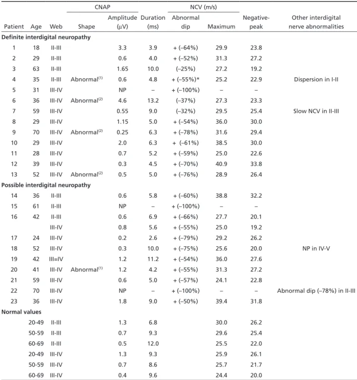
Table 2. Detailed near-nerve needle sensory nerve conduction findings in 23 patients with interdigital neuropathy of the foot.
Patient Age Web
CNAP
Duration (ms)
NCV (m/s)
Negative- peak
Other interdigital nerve abnormalities Shape
Amplitude (mV)
Abnormal
dip Maximum
Definite interdigital neuropathy
1 18 II-III 3.3 3.9 + (–64%) 29.9 23.8
2 29 II-III 0.6 4.0 + (–52%) 31.3 27.2
3 63 II-III 1.65 10.0 (–25%) 27.2 19.2
4 35 II-III Abnormal(1) 0.6 4.8 + (–55%)* 25.2 22.9 Dispersion in I-II
5 31 III-IV NP – + (–100%) – –
6 36 III-IV Abnormal(2) 4.6 13.2 (–37%) 27.3 23.3
7 59 III-IV 0.55 9.0 (–32%) 29.5 25.4 Slow NCV in II-III
8 29 III-IV 1.15 5.0 + (–54%) 36.0 30.0
9 70 III-IV Abnormal(2) 0.25 6.3 + (–78%) 31.6 29.4
10 29 III-IV 2.0 6.3 + (–61%) 38.5 30.0
11 28 III-IV 0.7 5.2 + (–59%) 25.0 22.6
12 39 III-IV 0.3 4.5 + (–70%) 40.9 33.8
13 52 III-IV Abnormal(2) 0.5 5.0 + (–76%) 28.9 26.4
Possible interdigital neuropathy
14 36 II-III 0.6 5.8 + (–60%) 38.8 32.2
15 61 II-III NP – + (–100%) – –
16 42 II-III 0.6 6.9 + (–66%) 27.7 20.1
III-IV 0.8 5.6 + (–55%) 25.0 19.2
17 24 III-IV 0.2 2.6 + (–79%) 29.2 26.2
18 52 III-IV 0.3 10.0 + (–75%) 25.6 20.0 NP in IV-V
19 42 III=IV 1.2 11.2 + (–54%) 36.0 27.6
20 41 III-IV Abnormal(1) 1.2 4.2 + (–55%) 31.3 27.2
21 59 III-IV 0.6 5.0 + (–57%) 24.1 22.8
22 70 III-IV NP – + (–100%) – – Abnormal dip (–78%) in II-III
23 36 III-IV 1.8 9.0 + (–50%) 39.4 31.8
Normal values
20-49 II-III 1.3 6.8 30.0 26.2
50-59 II-III 0.7 9.3 29.6 25.4
60-69 II-III 0.5 12.0 25.5 22.0
20-49 III-IV 1.3 9.3 25.9 26.1
50-59 III-IV 0.7 8.6 25.7 21.7
60-69 III-IV 0.4 9.6 24.4 20.0
Abnormal findings are bold lettered.* The lateral branch of II-III, amplitude decrease is 55%; diagnosis of lateral branch of II-III interdigital nerve is made. II-III interdigital nerve conduction: only 11% decrease in amplitude, but one branch study showed 55% decrease. (1)No initial positive peak is
Slow NCV and dispersion were seen in five (38%) and three cases (23%) respectively, either isolated or in combination. In one patient whose sensory loss was confined to one side of the interdigital nerve territo-ry, there was only an 11% decrease in amplitude of the sensory CNAP with interdigital nerve stimulation, but stimulation of the affected single digital nerve separately was able to document a 55% decrease in amplitude, confirming the diagnosis of IDN (Fig 3).
In 11 cases of possible IDN, the abnormal dip phe-nomenon was observed in all cases, a 50% or more decrease of CNAP amplitude criterion was seen in nine and absent CNAP in two cases. Low CNAP am-plitude was a common finding, seen in eight (73%) cases. Slow NCV and dispersion phenomenon were observed in five (45%) and four (40%) cases. Thus, in 11 cases of possible IDN, the abnormal dip phe-nomenon was the most helpful objective index for the diagnosis of IDN. In two cases with possible IDN, asymptomatic IDN was found by the abnormal dip phenomenon.
In summary, the near-nerve needle sensory nerve conduction of the plantar nerve showed a nerve con-duction abnormality in the involved nerve in all def-inite IDN cases and the abnormal dip phenomenon, the most common nerve conduction abnormality, confirmed the diagnosis of IDN in 77% of definite IDN cases. Thus, we conclude that the abnormal dip phenomenon is the most characteristic electrophysio-logical marker of IDN. Further, the abnormal dip phe-nomenon was able to confirm the diagnosis of IDN in 11 possible IDN cases and, identified asymptomat-ic IDN in two cases.
DISCUSSION
In our laboratory, we evaluated only 23 patients of suspected IDN over 22 years’ period. Most of these cases were referred to us to rule out tarsal tunnel syndrome, rather than to confirm the diagnosis of IDN. Thus, this small number represents most likely the referral bias.
The diagnosis of Morton’s neuroma is strongly suggested on the basis of clinical findings alone. As reported previously2,4, women were predominantly
affected in our series and III-IV IDN was most com-mon. Typically, patients complain of a precisely lo-calized pain between the affected metatarsal heads, which often radiates to the toes. The pain is aggra-vated classically by walking or standing and relieved by rest. Localized numbness is rarely complained of in the affected toes. In our series of definite IDN, 10 cases presented with localized pain over the affected
nerve and only five patients complained of numb-ness. Nearly all patients have localized tenderness on the interdigital nerve between the metatarsal heads. The most useful test in the clinical examination is the presence of localized tenderness of the involved web space demonstrated by manual compression from the plantar aspect, as noted in 10 of our cases. Wu recommended the “web space compression test” for the diagnosis of IDN4. This test is positive when
severe pain is produced by compressing the involved web space from the dorsal and plantar aspects with the thumb and index fingers and simultaneously squeezing the metatarsal heads together with the other hand. Sometimes this compression can cause a palpable click called Mulder’s sign, which is almost
specific for the diagnosis3. Sensory impairments are
often detectable in the affected interdigital web and toes but common in our series, being observed in 10 cases, because sensory loss in the affected interdigital web was one of required criterion in clinical diagno-sis of IDN. Tinel’s sign is extremely rare, being positive in only two cases in our series.
Although the clinical manifestations of Morton’s neuroma are quite typical to physicians experienced in this area, objective confirmation of diagnosis is required for most patients because TTS, distal sen-sory neuropathy, localized tendinitis, or arthritis can mimic IDN2,4. For this reason, the nerve conduction
study, ultrasound, CT, and MRI scans have been used. This is especially true in cases in which an objective sensory loss or Tinel’s sign was absent, as noted in possible IDN cases.
Electrophysiological evaluation of the interdigi-tal nerves was tried in vain with surface electrodes12.
The near-nerve needle sensory nerve conduction technique thus emerged as a more reliable test for this study10,13. In 1984, we described a method for
re-cording the sensory CNAP from the interdigital nerve using a special surface stimulating electrode between the toes and a needle recording electrode behind the medial malleolus10
. We were able to diagnose five patients with interdigital neuroma by this method. Abnormal dip phenomenon with a relatively normal nerve conduction velocity and normal duration of CNAP were the most typical findings, being observed in four of five cases. Although the dip phenomenon can be present in 18% of the normal population, the decrease in amplitude was always less than 49% of the preceding interdigital nerve in all 40 normal con-trols and 40-49% in two (5%) of 40 normal concon-trols. These two individuals were thought to have asymp-tomatic IDN because of abnormal CNAP shape. On the basis of this finding, abnormal dip phenomenon was defined in our study when there was a selective decrease by 50% or more in the amplitude of sensory CNAP in the involved interdigital nerve as compared to that in the preceding interdigital nerve.
Our previous study found abnormal dip phenom-enon in four of five IDN cases and concluded that it is the most characteristic electrophysiological finding in IDN10. The present study showed that the near-nerve
needle sensory nerve conduction of the interdigital nerves identified IDN in all of 13 definite IDN cases, and the electrophysiological marker for IDN, abnor-mal dip phenomenon, was present in 77% of cases. Thus, the present study confirmed again our belief
that the near-nerve needle sensory nerve conduction of the interdigital nerves is a sensitive diagnostic test and abnormal dip phenomenon is the most charac-teristic electrophysiological marker for IDN. In one case, the stimulation of the involved branch of the interdigital nerve was necessary to document abnor-mal dip phenomenon (Fig 3), emphasizing that the examiner may have to modify the stimulating tech-nique depending on the clinical finding. In three oth-er definite IDN cases, the diagnosis of IDN was made on the basis of non-specific nerve conduction abnor-malities: dispersion phenomenon and slow NCV. We also demonstrated that abnormal dip phenomenon can confirm the diagnosis of IDN in suspected IDN cases in which the localized pain and tenderness in the interdigital nerves are the sole findings as noted in all 11 possible IDN cases.
Abnormal dip phenomenon is not specific for IDN. We have observed it in a few patients with diffuse sensory neuropathy. Two of 12 patients with diabetic distal sensory neuropathy who showed interdigital nerve conduction abnormalities in more than three nerves (definite neuropathy pattern) also had abnor-mal dip phenomenon14
. In these cases, other inter-digital nerves showed abnormal nerve conduction in contrast to the isolated abnormal dip phenomenon in IDN.
The two most prominent nerve conduction ab-normalities in IDN in our study were an abnormal dip phenomenon, a selective decrease in the amplitude of sensory CNAP in the affected interdigital nerve and low CNAP amplitude. These findings are inter-preted to be indicative of a predominantly axonal neuropathy, which is consistent with the most ac-cepted theory that IDN is caused by repeated trau-ma rather than due to entrapment11. In entrapment
neuropathy, the classical nerve conduction findings are those of demyelinating neuropathy. In our study, electrophysiological evidence of demyelination was infrequent, being observed in only 3 cases.
Using the near-nerve needle technique for both stimulating and recording electrodes, Falck et al. confirmed the diagnosis of interdigital neuropathy in six patients13. All their patients had slow sensory
digital nerve more in isolation13. It is possible that
the branches of neighboring interdigital nerves may be stimulated in Oh’s method, because the surface electrodes are used as the stimulating electrodes in-stead of the needle10.
In recent years, non-invasive imaging tests have been used for demonstration of Morton’s neuroma. Shapiro reported that the ultrasound test identified the neuromas in 49 of 50 cases6. Turan et al.
demon-strated neuromas in 7 of 15 patients by the CT scan7.
Zannetti et al. reported MR imaging accuracy in 72% of 18 patients with Morton’s neuroma8. Thus, there
are cases of IDN without any enlargement which can not be detected by imaging tests. In these cases, the near-nerve needle sensory nerve conduction of the interdigital nerve is the only means of confirming IDN. Heise et al. showed a definite advantage of MRI over the near-nerve nerve conduction of the inter-digital nerve15. Among 17 patients with neuromas
disclosed by MRI, only ten showed a nerve conduc-tion abnormality. However, one must remember that “non-enlarged” interdigital neuropathy cannot be detected by imaging tests. In these cases, the nerve conduction test is the most reliable means for con-firming the diagnosis.
REFERENCES
1. Morton TG. A peculiar and painful affection of the fourth metatarso-phalangeal articulation. Am J Med Sci 1876;71:37-45.
2. Oh SJ, Meyer RD. Entrapment neuropathies of the tibial (posterior tib-ial) nerve. Neurol Clin 1999;17:593-615.
3. Miller DM. Forefoot pain: diagnosing metatarsophalangeal joint syno-vitis from interdigital neuroma. Foot Ankle 2001;22:914-915. 4. Wu KK. Morton’s interdigital neuroma: a clinical review of its
etiolo-gy, treatment, and results. J Foot Ankle Surg 1996;35:112-119. 5. Lassman, G. Morton’s toe: clinical, light and electron microscopy
in-vestigation in 133 cases. Clin Orthop 1979;142:73-84.
6. Shapiro PP, Shapiro SL. Sonographic evaluation of interdigital neuro-mas. Foot Ankle 1995;16:604-606.
7. Turan L, Lindgren U, Sahlstedt T. Computed tomography for diagno-sis of Morton’s neuroma. J Foot Ankle Surg 1991;30:244-245.
8. Zanetti M, Ledermann T, Zollinger H, Hodler J. Eficacy of MR imaging
in patients suspected of having Morton’s neuroma. Am J Rad 1997;168: 529-532.
9. Oh SJ. Electrophysiological diagnosis of the interdigital nerves of the foot. Muscle Nerve 1982;5:566-567.
10. Oh SJ, Kim HS, Ahmad BK. Electrophysiological diagnosis of interdig-ital neuropathy of the foot. Muscle Nerve 1984;7:218-225.
11. Oh SJ. Clinical electromyography: nerve conduction studies. Philadel-phia: Lippincott, Williams & Wilkins, 2003.
12. Guiloff RJ, Scadding JW, Klenerman L. Morton’s metatarsalgia: clini-cal, electrophysiological and histological observations. J Bone Joint Surg (Br) 1984;66:586-591.
13. Falk B, Hurme M, Hakkarainen S, Aario P. Sensory conduction veloci-ty of plantar digital nerves in Morton’s metatarsalgia. Neurology 1984; 34:698-701.
14. Park KS, Lee SH, Lee KW, Oh SJ. Interdigital nerve conduction study of the foot for an early detection of diabetic sensory polyneuropathy. Clin Neurophysiol 2003;114:894-897.