Jebmh.com
Original Article
J. Evid. Based Med. Healthc., pISSN- 2349-2562, eISSN- 2349-2570/ Vol. 3/Issue 75/Sept. 19, 2016 Page 4091
EFFECT OF HYPERTENSION ON MOTOR NERVE CONDUCTION VARIABLES
Aswathy Lloyds1, Sayam Subhash2, Delinda Linu Swornila3, Anusha Merline Justus41Assistant Professor, Department of Physiology, Dr. SMCSI Medical College, Karakonam. 2Assistant Professor, Department of Physiology, Dr. SMCSI Medical College, Karakonam. 3Associate Professor, Department of Physiology, Dr. SMCSI Medical College, Karakonam. 4Professor, Department of Physiology, Dr. SMCSI Medical College, Karakonam.
ABSTRACT
BACKGROUND
The most important medical and public health issue and the single cause of death worldwide is high blood pressure. Hypertension prevalence is on a rapid increase. Nerve conduction velocity test is an essential reliable clinical test for the diagnosis of the diseases of peripheral nerves that includes peripheral neuropathies. Nerve conduction study measures duration, latency, amplitude and conduction velocity. Conduction velocity and latency denote the speed of nerve impulse propagation. They are altered in disease, which cause demyelination. Amplitude denote the number of functioning fibers and it is altered in diseases causing axonal degeneration.
OBJECTIVES
The aim of the study is to assess the effect of hypertension on motor nerve conduction.
METHODS
The study was conducted in the Department of Physiology. The study protocol was approved by the Institutional Ethical Committee. A written informed consent was obtained from each participant. The study was done in 50 hypertensive patients and 50 normotensive subjects between the age group of 40-60 years.
STATISTICAL ANALYSIS
Unpaired ‘t’ test was used to find the statistical significance between both groups. The data was analysed using the Microsoft excel software. Group statistics was done and expressed as mean±SD.
RESULT
The results of motor nerve conduction variables were not statistically significant between control group and hypertensive group. (P >0.05).
CONCLUSION
Hypertension may produce axonal degeneration, but may not be affecting the myelination thus preserving nerve conduction velocity. Thus, hypertension itself may not affect the nerve conduction variables. Extensive studies are required to study the effect of hypertension in nerve conduction taking into consideration the duration, age, BMI and severity.
KEYWORDS
Nerve Conduction Study, Hypertension, Peripheral Neuropathy.
HOW TO CITE THIS ARTICLE: Lloyds A, Subhash S, Swornila DL et al.Effect of hypertension on motor nerve conduction
variables.J. Evid. Based Med. Healthc. 2016; 3(75), 4091-4093. DOI: 10.18410/jebmh/2016/874
INTRODUCTION: The most important medical and public
health issue and the single cause of death worldwide is high blood pressure.1 Hypertension prevalence is on a rapid
increase.2 Hypertension defines itself as a sustained
elevation of blood pressure >140/90 mm of Hg. Diagnosis is easy and simple to treat with surplus availability of medications, but sometimes it remains undetected, untreated and sometimes the treatment is not adequately
effective. Nerve conduction velocity test is an essential reliable clinical test for the diagnosis of the diseases of peripheral nerves that includes peripheral neuropathies.3,4
Nerve conduction study involves a noninvasive electrical stimulation of a peripheral nerve at one site and its noninvasive measurement of the evoked response at second site over the muscle innervated by the nerve (motor nerve conduction). Nerve conduction study measures duration, latency, amplitude and conduction velocity. Conduction velocity and latency denote the speed of nerve impulse propagation. They are altered in disease, which cause demyelination. Amplitude denote the number of functioning fibers and it is altered in diseases causing axonal degeneration.5
Financial or Other, Competing Interest: None. Submission 08-06-2016, Peer Review 18-06-2016, Acceptance 01-09-2016, Published 19-09-2016. Corresponding Author:
Dr. Aswathy Lloyds,
Assistant Professor, Department of Physiology, Dr. SMCSI Medical College, Karakonam, Kerala. E-mail: [email protected]
Jebmh.com
Original Article
J. Evid. Based Med. Healthc., pISSN- 2349-2562, eISSN- 2349-2570/ Vol. 3/Issue 75/Sept. 19, 2016 Page 4092
METHODS: The study was conducted in the Department of
Physiology. The study protocol was approved by the Institutional Ethical Committee. A written informed consent was obtained from each participant. The study was done in 50 hypertensive patients and 50 normotensive subjects between the age group of 40-60 years, which included both males and females.
Selection of Subjects: The criteria of considering patient hypertensive was BP >140/90 mm of Hg based on the average of 2 readings with a duration of less than 5 years on medication. The controls were healthy volunteers with BP <120/80 mm of Hg.6
Exclusion Criteria: The subjects with any associated diseases like diabetes, peripheral vascular diseases, pregnancy, alcohol, tobacco, smoking, terminally ill hypertensive patients, leprosy or other conditions, which are known to cause peripheral neuropathy were excluded from the study.
Establishment of Blood Pressure Status: All control volunteers and hypertensive patients underwent blood
pressure measurements. Standard mercury
sphygmomanometer with appropriate cuff size was used to measure blood pressure. The subject was asked to sit relaxed in a chair with her/his arm supported comfortably and the pressure cuff was applied closely to the upper arm. The cuff was rapidly inflated to pressure above the level at which the radial pulse could no longer be felt. The stethoscope was placed lightly over the brachial artery and the mercury column was immediately allowed to fall at the rate of 2 mmHg per second. The first perception of the sound was taken as the systolic pressure and then the mercury was allowed to fall further till the sound ceased to be tapping in quality, became fully muffled and finally disappeared. The level where it disappeared was taken as the diastolic pressure. The cuff was then deflated to zero pressure. The measurement was repeated twice with five-minute interval and the average taken for accuracy.
Establishment of Anthropometric Measurements:
Anthropometric measurements like height, weight and BMI were measured.
Recording of Nerve Conduction Velocity: The
instrument used for this study was computerised RMS ALERON 401. EMG (electromyography)/NCV (nerve conduction velocity)/EP (evoked potential) system designed for any neurophysiologic application. This machine includes nerve conduction study needle electromyography (EMG), F Wave, H reflex and all evoked potentials.
Nerve Conduction Study Evaluation: All subjects were
investigated by an electrophysiological study of the motor tibial nerve on both limbs by the RMS-EMG machine.
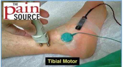
Motor Tibial Nerve Conduction Procedure: Recording
electrode/active electrode is placed on abductor hallucis or abductor digiti quinti slightly below and anterior to navicular tuberosity. Reference electrode is placed distally to active electrode over the muscle tendon near the metatarsal head. Ground electrode is placed between the recording electrode and stimulation site (S1). Stimulation is given on both sites such as distal site and proximal site. The distal site stimulation is given behind and proximal to the medial malleolus (S1) as shown in Figure 1. The proximal stimulation is given in the popliteal fossa along the flexor crease of the knee (S2) slightly lateral to midline in popliteal fossa shown in Figure 2. From the stimulation sites, Compound Muscle Action Potential (CMAP) duration, latency, amplitude and conduction velocity are determined in the motor tibial nerve. The conduction velocity is calculated by dividing the distance between the proximal (S2) and distal (S1) stimulating electrodes by the difference in proximal and distal latency.
Fig. 1: Pictorial Representation of Tibial Nerve Conduction Study: Distal Nerve Stimulation
Fig. 2: Pictorial Representation of Tibial Nerve Conduction Study: Proximal Nerve Stimulation
STATISTICAL ANALYSIS: Unpaired ‘t’ test was used to
find the statistical significance between both groups. The data was analysed using the Microsoft excel software. Group statistics was done and expressed as mean ±SD.
RESULT: The results of motor nerve conduction variables were not statistically significant between control group and hypertensive group (P >0.05).\
Amplitude Latency Conduction
Velocity
Controls 12.26±0.12 9.45±0.25 48.89±4.12 Hypertensives 13.12±0.13 9.56±0.15 47.9±4.10
Jebmh.com
Original Article
J. Evid. Based Med. Healthc., pISSN- 2349-2562, eISSN- 2349-2570/ Vol. 3/Issue 75/Sept. 19, 2016 Page 4093 DISCUSSION: This study aimed to investigate the effect of
motor nerve conduction variables of tibial nerve in patients with hypertension. No statistical significant differences were found in motor nerve conduction variables of hypertensives as compared to controls.
A study was done by Dhafir I. EI-Yassin et al7 to assess
the relationship between hypertension and peripheral neuropathy. The study assessed nerve conduction variables of sensory nerve function, motor nerve function and also F-wave measurement. They observed statistical significance of (p <0.05) for the association between hypertension patients and sensory nerve conduction that presented deterioration. However, the motor nerve conduction studies (Median, Ulnar, Tibial) did not show much changes; whereas, in their wave parameter assessment, the latency of the slowest F-wave was observed in the common peroneal nerve, which was prolonged. From their results, they interpret that smallest fibres were affected in hypertension.
Legrady P et al8 presented that nondiabetic
hypertensive patients also present the complications presented in diabetes. Patients who presented hypertension were undergoing antihypertensive therapy. In the study done by Viskoper et al,9 there is a reduction in nerve
conduction velocity in hypertensives. This is because hypertension cause vasospasm of blood vessels supplying the nerves. Popvtzer MM et al10 showed that motor nerve
conduction velocity is reduced in hypertensives when compared with controls. The results of our study is in accordance with the study done by Shubhangi D et al11 who
failed to demonstrate the effect of hypertension on nerve conduction velocity. Another study done by Negler et al12
also showed similar results of our study, which showed that there is no effect of hypertension on nerve conduction. They proposed that hypertension maybe producing axonal degeneration, but not affecting myelination thereby preserving nerve conduction velocity. Crowley SD13 and
Yasunari K et al14 have proved clinically that oxidative stress
is an outcome of chronic inflammation in hypertensive subjects. The onset of oxidative stress in hypertensive subjects depletes the levels of nitric oxide via the formation of peroxynitrite. This mechanism has been clinically proved by Moriel P et al.15 But, our study is in relation with the study
done by Negler et al and Shubhangi D et al, which showed a negative correlation between nerve conduction and hypertension.
CONCLUSION: Hypertension may produce axonal
degeneration, but may not be affecting the myelination, thus preserving nerve conduction velocity. Thus, hypertension itself may not affect the nerve conduction variables. Associated factors such as age, BMI and other diseases may cause variations in nerve conduction defects. Extensive studies are required to study the effect of hypertension in nerve conduction taking into consideration the duration, age, BMI and severity of the disease.
REFERENCES
1. Thakur BB. Management of hypertension in Diabetes. Med update 2010;20:407-412.
2. Kumar P, Desai VK, Kosambia JK. Prevalence of hypertension amongst the employees of a mega industry of south Gujarat. Indian J common Med 2012;27:19-25.
3. Fransen H, Van den Bergh PY. Nerve conduction studies in peripheral neuropathy: practical physiology and patterns of abnormality. Acta Neurol Berg 2006;106(2):73-81.
4. Kimura J. Principles and pitfalls of nerve conduction studies. Ann Neurol 1984;16(4):415-429.
5. Kong X, Lesser EA, Potts FA, et al. Utilisation of nerve conduction studies for the diagnosis of polyneuropathy in patients with diabetes. J Diabetes Sci Technol 2008;2(2):268-274.
6. Chobanian AV, Bakris GL, Black HR, et al. The seventh report of the Joint National Committee on detection evaluation and treatment of high blood pressure. Hypertension 2003;42(6):1206-1252.
7. Yassin DIE, Shamma YMA, Ajeena IM.
Electrophysiological study of peripheral nerves in hypertensive patients. Kurfa Med Journal 2009;12(1):367-379.
8. Legrady P, Bajcsi D, Lengyel C, et al. Investigation of cardiac autonomic and peripheral sensory neuropathy in diabetic and nondiabetic patients with hypertension. Clin Exp Hypertens 2013;35(6):465-469.
9. Viskoper RJ, Chaco J, Aviram A. Nerve conduction velocity in assessment of hypertension. Arch Intern Med 1971;128(4):574-575.
10. Popvtzer MM, Rosenbaum BJ, Gordon A, et al. Relief of uremic polyneuropathy after bilateral nephrectomy. N Engl J Med 1969;281:949-950. 11. Shubhangi D, Jaya M, Khan ST, et al. A prospective,
randomized parallel control study to evaluate the nerve conduction velocity in patients with essential hypertension. International Journal Recent Trends in Science and Technology 2013;7(3):127-128.
12. Negler W. Motor nerve conduction velocity in systemic diseases JAMA 1972;219(12):1632. 13. Crowley SD. The cooperative roles of inflammation
and oxidative stress in the pathogenesis of hypertension. Antioxid Redox Signal 2014;20(1):102-120.
14. Yasunari K, Maeda K, Nakamura M, et al
.
Oxidative stress in leukocytes is a possible link between blood pressure, blood glucose, and C-reacting protein. Hypertension 2002;39(3):777-780.15. Moriel P, Sevanian A, Ajzen S, et al. Nitric oxide, cholesterol oxides and endothelium-dependent vasodilation in plasma of patients with essential hypertension. Braz J Med Biol Res 2002;35(11):1301-1309.