Arq Neuropsiquiatr 2007;65(4-B):1245-1248
1245
TRUE NEUROGENIC THORACIC OUTLET
SYNDROME IN A COMPETITIVE SWIMMER
A case report of this rare association
Diogo Fraxino de Almeida
1, Richard D. Meyer
2, Shin J. Oh
1ABSTRACT - True neurogenic thoracic outlet syndrome (TOS) is an uncommon disorder despite of be a fre-quent reason for referral to the EMG laboratories. We describe the second case in the literature of true TOS in a competitive swimmer with progressive weakness and severe atrophy of the left thenar eminence. EMG showed lower trunk plexopathy. X-ray and MRI of the cervical spine and brachial plexus were normal. Sur-gical exploration evidenced the lower trunk retracted and pulled by a fibrous band. It was excised and neu-rolysis of the plexus was done with gradual improvement of function. We discuss the possible pathophysi-ology of this association.
KEY WORDS: thoracic outlet syndrome, swimming, brachial plexopathy.
Síndrome do desfiladeiro torácico verdadeiro em um nadador competitivo: relato de caso desta rara as-sociação
RESUMO - A síndrome do desfiladeiro torácico verdadeiro é condição incomum, apesar de ser uma razão freqüente de encaminhamento aos laboratórios de eletroneuromiografia. Nós descrevemos o segundo caso na literatura desta doença rara em um nadador competitivo com fraqueza e atrofia severa da eminência tenar esquerda. A ENMG mostrou plexopatia do tronco inferior. O RX e as ressonâncias magnéticas da co-luna cervical e do plexo braquial foram normais. Exploração cirúrgica mostrou o tronco inferior tracionado por uma banda fibrosa. Ela foi ressecada e procedeu-se a neurólise do plexo com melhora gradual da fun-ção. Nós discutimos a possível fisiopatologia desta associafun-ção.
PALAVRAS-CHAVE: síndrome do desfiladeiro torácico, natação, plexopatia braquial.
University of Alabama at Birmingham, UAB University Hospital and Veterans Affairs Medical Center, Birmingham, Alabama, USA:
1MD, Department of Neurology; 2MD, Department of Surgery, Division of Orthopedic Surgery.
Received 15 June 2007, received in fi nal form 24 August 2007. Accepted 27 September 2007.
Dr. Shin J. Oh - Department of Neurology / The University of Alabama at Birmingham/ UAB Station - Birmingham, Alabama 35294, USA. E-mail: [email protected]
Thoracic outlet syndrome (TOS) is a term coined by Peet et al in 1956 to a wide variety of symptoms orig-inated by compression of the neurovascular bundle
at the transition between the neck and axilla1. There
are very few diseases so controversial in the medical
literature such as TOS2,3. Based on these controversies,
the TOS can be divided in vascular and neurogenic3.
The vascular TOS can be subdivided in arterial and ve-nous according to the compression of the subclavian artery or vein respectively. They result in ischemia of the digits and hand or swelling of the arm and
rep-resent only 5% of the patients with TOS4. The
neuro-genic TOS represent the rest 95% and can be subdi-vided in true and disputed.
Following the ongoing debate in regard to the entity of TOS over the past two decades, a consen-sus has emerged that classic (true) neurogenic TOS is
uncommon and is usually caused by compression of the lower trunk of the brachial plexus due to a
cervi-cal rib or band and enlarged scervi-calenus muscles5-7. This
compression results in arm pain, numbness of the in-ner surface of the hand and forearm, and character-istic wasting and weakness of the thenar and intrinsic hand muscles. Distinct electrophysiological abnormal-ities (low compound muscle action potential in the thenar and intrinsic muscles, abnormal sensory nerve conduction in the ulnar nerve, prolonged F-wave la-tency in the ulnar nerve, and abnormal medial ante-brachial cutaneous sensory nerve conduction) are not-ed in classic neurogenic TOS8-10.
TOS has been described in aquatic athletes includ-ing competitive swimmers, divers, water polo players,
and synchronized swimmers11,12. Most of these cases
non-Arq Neuropsiquiatr 2007;65(4-B)
1246
Neurogenic thoracic outlet syndrome Almeida et al.
classic or disputed TOS. There has been just one re-ported case of classic (true) neurogenic TOS in
com-petitive swimmers13, and we are reporting another
such case.
CASE
Seven weeks prior to the initial evaluation, a 17-year-old right-handed male suddenly developed dragging of the
fi fth fi nger of left hand followed by progressive weakness and atrophy involving the whole left hand, after participa-tion in competitive swimming. He denied any sensory symp-toms and reported no back, arm or hand pain. There was no recent history of acute trauma, immunization, or fl u-like symptoms. He had been a competitive freestyle swim-mer for several years. Family history was positive for diabe-tes and coronary artery disease.
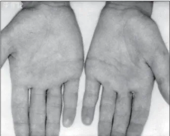
The general physical examination was normal. On neu-rological examination the patient was alert and orient-ed, with normal cognitive function and cranial nerves. No Horner’s sign was seen. There was weakness (grade 3-4 on MRC scale) of thenar, hypothenar and intrinsic muscles of the left hand. Moderate atrophy in the left thenar, hypoth-enar and interosseous muscles was noted, giving an impres-sion of smaller hand (Figure). Mild atrophy of the left fore-armfl exor muscles was noted as well. Deep tendon refl exes were normal and symmetrical. Sensation was intact to pin-prick, light touch, temperature and proprioception. There was evidence of decreased vibratory sensation in the left
fi ngers. At the supraclavicular fossa, no palpable mass or tender spot was noted, but inconsistent Tinel’s sign with ra-diating sensation to the digit V was present.
X-rays of the cervical spine and thorax did not show any cervical rib. MRI scans of the brain and left brachial plexus were unremarkable, and MRI of the cervical spine showed insignifi cant left paraspinal disc protrusion at the C5-C6 lev-el without spinal canal or foraminal compromise.
The nerve conduction study showed prolonged F-wave latencies in the left median and ulnar motor nerves, pro-longed terminal latency and low CMAP amplitude in the left ulnar nerve, and normal NCS in the left ulnar and me-dian sensory nerves (Table 1). No sensory compound nerve action potential (CNAP) was recorded from the left medi-al antebrachimedi-al cutaneous nerve. The needle EMG study
re-Figure. Atrophy of left thenar and hypothenar muscles.
Table 1. Nerve conduction data.
11/16/01 4/8/03 (PO 10 m) Normal
L/NCV* Amp** L/NCV* Amp** L/NCV* Amp**
Sensory nerve conduction Media
Ulnar Medial ABC
46.4 44.0
NP***
27.0 18.0
46.4 44.2 48.1
31.0 19.8
3.3
41.3 39.3 41.7
10 10 10 Mixed nerve conduction
Median: Wrist-elbow Elbow-axilla Ulnar: Wrist-elbow Elbow-axilla
55.6 58.1 61.0 69.0
64.0 89.0 18.0 56.0
54.5 57.7 53.4 52.4
51.0 64.0 40.0 42.6
49.4 53.4 47.5 48.1
10 10 10 10 Motor nerve conduction
Median: Terminal latency Elbow-wrist Axilla-elbow F-wave
Ulnar: Terminal latency Elbow-wrist Axilla-elbow F-wave
2.9 60.0 56.3
32.3 3.0
58.9 81.0
36.3
6.6 6.4 5.6
5.0
4.5 4.5
2.9 54.1 54.2
29.8 3.3
51.4 52.4
34.2
11.8 11.3 11.2
4.6 4.0 3.7
3.6 50.0 56.0 29.7 2.5 50.6 52.3 30.3
5 5 5
5 5 5
*L/NCV, Latency (msec)/ NCV (m/s); **Amplitude of compound nerve action potential (µV) for sensory and mixed nerve
con-duction and amplitude of compound muscle action potential (mV) for motor nerve concon-duction; ***Normal side: 52.2 m/s in NCV and 12 µV in amplitude. PO, post-operative; ABC, antebrachial cutaneous; NP, no potential. Bold numbers represent
Arq Neuropsiquiatr 2007;65(4-B)
1247 Neurogenic thoracic outlet syndrome Almeida et al.
vealed positive sharp waves and fi brillations with high-am-plitude, long-duration motor unit potentials in the left fi rst dorsal interosseous and abductor pollicis brevis muscles. The left biceps, deltoid, triceps and paraspinal muscles showed normal EMG fi ndings.
Over the next three months, there was minimal improve-ment in strength in the intrinsic hand muscles after the pa-tient stopped competitive swimming. In view of such mini-mal improvement, surgical exploration was recommended for classic neurogenic TOS. It showed entrapment of the lower trunk of the brachial plexus by a pleural band caus-ing retraction of this segment over the middle and upper trunks. The abnormal band was excised and neurolysis of the plexus was done to avoid further entrapment and scar-ring around the plexus. There was mild improvement in the strength of intrinsic hand muscles (fi nger-spreading in-creased from 3 to 4 by MRC scale and hand dynamometry from 12 kg to 17 kg), but no improvement was noted in mus-cle atrophy over a 2-year follow-up period. Nerve conduc-tion study 10 months after the surgery showed defi nite im-provement: the CMAP amplitude from the abductor pollicis brevis muscle was increased from 6.6 to 11.6 mV and the sen-sory CNAP, though the amplitude was low, was obtained in the left medial antebrachial cutaneous nerve (Table 1).
DISCUSSION
Our patient had progressive muscle weakness and wasting in the left hand, which are the hallmarks of
classic (true) neurogenic TOS. There was no history of pain or sensory complaints as typically seen in non-neurogenic TOS. Imaging work-up was not able to demonstrate any cervical rib or enlarged scalenus muscle. Nerve conduction studies showed almost all the typical electrophysiological abnormalities of neurogenic TOS (low CMAP amplitude, more often in the median than in the ulnar nerve, prolonged ulnar F-wave latency, abnormal ulnar sensory nerve con-duction, and absent medial antebrachial cutaneous
sensory nerve conduction)9,10. Low CMAP amplitude
in the median nerve was not evident until the post-operative second NCS which showed almost doubling of the CMAP amplitude. One exception was the nor-mal sensory nerve conduction of ulnar nerve. In re-cent years, the medial antebrachial cutaneous sensory CNAP seems to be the most sensitive nerve
conduc-tion parameter to confi rm classic neurogenic TOS14,15.
It is particularly helpful when the ulnar sensory CNAP is normal and TOS is clinically suspected. Kothari et al. believe that the medial antebrachial cutaneous is more sensitive than ulnar sensory nerve conduction study because the former carries predominantly T1
fi bers instead of C8 fi bers carried by the ulnar sensory
nerve14. It is widely accepted that the T1 fibers are
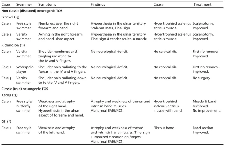
Table 2. Cases with thoracic outlet syndrome among swimmers.
Cases Swimmer Symptoms Findings Cause Treatment
Non classic (disputed) neurogenic TOS
Frankel (17) Case 1 Free style
swimmer
Numbness over the right forearm and hand.
Hypoesthesia in the ulnar territory. Scalenus mass, Tinel sign.
Hypertrophied scalenus anticus muscle.
Scalenotomy. Improved. Case 2 Varsity
swimmer
Aching in the right forearm and hand ulnar aspect.
Hypoesthesia in the ulnar territory. Tinel sign & tender scalenus muscle.
Hypertrophied scalenus anticus muscle.
Scalenotomy. Improved. Richardson (11)
Case 1 Varsity swimmer
Shoulder numbness and tingling radiating to the IV and V fi ngers.
No neurological defi cit. No cervical rib. First rib removal. Improved.
Case 2 Waterpolo player
Shoulder pain radiating to the forearm, the IV and V fi ngers.
No neurological defi cit. No cervical rib. First rib removal. Improved. Case 3 Varsity
swimmer
Shoulder pain radiating down to to the IV and V fi ngers.
No neurological defi cit. No cervical rib. No surgery.
Classic (true) neurogenic TOS
Katirji (13) Case 1 Free style/
butterfl y swimmer
Weakness and atrophy of the right hand. Hypoesthesia in the ulnar aspect of forearm and hand.
Atrophy and weakness of thenar and intrinsic hand muscles.
Abnormal EMG/NCS.
Hypertrophied scalenus anticus muscle with band.
Muscle & band sectioned. No improvement.
Oh (*)
Case 1 Free style swimmer
Weakness and atrophy of the left hand.
Atrophy and weakness of thenar and intrinsic hand muscles; Tinel sign ± impaired vibration on fi ngers. Abnormal EMG/NCS.
Fibrous band. Band section. Improved.
Arq Neuropsiquiatr 2007;65(4-B)
1248
Neurogenic thoracic outlet syndrome Almeida et al.
predominantly involved in true neurogenic TOS. Ana-tomical preference would explain why the abductor pollicis brevis muscle is usually more atrophied than
thefi rst dorsal interosseous and the median CMAP is
more severely affected than the ulnar CMAP. Shoulder pain has always been a common com-plaint of athletes who use their arms extensively for
sports12. Swimmers are the most frequently affected
group of competitive athletes: 42% of America’s best
swimmers have “swimmer’s shoulder”11. Swimmer’s
shoulder is thought to be an impingement syndrome due to chronic irritation of the humeral head and rotator cuff on the coracoacromial arch during
ab-duction of the shoulder11,16. This is said to be due to
the controlled, repetitive power motion at the very extreme of abduction and external rotation of the shoulder which is required in freestyle, butterfly,
and backstroke swimming11. TOS has been included
in the differential diagnosis of shoulder pain in the
swimmer11. Seven cases of TOS including our case
have been reported among swimmers (Table 2). Five cases were non-classic or disputed neurogenic TOS
due to lack of any objective neurological defi cit. Two
patients of Frankel et. al. had hypertrophied scale-nus anticus muscle and have improved after
scaleno-tomy17. Two of three patients of Richards et. al had
symptomatic relief with removal of the fi rst rib and
resumed competitive swimming11. Only one patient of
the literature had classic neurogenic TOS13. During the
surgical exploration there was no cervical rib but a
hypertrophied scalenus anticus muscle with a fi brous
band. There was no improvement after section of the
band and neurolysis13. There is also a report of one
case of “effort thrombosis” of the subclavian vein in
a competitive swimmer18, without any evidence of
neurogenic TOS.
The mechanism of classic neurogenic TOS in the swimmer was thought to be due to hypertrophy of the scalenus anticus muscle as an expression of over-development of the neck and shoulder muscles, which
results from many years of training in this sport17.
Hy-pertrophy of the scalenus anticus muscle was also
seen in Katirji and Hardy’s case13. However, they also
described a fi brous band within the scalenus anticus
muscle, which could be the cause of the entrapment. In our patient, there was no hypertrophic scalenus muscle but a fibrous band compressing the lower trunk of the brachial plexus was present. We believe
that congenital fi brous bands were the cause of
clas-sic neurogenic TOS in these two patients, and that competitive swimming aggravated or precipitated TOS symptoms.
REFERENCES
1. Peet RM, Henriksen MD, Anderson TP. Thoracic outlet syndrome: evalu-ation of a therapeutic exercise program. Mayo Clin Proc 1956;31:281-287. 2. Roos DB. The thoracic outlet syndrome is underrated. Arch Neurol
1990;47:327-328.
3. Wilbourn AJ. The thoracic outlet syndrome is oversiagnosed. Arch Neu-rol 1990;47:328-330.
4. Scola RH, Werneck LC, Iwamoto FM, Maegawa GH, Faoro LN, Calde-ira FH. True neurogenic thoracic outlet syndrome: report of two cases. Arq Neuropsiquiatr 1999;57:659-665.
5. GilliaĴ RW, LeQuesne PM, Longue V, Sumner AJ. Wasting of the hand associated with cervical band. J Neurol Neurosurg Psychiatry 1970; 33:615-624.
6. SwiĞ TR, Nichols FT. The droopy shoulder syndrome. Neurology 1984; 34:212-215.
7. Wilbourn AJ. Thoracic outlet syndrome. Neurol Clinics 1999;17:477-497. 8. Oh SJ. Clinical Electromyography. Nerve Conduction Studies, 3rd Ed.
Philadelphia: LippincoĴ , Williams & Wilkens, 2003.
9. Smith T, Trojaborg W. Diagnosis of thoracic outlet syndrome: value of sensory and motor conduction studies and quantitative electromyogra-phy. Arch Neurol 1987;44:1161-1163.
10. Wilbourn AJ, Hansen M, Hardy R. Neurogenic true thoracic out-let syndrome: electrodiagnostic features in 11 patients. Muscle Nerve 1982;5:558.
11. Richardson AB. Thoracic outlet syndrome in aquatic athletes. Clin Sports Med 1999;18:361-378.
12. Struckel RJ, Garrick JG. Thoracic outlet compression in athletes: a report of four cases. Am J Sports Med 1978;6:35-39.
13. Katirji B, Hardy Jr. RW. Classic neurogenic thoracic outlet syndrome in a competitive swimmer: a true scalenus anticus syndrome. Muscle Nerve 1995;18:229-233.
14. Kothari MJ, Macintosh K, Heistand M, Logigian EL. Medial antebrachi-al cutaneous sensory studies in the evantebrachi-aluation of neurogenic thoracic outlet syndrome. Muscle Nerve 1998;21:647-649.
15. Nishida T, Price SJ, Minieka MM. Medial antebrachial cutaneous nerve conduction in true neurogenic thoracic outlet syndrome. Electromyogr Clin Neurophysiol 1993;33:285-288.
16. Richardson AB, Jobe FW, Collins HR. The shoulder in competitive swim-ming. Am J Sports Med 1980;8:159-163.
17. Frankel SA, Hirata Jr. I. The scalenus anticus syndrome and competitive swimming: report of two cases. JAMA 1971;215:1796-1798.