Multiple forms of cotyledonary
β
-galactosidases from
Vigna unguiculata
quiescent seeds
JOAQUIM ENÉAS-FILHO
1,2, FABRÍCIO BONFIM SUDÉRIO
1, ENÉAS GOMES-FILHO
1and JOSÉ TARQUÍNIO PRISCO
1(received: June 6, 1999; accepted: November 22, 1999)
ABSTRACT- (Multiple forms of cotyledonaryβ-galactosidases fromVigna unguiculataquiescent seeds).
Cotyledon-aryβ-galactosidases were isolated and partially purified from Pitiúba cowpea (Vigna unguiculata(L.) Walp.) quiescent
seeds. The purification steps consisted of precipitation of the crude extract with ammonium sulphate in the range of 20-60% saturation, acid precipitation, DEAE-Sephadex ion-exchange chromatography and Lactosyl-Sepharose affinity chromatography. This purification process gave rise to threeβ-galactosidases-rich fractions:β-gal I,β-gal II andβ-gal III, which were purified about 5, 509, and 62 fold, respectively. They reached maximal enzyme activity at different pH ranges: 3.5-4.5 forβ-gal I, 3.0-3.5 forβ-gal II, and 3.0-4.0 forβ-gal III. Their maximal activities were reached when the temperature of the assay medium was 60 °C, and preincubation of the enzymes at different temperatures has shown that they were heat-stable up to 50 °C. There were no significant differences among the partially purified enzymes as to their response to the different effectors tested, except for Mn2+and EDTA, which affectedβ-gal I,β-gal II, andβ-gal III differ-ently. They were slightly affected by Mg2+, Ca2+, Zn2+, Co2+, tartarate, molybdate, glucose, and lactose, strongly inhib-ited by Cu2+and galactose, and inactivated by Hg2+. These chemical and physical properties are similar to those found for other plantβ-galactosidases. Although three isoforms of this enzyme were obtained through this purification process, isoelectric focusing in polyacrylamide slab gel of these enzyme-proteins suggests that cotyledons of Pitiúba cowpea qui-escent seeds possess four isoforms ofβ-galactosidases.
RESUMO- (Múltiplas formas de β-galactosidases cotiledonares de sementes quiescentes de Vigna unguiculata). β-galactosidases cotiledonárias foram isoladas e purificadas, parcialmente, de sementes quiescentes de feijão-de-corda (Vigna unguiculata(L.) Walp.) Pitiúba. As etapas de purificação consistiram de precipitação do extrato bruto com sulfato
de amônio na faixa de 20-60% de saturação, precipitação ácida, cromatografia de troca-iônica em DEAE-Sephadex e cromatografia de afinidade em Lactosil- Sepharose. Esse processo de purificação deu origem a três frações ricas em
β-galactosidases:β-gal I,β-gal II eβ-gal III, as quais foram purificadas cerca de 5, 509 e 62 vezes, respectivamente. Elas atingiram máxima atividade enzimática em diferentes faixas de pH: 3,5-4,5 paraβ-gal I, 3,0-3,5 paraβ-gal II e 3,0-4,0 paraβ-gal III. Suas atividades máximas foram alcançadas quando a temperatura do meio de ensaio era 60 °C, e a preincubação das enzimas em diferentes temperaturas mostrou que elas eram termoestáveis até 50 °C. Não houve diferenças significativas entre as enzimas parcialmente purificadas no que respeita à resposta dos diferentes efetores testados, exceto para Mn2+e EDTA, que afetaram, diferentemente,β-gal I,β-gal II eβ-gal III. Elas foram ligeiramente afetadas por Mg2+, Ca2+, Zn2+, Co2+, tartarato, molibdato, glicose e lactose, fortemente inibidas por Cu2+e galactose, e inativadas por Hg2+. Essas propriedades químicas e físicas são semelhantes às encontradas para outrasβ-galactosidases de plantas. Embora três isoformas dessa enzima tenham sido obtidas através desse processo de purificação, a focalização isoelétrica em placa de gel de poliacrilamida dessas proteinas enzimáticas sugere que cotilédones de sementes quiescentes de feijão-de-corda Pitiúba possuem quatro isoformas deβ-galactosidases.
Key words - Cotyledons, cowpea, enzyme purification, quiescent seeds
Introduction
β
-galactosidases (EC 3.2.1.23) are hydrolytic
enzymes acting at
β
-galactosydic bonds of both
oligo- and polysaccharides. Although they are
widely distributed in plants (Wallenfels & Malhotra
1961) their physiological role is still not well
under-stood (Li et al. 1975, Matheson & Saini 1977,
1. Laboratório de Fisiologia Vegetal, Departamento deBioquímica e Biologia Molecular, Centro de Ciências, Universidade Federal do Ceará. Caixa Postal 1065, 60001-970 Fortaleza, CE, Brasil. 2. Corresponding author. Rua Coronel Linhares, 280
Sekimata et al. 1989, Simos et al. 1989, Kundu et al.
1990, Giannakouros et al. 1991, Enéas-Filho et al.
1995). It has been suggested that these enzymes are
associated to the depletion of oligo- and
polysaccha-rides during the initial phases of seed germination
(Matheson & Saini 1977, Corchete & Guerra 1986,
Biswas 1987, Enéas-Filho et al. 1995), involved in
the metabolism of cell wall constituints (Corchete &
Guerra 1987a, Edwards et al. 1988, Dopico et al.
1989, Konno & Katoh 1992, Konno & Tsumuki
1993, Gómez et al. 1995, Kitagawa et al. 1995), and
in the process of fruit ripening (Pressey 1983,
Ogawa et al. 1990, Veau et al. 1993, Ali et al. 1995).
β
-galactosidase activity has been detected in
quies-cent seeds (Dey 1984) as well as during germination
and seedling establishment (Agrawal & Bahl 1968,
Corchete & Guerra 1987b, Kundu et al. 1990,
Buckeridge & Reid 1994, Enéas-Filho et al. 1995).
Isolation and purification studies suggest that they
occur under multiple forms (Biswas 1986, 1987,
Corchete & Guerra 1987a, Kundu et al. 1990).
How-ever, it is not clear if these multiple forms come from
the same part or organ of the seed or seedling or if
each one of them come from a different parts of the
seed or seedling. This knowledge is of fundamental
importance for the study of their physiological role.
Therefore, the objective of the present work was to
isolate, partially purify, and characterize
cotyledon-ary
β
-galactosidases extracted from quiescent
Pitiúba cowpea seeds.
Material and methods
Pitiúba cowpea (Vigna unguiculata(L.) Walp.) seeds were obtained from the Centro de Ciências Agrárias, Universidade Federal do Ceará, Fortaleza, Ceará, Brazil. All seeds were stored in sealed glass bottles containing sil-ica gel and kept at approximately 10 °C until used in the experiments.
The extraction ofβ-galactosidases was performed according to Corchete & Guerra (1987b) with small modi-fications. Cotyledons of quiescent seeds were macerated and homogenized in cold 25 mM citrate -50 mM phos-phate buffer (McIlvaine 1921), pH 5.5, for 1h. The propor-tion of tissue to grinding medium (McIlvaine buffer) was 1:10 (m/v). All procedures were performed at 4 °C unless otherwise stated. The suspension was filtered through a nylon net, centrifuged at 10,000 g for 30 min, and the pre-cipitate discarded. The supernatant (crude extract) was
precipitated with ammonium sulphate in the range of 20-60% saturation. After centrifugation, the precipitate was resuspended in McIlvaine buffer, pH 5.5 and then lowered to 3.5 with gradual additions of 1 M citric acid. This new precipitate was discarded after centrifugation at 10,000 g for 30 min, and the pH of the supernatant was ad-justed to 5.5 with 0.8 M dibasic sodium phosphate, dia-lyzed against distilled water at 8 °C for 24 h, and lyophilized (F20-60) for further use. This lyophilized
pow-der was resuspended in 25 mM Tris - HCl buffer, pH 7.2 and applied on a DEAE-Sephadex A-50 ion-exchange col-umn (17 x 180 mm) equilibrated with the same buffer at 10 °C, and the flow rate adjusted to 31.5 mL.h-1. The auto-matic fraction collector was adjusted to collect fractions of 4.2 mL per tube, and the column was eluted with the equi-librium buffer. The retained peak was eluted with a linear NaCl gradient (0.2 to 1.0 M). Absorbance at 280 nm and
β-galactosidase activity were determined in each fraction , and the ones that showed the highest activities (peaks DS-I and DS-II) were dialyzed against distilled water for 24 h, concentrated and applied on a Lactosyl-Sepharose affinity column (16 x 190 mm) equilibrated with McIlvaine buffer, pH 4.0, diluted 1:4, containing 0.1 mM EDTA and 1.0 mM 2-ME at 4 °C (Campillo & Shannon 1982). The flow rate was adjusted to 36 mL.h-1; fractions of 4.8 mL were eluted with the equilibrium buffer and the retained fractions were eluted with the same buffer containing 100 mM lactose and 0.5 M NaCl. After each chromatography the column was regenerated with 6 M urea (Simos et al. 1989).
β-Galactosidase activity was measured according to Kanfer et al. (1973) as modified by Enéas-Filho et al. (1995). A 3 mM solution of the substrate was prepared by dissolving p-nitrophenylβ-D-galactopyranoside (Sigma Co.) in McIlvaine buffer, pH 4.0. A 0.5 mL aliquot of this substrate solution was added to 0.5 mL of appropriately di-luted enzyme extract, and the mixture was incubated at 37 °C for 15 min. The reaction was stopped by the addition of 1.5 mL of 0.1M Na2CO3. Enzyme activity was determined
by measuring absorbance at 400 nm (∆A400) and
subtract-ing this value from A400of the blank. It was also expressed
in units of activity (UA), one UA being defined as a differ-ence in absorbance (∆A400) of 0.01 (Enéas-Filho et al.
1995). Protein was determined by absorbance at 280 nm or according to Bradford (1976), using bovine serum albu-min 2x crystalline (Sigma Co.) as standard.
forβ-galactosidase. The effects of EDTA, galactose, glu-cose, lactose, tartarate, molybdate and several bivalent cations on enzyme activity were tested by taking 0.5 mL aliquots of the enzyme preparation, preincubating them at 37 °C for 10 min in absence and presence of the effectors, and then assaying the mixture for enzyme activity. The fi-nal concentration of all effectors in the assay medium was 4 mM, except for molybdate that was 0.1 M and for galactose, glucose and lactose that were 8 mM.
Polyacrylamide gel isoelectric focusing in slab gels was carried out according to Robertson et al. (1987) in the pH range from 3 to 10. After isoelectric focusing the gel was separate into two parts, and visualization of protein markers and enzyme bands were carried out according to Simos & Georgatsos (1988). A pI kit containing markers with pI ranging from 3.55 to 9.30 (Sigma Co.) was used.
Results and Discussion
A summary of the purification procedure used
for the cotyledonary
β
-galactosidases isolated from
quiescent seeds is presented in table 1. The initial
step (F
20-60) was included to eliminate compounds of
small mass and to concentrate the preparation that
was
then
subjected
to
ion-exchange
DEAE-Sephadex followed by affinity on
Lactosyl-Sepharose chromatography. The elution
pattern of the ion-exchange chromatography
showed two peaks of protein and of
β
-galactosidase
activity (figure 1A): DS-I and DS-II, which were
pu-rified 24.2 and 2.4 fold, respectively. The first peak
(DS-I), corresponded to the material that was not
re-tained by the column, suggesting that the proteins of
DS-I are relatively basic in nature. A similar
β
-galactosidase has been reported in
Vigna radiata
seeds (Kundu et al. 1990). The second peak (DS-II),
eluted with the equilibrium buffer containing 0.7 M
sodium chloride, corresponded to a
β
-galactosidase
strongly bound to the column. The existence of
frac-tions eluted from similar columns which showed
cotyledonary
β
-galactosidase activity were also
ob-served <%-2>in
Phaseolus vulgaris
(Agrawal &
Bahl 1968),
Vigna
sinensis (Biswas 1987), and
Vigna radiata
(Kundu et al. 1990) seeds. However,
these
β
-galactosidases were not strongly bound to
the ion-exchange column. The elution pattern of
Lactosyl-Sepharose affinity chromatography of
DS-I (figure 1B) showed two peaks of enzyme
activ-ity:
β
-gal I and
β
-gal II, which were purified 4.7 and
508.6 fold, respectively. The first peak (
β
-gal I),
cor-responded to the fraction that was not retained by the
column, and the other peak (
β
-gal II), eluted with the
equilibrium buffer containing 100 mM lactose and
0.5 M NaCl, corresponded to the enzyme fraction
re-tained by the column. Lactosyl-Sepharose affinity
chromatography columns have been used for plant
β
-galactosidases isolation and purification (Simos et
al. 1989). According to these authors several
β
-galactosidases from different plant species have
been studied and all of them are retained by these
columns. The high enzyme activity observed for the
second peak (
β
-gal II) associated to the fact that it
Table 1. Purification of cotyledonaryβ-galactosidases from Pitiúba cowpea,Vigna unguiculata(L.) Walp., quiescent
seeds (F20-60= ammonium sulphate precipitation).
Step of purification Volume (mL)
Total activity (UA.min-1)
Total Protein (mg)
Specific Activity (UA.mgP-1.min-1)
Factor of Purifi-cation
Crude Extract 270.0 44,352 1,158.0 38 1
F20-60 53.5 42,372 219.0 194 5.1
DEAE-Sephadex chromatography8
DS-I 153.0 26,020 28.3 919 24.2
DS-II 99.0 14,993 164.0 91 2.4
Lactosyl-Sepharose affinity chromatography8
β-gal I 37.0 4,172 23.2 180 4.7
β-gal II 32.5 9,663 0.5 19,325 508.6
was retained by this type of affinity chromatography
column reinforces the idea that
β
-gal II corresponds
to a
β
-galactosidase. Even though
β
-gal I has shown
a much lower enzyme activity than
β
-gal II it can be
also considered as a fraction containing
β
-galactosidase because the binding to the
Lactosyl-Sepharose affinity column is not an
abso-lute criteria for plant
β
-galactosidase identification.
The elution pattern of Lactosyl-Sepharose affinity
chromatography of DS-II (figure 1C) showed only
one peak of enzyme activity (
β
-gal III), which was
purified 62 fold. This peak, eluted with the
equilib-rium buffer containing 100 mM lactose and 0.5 M
NaCl, probably corresponded to a
β
-galactosidase
because it showed high enzyme activity and it was
bound to the Lactosyl-Sepharose affinity colunm
(Simos et al. 1989). Therefore, the cotyledonary
ex-tract from quiescent seeds of Pitiúba cowpea
con-tained three fractions showing high
β
-galactosidase
activity:
β
-gal I,
β
-gal II, and
β
-gal III, which were
purified about 5, 509, and 62 fold, respectively (table
1).
The effect of pH on the cotyledonary
β
-galactosidases (
β
-gal I,
β
-gal II, and
β
-gal III)
ac-tivities is shown in figure 2. In the pH range 2.5-6.5
the maximum enzyme activity varied among the
three
β
-galactosidase rich fractions: 3.5-4.5 for
β
-gal I, 3.0-3.5 for
β
-gal II, and 3.0-4.0 for
β
-gal III
(figure 2). These results are similar to those found
for cotyledonary
β
-galactosidases from
Vigna
sinensis
(Biswas 1987) and
Vigna radiata
(Kundu et
al. 1990), as well as for seeds (Agrawal & Bahl 1968,
Li et al. 1975, Matheson & Saini 1977, Edwards et
Figure 1. Purification of cotyledonaryβ-galactosidasesextracted from Pitiúba cowpea,Vigna unguiculata(L.)
Walp., quiescent seeds. A. DEAE-Sephadex chromatog-raphy of cotyledonary β-galactosidase rich fraction (F20-60). B. Lactosyl-Sepharose affinity chromatography
of the cotyledonaryβ-galctosidase rich peak (DS-I). C. Lactosyl-Sepharose affinity chromatography of the coty-ledonaryβ-galctosidase rich peak (DS-II). Protein (A280, ___) andβ-galctosidase activity (∆A
400x 15 min-1, —-).
The arrows represent the start of the addtion of the elution buffer containing 100 mM lactose and 0.5 M NaCl as de-scribed under Material and methods.
Figure 2. Enzyme activity as a function of the assay me-dium pH of partially purified cotyledonary
β-galactosidases extracted from Pitiúba cowpea,Vigna unguiculata(L.) Walp., quiescent seeds.β-gal I (o),β-gal
al. 1988, Sekimata et al. 1989, Giannakouros et al.
1991, Buckeridge & Reid 1994), shoots (Konno &
Tsumuki 1993), leaves (Sawicka & Kacperska
1995) and fruits (Pressey 1983, Ogawa et al. 1990,
Ranwala et al. 1992) of different species.
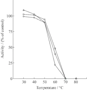
The effect of assay temperature on the activities
of partially purified
β
-galactosidases is shown in
fig-ure 3. In the temperatfig-ure range studied (30-80 °C)
the activities of all
β
-galactosidases increased with
temperature up to 60 °C after which they decreased
reaching at 80 °C values that corresponded to
ap-proximately 10% of their maximal activities.
Simi-lar temperature optima have been reported for other
plant
β
-galactosidases (Biswas 1987, Edwards et al.
1988, Kundu et al. 1990, Ogawa et al. 1990,
Ranwala et al. 1992, Konno & Tsumuki 1993,
Sawicka & Kacperska 1995).
The effect of temperature on partially purified
cotyledonary
β
-galactosidases is shown in figure 4.
The enzymes preincubated at temperature ranging
from 30 to 80 °C for 10 min maintained a quite stable
activity up to 50 °C when there was an abrupt
de-crease in enzyme activity, and at 70 °C they were
completely inactivated. This rapid decrease in
en-zyme activity above 50 °C was observed for all
β
-galactosidases studied. These data indicate that
the partially purified cotyledonary
β
-gal I,
β
-gal II
and
β
-gal III extracted from Pitiúba cowpea were
heat stable up to 50 °C, and that their thermostability
is similar to the
β
-galactosidases isolated from
coty-ledons of
Vigna sinensis
(Biswas 1987) and
Vigna
radiata
(Kundu et al. 1990), as well as from
β
-galactosidases isolated from other plants sources
(Konno & Katoh 1986, Simos & Georgatsos 1988).
However, they differ from the ones obtained with
β
-galactosidases isolated from radish (Sekimata et
al. 1989) and tomato seeds (Pressey 1983), which
were inactive at 55 °C or had only 50% of their initial
activities at temperature ranging from 48 to 52 °C,
respectively.
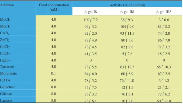
The effects of bivalent cations and other
effectors on the activities of the partially purified
cotyledonary
β
-galactosidases are shown in table 2.
These effects did not vary among the partially
puri-fied cotyledonary
β
-galactosidases, except for Mn
2+and EDTA, which affected differently
β
-gal I,
β
-gal
II, and
β
-gal III. Mn
2+did not affect the activity of
β
-gal I, and strongly inhibited the activities of
β
-gal
II and
β
-gal III, while EDTA has shown a weak
inhi-Figure 3. Enzyme activity as a function of assay medium temperature of partially purified cotyledonary
β-galctosidases extracted from Pitiuba cowpea, Vigna unguiculata(L.) Walp., quiescent seeds.β-gal I (o),β-gal
II ( ) andβ-gal III (∆). Values represent the mean of three different experiments with three replicates each.
Figure 4. Enzyme thermostability of partially purified cot-yledonaryβ-galctosidases extracted from Pitiúba cowpea,
Vigna unguiculata(L.) Walp., quiescent seeds.β-gal I (o),
bition of both
β
-gal I and
β
-gal II, but strongly
inhib-ited
β
-gal III. The analysis of each of the effectors on
the activities of the three partially purified
cotyle-donary
β
-galactosidases has shown that while Mg
2+,
Ca
2+, Zn
2+, Co
2+, tartarate, molybdate, glucose, and
lactose weakly inhibited their activities (less than
40%), the inhibition due to Cu
2+and galactose were
strong (higher than 60%), and Hg
2+completely
in-hibited the activities of all Pitiúba cowpea partially
purified cotyledonary enzymes. Studies concerning
the response of plant
β
-galactosidases to effectors
are scarce, especially those dealing with the effects
of glucose, lactose, molybdate and tartarate.
Never-theless, the effects of Ca
2+, Co
2+, Hg
2+, Mg
2+, Zn
2+,
galactose and glucose on different plant
β
-galactosidases were similar to those obtained here
(Li et al. 1975, Biswas 1987, Corchete & Guerra
1987a, Kundu et al. 1990, Ogawa et al. 1990, Konno
& Katoh 1992, Ranwala et al. 1992). However,
Co
2+, which behaved as a weak inhibitor of the
coty-ledonary
β
-galactosidases from Pitiúba cowpea
qui-escent seeds, has been described as a strong inhibitor
of 4-day-old
Vigna sinensis
cotyledonary
β
-galactosidases (Biswas 1987), and did not affect
the activities of cotyledonary
β
-galactosidases
ex-tracted from 4-day-old
Vigna radiata
(Kundu et al.
1990). Molybdate, which was a weak inhibitor of
the cotyledonary
β
-galactosidases studied in this
pa-per, has been described as a strong inhibitor of both
4-day-old
Vigna sinensis
(Biswas 1987) and
Vigna
radiata
(Kundu et al. 1990) cotyledonary
β
-galactosidases. These results suggest that the
re-sponse of cotyledonary
β
-galactosidases is a
func-tion of both species differences and developmental
stage. The strong inhibition of galactose (table 2),
which is one of the products of the catalytic action of
β
-galactosidases on
β
-galactosides has also been
ob-served by others (Li et al. 1975, Biswas 1987,
Corchete & Guerra 1987a, Kundu et al. 1990). These
results suggest that galactose might be acting as an
end product inhibitor of
β
-galactosidases.
Isoelectric focusing in polyacrylamide slab gels
was performed with the partially purified
cotyledon-ary
β
-galactosidases (
β
-gal I,
β
-gal II and
β
-gal III).
Table 2. Effect of bivalent cations and other effectors on the activity of partially purified cotyledonaryβ-galactosidases from Pitiúba cowpea,Vigna unguiculata(L.) Walp., quiescent seeds. The values are expressed in percentage of the con-trol (without the addition of effectors); they represent the meanstandard deviation of three different experiments with three replicates each.
Addition Final concentration
(mM)
Activity (% of control)
β-gal I8 β-gal II8 β-gal III8
MnCl2 4.0 1097.2 389.3 30.6
MgCl2 4.0 943.2 1049.0 818.2
CaCl2 4.0 922.0 9311.5 742.0
ZnCl2 4.0 784.9 803.6 867.0
CoCl2 4.0 734.5 829.8 713.2
CuCl2 4.0 413.5 52.6 182.5
HgCl2 4.0 0 0 0
Tartarate 4.0 735.5 6312.3 6510.3
Molybdate 0.1 646.0 608.9 672.5
EDTA 4.0 783.2 7611.8 31.2
Galactose 8.0 357.5 121.5 212.1
Glicose 8.0 853.2 766.1 726.2
Both
β
-gal I and
β
-gal III showed only one band of
enzyme activity corresponding to pIs 8.7 and 6.6,
while
β
-gal II showed two distinct bands of enzyme
activity corresponding to pIs 8.4 and 8.1 (data not
shown). Although the results of Lacosyl-Sepharose
affinity chromatography (figure 1B and 1C) suggest
the existence of three isoforms of cotyledonary
β
-galactosidases (
β
-gal I,
β
-gal II and
β
-gal III),
isoelectric focusing in polyacrylamide slab gel of
these proteins (data not shown) indicated that the
quiescent seeds of this cultivar of
Vigna unguiculata
possess four isoforms of cotyledonary
β
-galactosidases. The presence of four isoforms of
cotyledonary
β
-galactosidases has also been
ob-served in 4-day-old seedlings of
Vigna sinensis
(Biswas 1987) and
Vigna radiata
(Kundu et al.
1990), using different methods of enzyme
purifica-tion. The presence of multiple forms of the same
en-zyme in cotyledons suggest that the isoforms might
have different metabolic roles in different tissues or
cells. Therefore, their location within the cotyledons
as well as their changes along germination and
seed-ling establishment should be a prerequisite for the
determination of their biochemical and
physiologi-cal roles, at least during this developmental process.
Acknowledgements - To Financiadora de Estudos e Projetos (FINEP) and Conselho Nacional de Desenvolvimento Científico e Tecnológico (CNPq) for their financial support.
References
AGRAWAL, K.M.L. & BAHL, O.P. 1968. Glycosidases ofPhaseolus vulgaris. II. Isolation and general
prop-erties. The Journal of Biological Chemistry 243:103-111.
ALI, Z.M., ARMUGAM, S. & LAZAN, H. 1995.
β-Galactosidase and its significance in ripening mango fruit. Phytochemistry 38:1109-1114. BISWAS, T.K. 1986. Cationic form ofβ-galactosidase in
the germinating seeds ofVigna sinensis(Linn) Savi. Archives of Biochemistry and Biophysics 251:379-384.
BISWAS, T.K. 1987. Characterization of
β-galactosidases from the germinating seeds of
Vigna sinensis. Phytochemistry 26:359-364.
BRADFORD, M.M. 1976. A rapid and sensitive method for the quantitation of microgram quantities of pro-tein utilizing the principle of propro-tein-dye binding. Analytical Biochemistry 72:248-254.
BUCKERIDGE, M.S. & REID, J.S.G. 1994. Purification and properties of a novel β-galactosidase or exo-(1-4)-β-D-galactanase from the cotyledons of germinatedLupinus angustifolius L. seeds. Planta
192:502-511.
CAMPILLO, E.D. & SHANNON, L.M. 1982. An
α-galactosidase with hemagglutinin properties from soybean seeds. Plant Physiology 69:628-631. CORCHETE, M.P. & GUERRA, H. 1986. Effect of NaCl
and polyethylene glycol on solute content and glycosidase activities during germination of lentil seeds. Plant Cell and Environment 9:589-593. CORCHETE, M.P. & GUERRA, H. 1987a. α- and
β-galactosidase activities in proteins bodies and cell walls of lentil seed cotyledons. Phytochemistry 26:927-932.
CORCHETE, M.P. & GUERRA, H. 1987b. α- and
β-galactosidase activities during germination of len-til seeds. Plant Physiology and Biochemistry 25:105-109.
DEY, P.M. 1984. Occurrence of glycoprotein glycosidases in mature seeds of mung bean (Vigna radiata). Phytochemistry 23:257-260.
DOPICO, B., NICOLÁS, G. & LABRADOR, E. 1989. Partial purification of cell wallβ-galactosidases from
Cicer arietinum epicotyls. Relationship with cell
wall autolytic processes. Physiologia Plantarum 75:458-464.
EDWARDS, M., BOWMAN, Y.J.L., DEA, I.C.M. & REID, J.S.G. 1988. Aβ-galactosidase from nastur-tium (Tropaeolum majusL.) cotyledons. The Journal
of Biological Chemistry 263:4333-4337.
ENÉAS-FILHO, J., OLIVEIRA-NETO, O.B., PRISCO, J.T., GOMES-FILHO, E. & NOGUEIRA, C.M. 1995. Effects of salinityin vivoandin vitroon
cotyle-donary galactosidases fromVigna unguiculata(L.)
Walp. during seed germination and seedling estab-lishment. Revista Brasileira de Fisiologia Vegetal 7:135-142.
GÓMEZ, L.D., CASANO, L.M., BRAGA, M.R. & BUCKERIDGE, M.S. 1995. Changes in extracellularβ-galactosidase and protease activities during bean hypocotyl growth. Revista Brasileira de Fisiologia Vegetal 7:1-6.
KANFER, J.N., PETROVICH, R. & MUMFORD, R.A. 1973. Purification ofα- andβ-galactosidases by af-finity chromatography. Analytical Biochemistry 55:301-305.
KITAGAWA, Y., KANAYAMA, Y. & YAMAKI, S. 1995. Isolation ofβ-galactosidase fractions from jap-anese pear: Activity against native cell wall polysac-charides. Physiologia Plantarum 93:545-550. KONNO, H. & KATOH, K. 1992. An extracellular
β-galactosidase secreted from cell suspension cul-tures of carrot. Its purification and involvement in cell wall-polysaccharide hydrolysis. Physiologia Plantarum 85:507-514.
KONNO, H. & TSUMUKI, H. 1993. Purification of a
β-galactosidase from rice shoots and its involvement in hydrolysis of the natural substrate in cell walls. Physiologia Plantarum 89: 40-47.
KUNDU, R.K., DE-KUNDU, P. & BANERJEE, A.C. 1990. Multiple formsβ-galactosidase from the ger-minating seeds of Vigna radiata. Phytochemistry
29:2079-2082.
LI, S-C., MAZZOTTA, M.Y., CHIEN, S-F. & LI, Y-T. 1975. Isolation and characterization of jack bean
β-galactosidase. The Journal of Biological Chemis-try 250:6786-6791.
McILVAINE, T.C. 1921. A buffer solution for colorimetric comparison. The Journal Biological Chemistry 49:185-186.
MATHESON, N.K. & SAINI, H.S. 1977.
α-L-Arabinofuranosidases and β-D-galactosidases in germinating-lupin cotyledons. Carbohydrate Re-search 57:103-116.
OGAWA, H., FUKUMOTO, H., YANO, T., YAMAMOTO, K. & TOCHIKURA, T. 1990. Purifi-cation and characterization ofβ-galactosidase from kiwifruit. Nippon Shokuhin Kogyo Gakkaishi 37:298-305.
PRESSEY, R. 1983.β-Galactosidases in ripening toma-toes. Plant Physiology 71:132-135.
RANWALA, A.P., SUEMATSU, C. & MASUDA, H. 1992. The role ofβ-galactosidase in the modification of cell wall components during muskmelon fruit rip-ening. Plant Physiology 100:1318-1325.
ROBERTSON, E.F., DANNELLY, H.K., MALLOY, P.J. & REEVES, H. 1987. Rapid isoelectric focusing in a vertical polyacrylamide minigel system. Analytical Biochemistry 167: 290-294.
SAWICKA, T. & KACPERSKA, A. 1995. Soluble and cell wall-associated β-galactosidases from cold-grown rape (Brassica napusL., var.oleiferaL.) leaves. Journal of Plant Physiology 145:357-362. SEKIMATA, M., OGURA, K., TSUMURAYA, Y.
HASHIMOTO, Y. & YAMAMOTO, S. 1989. A
β-galactosidase from radish (Raphanus sativusL.)
seeds. Plant Physiology 90:567-574.
SIMOS, G. & GEORGATSOS, J.G. 1988. Lac-tose-hydrolyzing β-glycosidases of barley meal. Biochimica et Biophysica Acta 967:17-24. SIMOS, G., GIANNAKOUROS, T. & GEORGATSOS,
J.G. 1989. Plantβ-galactosidases: purification by af-finity chromatography and properties. Phytochemistry 28:2587-2592.
VEAU, E.J.I.D., GROSS, K.C., HUBER, D.J. & WATADA, A.E. 1993. Degradation and solubilization of pectin byβ-galactosidases purified from avocado mesocarp. Physiologia Plantarum 87:279-285.