Purification and characterization of cytosolic
and cell wall
β
-galactosidases from
Vigna unguiculata
stems
Fabrício Bonfim Sudério
1,2, Gislainy Karla da Costa Barbosa
1, Enéas Gomes-Filho
1and Joaquim Enéas-Filho
1*1 Laboratório de Fisiologia Vegetal, Departamento de Bioquímica e Biologia Molecular, CC/UFC, CP 6039, 60.455-970, Fortaleza, CE, Brazil.
2 Universidade Estadual do Ceará, Faculdade de Educação de Crateús, Rua Dr. José Furtado S/N – Prédio do CAIC, Bairro Cidade Nova – Crateús – CE, CEP 63.700-000, Brasil.
* Corresponding author. tel.: (+55) 85 3366 9827; fax (+55) 85 3366 9829; e-mail: [email protected] Received: 31 March 2010; Accepted: 05 November 2010.
ABStrACt
Three β-galactosidase isoforms, β-gal I and β-gal II (cytosolic) and β-gal III (cell wall-associated), were isolated from stems of Vigna unguiculata (L.) Walp. cv. Pitiúba seedlings. Purification consisted of ammonia sulfate fractionation followed by chromatography in DEAE-Sephadex and Lactosyl-Sepharose columns. The two cytosolic isoforms showed the same chromatography pattern, which differed from that of β-gal III. Electrophoresis revealed a single band of protein for β-gal II and β-gal III which also expressed
β-galactosidase activity in gel. The apparent molecular mass of the β-gal I, II and III was 89, 146 and 124 kDa, respectively. The three isoforms revealed the same optimal pH (4.0) and the same optimal assay temperature (55°C) for enzyme activity. The three isoforms were stable at temperatures up to 50°C, and incubation with glucose and galactose expanded their thermal stability as well as inhibited their activities. Galactose was the most effective in promoting these effects and β-gal I and II were competitively inhibited by this sugar. Kinetic analysis using β-PNPG as substrate, revealed KM of 1.69, 1.76 and 1.43 for β-gal I, β-gal II and
β-gal III, respectively. The β-gal I was able to hydrolyze all synthetic substrates tested, whereas β-gal II exhibited only β-fucosidase and a-arabinosidase activities, and β-gal III was limited to the a-galactosidase, β-fucosidase and a-arabinosidase activities. These results are consistent with three distinct β-galactosidases exhibiting quite similar kinetic features, but endowed with different functional specificities probably related to their specific roles in the plant cell physiology.
Key words: enzymatic kinetics, cowpea, optimal pH, enzyme purification, thermal stability, thermal inactivation
INtrODUCtION
The degradation of cell wall polysaccharides depends on action by various enzymes, which were classified by Fry (2004) in exopolysaccharides, endopolysaccharides and other hydrolases. After synthesis and deposition of wall polysaccharides, these enzymes participate in the degradation of different cell wall polysaccharides and may result in changes in their structure and composition. The polysaccharides thus act in the regulation of cell wall alterations and expansions.
However, no information has been reported on the exact cell location of these enzymes (Minic and Jouanin, 2006).
Glycoside hydrolases form an ample group of enzymes that hydrolyze glycoside bonds between two or more carbohydrates or between one carbohydrate and another type of macro molecule (Minic and Jouanin, 2006; Pérez-Almeida and Carpita, 2006). Various studies report that
elongating of the cell (Minic and Jouanin, 2006; Pérez-Almeida and Carpita, 2006). These enzymes act as lactases and transglycosylases (Mathews, 2005) and catalyze the enzymatic hydrolysis between the carbohydrates glycosidic bonds (Pérez-Almeida and Carpita, 2006).
For understanding the physiological roles of new
β-galactosidases, the proteins need to be purified previously in order to perform studies on their physical and catalytic properties. Besides the purification and characterization studies of β-galactosidases of vegetative organs (Konno and Tsumuki, 1993; Sawicka and Kacperska, 1995), several studies have also been conducted on the characterization of β-galactosidases isolated from storage organs (Enéas-Filho et al., 2000; Balasubraniam et al., 2005; Alcântara et al., 2006). The differences observed among these
β-galactosidases should reflect differences between cultivars and species, different organs, different locations in the cell, possible modifications during plant development and different conditions to which the plants were submitted. Characterize the protein molecular mass, the optimal pH and temperature for catalysis, the thermo stability in the presence of sugars, effects of some sugars on enzymatic activity, kinetic analysis including determination of the enzyme affinity and specificity to different substrates. The β-galactosidases act on mobilization of cell wall constituents during plant germination and growth. According to Chilaka et al. (2002), β-galactosidase stabilizes against thermo inactivation in the presence of glucose and galactose, suggesting that the thermo inactivation route involves changes in the active enzyme site, so that exposure of such to high temperatures may not necessarily deactivate it due to the protection offered by these sugars, making it more resistant to high temperatures (Chilaka et al., 2002). On the other hand, some sugars can also bind to active sites or any other enzyme region, acting as competitive, uncompetitive or non-competitive inhibitors, reducing the product formation rate in the reaction (Nelson and Cox, 2000).
Cowpea stems were object of this study due to the lack of information on β-galactosidases from this kind of plant tissue. Towards this end it was purified and characterized
β-galactosidases derived from the cytosol and cell walls isolated from cowpea seedlings hypocotyls. A comparative analysis was carried out among the cytosolic β-galactosidases and those from cell walls in order to provide information about the biochemical identity of these enzymes.
MAtErIAlS AND MEthODS
Vegetal material, germination and collection conditions: Cowpea [Vigna unguiculata (L.) Walp.], cultivar Pitiúba seeds were used. Seeds treated with sodium hypochlorite for five minutes (Gomes-Filho and Prisco, 1978) were sowed in distilled water in sheets of filter paper, for a 12 h photoperiod, irradiance of 16 mmol m-2 s-1, temperature of 25 ± 2°C and relative humidity near 100%. After sowing, the seedlings were selected and collected in development stage VIII (radicle longer than 5 cm, cotyledons outside the paper, straight hypocotyl and open, cotyledonal leaves), according to Sousa et al. (2004). After collection, the stems were lyophilized, macerated and stored at 10°C.
Extraction and purification of cytosol and cell wall
β-galactosidases: Lyophilized stems were macerated in a sodium citrate buffer 25 mM – potassium phosphate 50 mM, pH 5.5 (McIlvaine buffer) with polyvinylpyrrolidone (PVP) 0.1%, at a proportion of 1:50 (m/v), for one hour in an ice bath. The homogenate was centrifuged at 16,000 g for 30 minutes, at 4oC. The supernatant (containing the cytosolic proteins) was precipitated with ammonia sulfate 20-80%, centrifuged and dialyzed against deionized water for 24 hours, at 4oC, obtaining a cytosolic protein fraction (F20-80) (Enéas-Filho et al., 1995). The precipitate was resuspended and washed with a sodium phosphate buffer 10 mM, pH 7.0, at 10°C. The residue was then washed with acetone at -20°C, and then washed with the same buffer. The residue was resuspended in a sodium citrate buffer 10 mM/sodium phosphate 10 mM, pH 5.5, with 3 M NaCl and PVP 0.1% (m/v), at 4°C, for 24 hours, under agitation. After centrifugation at 16,000 x g, for 15 minutes, the supernatant was filtered, dialyzed against a sodium citrate buffer 10 mM/sodium phosphate 10 mM, pH 5.5, for 24 hours, at 4°C and centrifuged at 16,000 x g, for 15 minutes. The supernatant was precipitated with ammonia sulfate at a saturation range of 40-100%, centrifuged, resuspended and dialyzed against deionized water for 24 hours, at 4oC, obtaining a (F
40-100) protein fraction ionically cell wall-bound (Seara et al., 1988).
the cytosolic glucose-6-phosphate and the vacuolar acid phosphatase dehydrogenase. Glucose-6-phosphate dehydrogenase activity was determined according to Molina et al. (2003) and vacuolar acid phosphatase activity was determined according to the Granjeiro et al. (2003) method. In general, each ion exchange chromatography (DEAE Sephadex A-50) received 150,000 UA×h-1 of the 20-80% fraction (F
20-80 – cytosolic proteins), lyophilized and dissolved in 5.0 mL of a Tris-HCl 25 mM buffer, pH 7.2, with chromatography performed at a flow of 31.5 mL×h-1, at 4°C. Elution was conducted with a saline gradient (0.2-1.0 M NaCl, in a Tris-HCl 25 mM pH 7.2 buffer; 100 mL) (Enéas-Filho et al., 2000).
β-galactosidase activity peaks were gathered and dialyzed against deionized water for 24 hours, at 4°C, concentrated separately by partial lyophilization and applied to an affinity column (Lactosyl-Sepharose). The 40-100% lyophilized fraction (F40-100 – proteins ionically cell wall-bound) was applied directly to the affinity column at a flow of 36.0 mL×h -1 at 4°C. Elution was conducted with the equilibrium buffer,
containing lactose 100 mM and NaCl 0.5 M. Fractions rich in β-galactosidase activity were united, dialyzed against deionized water for 24 hours, at 4°C, and stored at -20ºC (Enéas-Filho et al., 2000).
Determinations of β-galactosidase activity and protein content:β-galactosidase activity was determined according to Kanfer et al. (1973), with modifications. Reaction mixture was composed of 0.5 mL of extract and the synthetic 3 mM p-nitrophenyl-β-D-galactopyranoside substrate (β-PNPG) in a McIlvaine pH 4.0 buffer. After 15 minutes at 55ºC in a double boiler, the reaction was interrupted by adding 1.5 mL of 0.1 M sodium carbonate. Enzymatic activity was determined by the quantity of product formed, measuring absorbance in 400 nm (A400), where one UA corresponds to one 0.01 DA400. The determination of protein concentrations was performed according to Bradford (1976) using bovine serum albumin as standard.
Electrophoresis in polyacrylamide gel (pAGE-native)
in acid conditions: Electrophoresis was conducted in
native conditions (PAGE-native) and at acidic pH according to Goldenberg (1997), with modifications. Purified
β-galactosidases were equilibrated with a potassium acetate buffer 1 M, pH 6.8, containing glycerol (50%) and basic fuchsine (0.005%). Purified enzymes (30 mL) were applied in duplicate, separately, in the wells and the electrophoretic
pattern developed in 0.14 M acetate buffer - 0.35 M β-alanine, pH 4.3, at a current of 30 mA in the direction of the cathode for approximately five hours. In the end, the polyacrylamide gel was divided in half. One half was used for the specific staining of the β-galactosidase activity (Simos and Georgatsos, 1988). For this, the synthetic substrate 6-bromo-2-naphthyl-β -D-galactopyranoside (4.0 mM) was used. The other half was used to stain proteins with silver nitrate according to Blum et al. (1987).
Characterization and properties of β-gal I, β-gal II and
β-gal III: Molecular mass was determined in G-150 Sephadex gel filtration chromatography (Biswas, 1987) at a flow of 18 mL×h-1, which was equilibrated with 25 mM Tris-HCl, pH 7.2 buffer. Enzymatic activities were studied in a pH range that varied between 2.5 and 6.5, at 0.5 intervals. The activity assay for determining the optimal temperature of enzymes was run at temperatures of 30, 40, 50, 55, 60, 70 and 80 °C. In order to determine β-galactosidase thermal stability, 0.5 mL aliquots of the enzymatic solution were incubated for 10 minutes at different temperatures (30, 40, 50, 60, 70 and 80°C), followed by cooling in an ice bath and subsequent determination of enzymatic activity in standard assay conditions. The thermal inactivation of enzymes was verified through the pre-incubation of 0.5 mL aliquots at 60°C for 20, 40, 60 and 80 min, in the absence and presence of 50 mM galactose or glucose. After incubation, the aliquots were placed in ice baths and then the enzymatic activity was determined in standard conditions and expressed in percentages of control (control 1- sample without thermal treatment and in the absence of sugars; control 2 – sample without thermal treatment in the presence of 50 mM galactose; control 3 – sample without thermal treatment in the presence of 50 mM glucose). In order to analyze the effect of some sugars in β-galactosidase activity, assays were conducted in standard conditions, in the absence (control) and presence of lactose, galactose, arabinose and fucose sugars (all at concentrations of 50 mM).
specificity of each purified β-galactosidase was estimated by their capacity to hydrolyze the synthetic substrates: p-nitrophenyl
β-D-xylopyranoside, p-nitrophenyl β-D-fucopyranoside, p-nitrophenyl β-D-glucopyranoside, p-nitrophenyl a -L-arabinopyranoside, p-nitrophenyl a-D-mannopyranoside, p-nitrophenyl a-D-galactopyranoside and p-nitrophenyl a -L-fucopyranoside (all at concentrations of 3 mM). Enzymatic activity values were expressed in percentages of control (β -D-galactosidase activity), using the β-PNPG substrate.
rESUltS
Fractions containing cytosolic proteins presented almost all β-galactosidase activity in the 20-80% range, while fractions containing cell wall proteins presented most of their activity in the 40-100% range (Table 1). The F20-80 fraction for cytosolic protein presented a 98.7% recovery in β-galactosidase activity and 1.8 times purification, while F40-100 for cell wall proteins was obtained with a 40.1 recovery of β-galactosidase activity and purification of 1.1 times (Table 1).
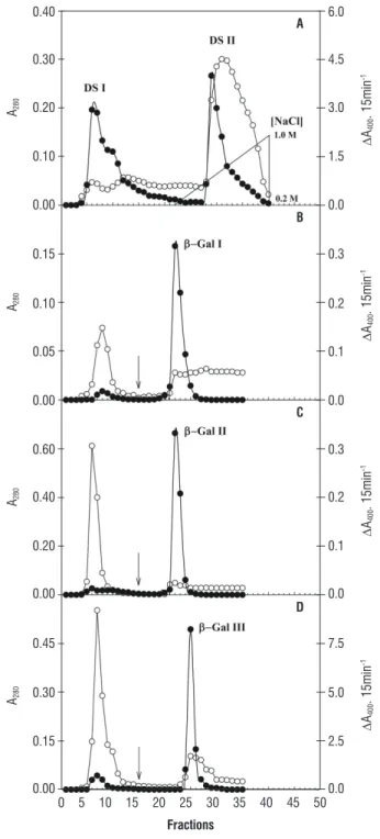
The DEAE-Sephadex A-50 column chromatography profile for F20-80 of cytosolic proteins revealed the existence of two protein peaks (A280), one non-retained (DS I) and the other adsorbed to DEAE-Sephadex (DS II) (Figure 1A). Both the DS I and the DS II peaks presented β-galactosidase activity. DS I and DS II protein fractions showed purification of 10.1 and 1.5 times, respectively (Table 1).
DS I and DS II fractions were applied on a Lactosyl-Sepharose affinity column. Application of DS I in the affinity
column resulted in a big non-retained protein peak (A280) in the column, with residual β-galactosidase activity that was not used for subsequent analyses. On the other hand, the peak that was retained, called β-gal I, presented almost all β-galactosidase activity (Figure 1B) and purification of 133.5 times (Table 1). When DS II was applied to affinity chromatography, the chromatography profile also resulted in two peaks at A280, one non-retained, very evident, but with very low β-galactosidase activity that was not used in the analyses, and the other retained, with expressive β-galactosidase activity, which was called β-gal II (Figure 1C) and a purification of 21.4 times (Table 1).
The chromatography profile of cell wall proteins for F
40-100 derived from Lactosyl-Sepharose column revealed the existence of a non-retained protein peak (A280), with residual
β-galactosidase activity that was not used in subsequent analyses, and the other, retained, with a considerable percentage of β-galactosidase activity, which was called β-gal III (Figure 1D) and which presented purification of 52.3 times (Table 1).
Electrophoretic analysis β-galactosidases isolated revealed that β-gal II and β-gal III presented a single protein band that corresponded to the same band with β-galactosidase activity (Figure 2). However, due to little quantity of protein and enzymatic activity of β-gal I, the revelation of protein with silver and the determination of enzymatic activity were not effective and identification was thus not possible.
The Sephadex G-150 column chromatography profile for the three β-galactosidase isoforms revealed apparent molecular masses of 89, 146 and 124 kDa for β-gal I, β-gal II and β-gal III, respectively (Figure 3).
table 1. Purification of cytosolic and cell wall β-galactosidases of cowpea seedling stems [Vigna unguiculata (L.) Walp.] cv. Pitiúba.
Cytosolic β-galactosidases purification
step
Activity
(UA.h-1) protein (mg) Specific Activity (UA.mgp-1.h-1) recovery (%) purification factor (fold)
Crude Extract 3,003,000 194.4 15,448 100 1
F20-80 2,966,080 106.4 27,877 98.8 1.8
DS-I 1,174,333 7.5 156,578 39.1 10.1 DS-II 744,785 31.5 23,644 24.8 1.5
β-gal I 453,696 0.2 2062,255 15.1 133,5
β-gal II 245,180 0.7 331,324 8.2 21,4
Cell wall β-galactosidase purification
step
Activity (UA.h-1)
protein (mg)
Specific Activity (UA.mgp-1.h-1)
recovery (%)
purification factor (fold)
Crude Extract 209,040 11.04 18,935 100 1
F40-100 84,014 3.87 21,709 40.2 1.1
0.40 0.30 0.20 0.10 0.00 0.15 0.10 0.05 0.00 0.60 0.40 0.20 0.00 0.45 0.30 0.15 0.00 6.0 4.5 3.0 1.5 0.0 0.3 0.2 0.1 0.0 0.3 0.2 0.1 0.0 7.5 5.0 2.5 0.0 A280 A280 A280 A280 D A400 . 15min -1 D A400 . 15min -1 D A400 . 15min -1 D A400 . 15min -1 A B C D
0 5 10 15 20 25 30 35 40 45 50
Fractions
Figure 1. Purification of cytosolic (A, B and C) and cell wall (D)
β-galactosidases of cowpea seedling stems [Vigna unguiculata (L.) Walp.] A. DEAE-Sephadex chromatography of the fraction rich in
β-galactosidase activity (F20-80). B. and C. Lactosyl-Sepharose affinity
chromatography of non-retained (DS I) and retained (DS II) DEAE-Sephadex peaks of cytosolic β-galactosidases, respectively. D. Lactosyl-Sepharose affinity chromatography of the fraction rich in β-galactosidase activity (F40-100). Protein (A280 ) and β-galactosidase activity (DA400
15min-1 ). The arrow indicates the beginning of the use of the elution
buffer containing 100 mM lactose and 0.5 M NaCl.
1 2 3 4 5 6
Figure 2. Electrophoresis in polyacrylamide gel in native conditions (gradient 5-15%), in acidic pH. The bands correspond to the β-galactosidases of cowpea seedling stems [Vigna unguiculata (L.) Walp.] cv. Pitiúba. 1.β-gal II (cytosolic) - 10 mg of protein; 2.β-gal II (cytosolic) - 5 mg of protein; 3. β-gal II (cytosolic) - 1.25 mg of protein; 4.β-gal II (cytosolic) – 2.5 mg of protein; 5.β-gal III (cell wall) - 10 mg of protein; 6.β-gal III (cell wall) – 1.25 mg of protein. The lanes 1, 2 (β-gal II) and 5 (β-gal III) represents the specific staining of β-galactosidase activity. The lanes 3, 4 (β-gal II) and 6 (β-gal III) represents the protein staining.
0.12 0.09 0.06 0.03 0.00 0.15 0.10 0.05 0.00 0.09 0.06 0.03 0.00 A280 A280 A280 D A400 . 15min -1 D A400 . 15min -1 D A400 . 15min -1 0.8 0.6 0.4 0.2 0.0 0.9 0.6 0.3 0.0 0.9 0.6 0.3 0.00
Evolution volume (ml)
0 10 20 30 40 50 60 70 80
A
B
C
Figure 3. Sephadex G-150 elution profile for determining the molecular
mass of β-gal I, β-gal II and β-gal III of cowpea seedling stems [Vigna unguiculata (L.) Walp.] cv. Pitiúba. Protein (A280 ) and β-galactosidase
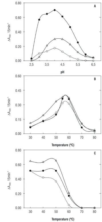
Enzymatic activities of cytosolic (β-gal I and β-gal II) and cell wall (β-gal III) β-galactosidases as a function of pH in the reaction medium can be observed in figure 4A. In all samples analyzed, there was an increase in enzymatic activity starting at pH 2.5 until reaching maximum activity at pH 4.0, with a progressive decrease until pH 6.5.
Optimal assay temperature for β-gal I, β-gal II and
β-gal III enzymes was 55°C, and in all there was very little difference in activity between 55 and 60°C and a considerable drop in activity after 60°C (Figure 4B). However, in β-gal I and β-gal II there was considerable difference in activity between 50 and 55°C, contrary to β-gal III, where there is very little difference in activity between these temperatures (Figure 4B).
In the thermal stability study of β-gal I, II and III enzymes (Figure 4C), β-gal I presented a reduction in activity when incubated at a temperature of 40°C. At 60°C, they all presented a pronounced drop until complete inactivation at 70°C. In general, thermal inactivation curves (pre-incubation of enzymes at 60°C) as a function of time (20, 40, 60 and 80 minutes) for
β-gal I, II and III (Figure 5) showed that in the absence of any sugar (control) the curves were biphasic, with an initial rapid phase, with great loss of activity, and a second phase that was slower. Pre-incubation of β-gal III at 60°C, for 40 minutes, resulted in the almost complete inactivation of enzyme activity. However, for β-gal I and II enzymes, this only occurred with 80 minutes of pre-incubation. Figure 5 also shows the thermal inactivation study of β-gal I, II and III in the presence of glucose and galactose. Glucose and galactose increased the thermal stability of enzymes, where activity loss was less in the presence of galactose than in the presence of glucose. Of all those sugars tested for their effects on β-gal I, II and III activities (Table 2), in the presence of β-PNPG, the higher inhibition was caused by galactose, followed by lactose and fucose. In general, arabinose had the least effect on β-gal I, II and III activity.
D
A400
. 15min
-1
D
A400
. 15min
-1
D
A400
. 15min
-1
0.80
0.60
0.40
0.20
0.00
2,5 3,5 4,5
ph
5,5 6,5
A
0.60
0.45
0.30
0.15
0.00
30 40 50
temperature (ºC)
60 70 80
B
0.80
0.60
0.40
0.20
0.00
C
30 40 50
temperature (ºC)
60 70 80
Figure 4. A. β-galactosidase activity as a function of pH; B. Effect of assay medium temperature on β-galactosidase activity; C. β-galactosidase activity as a function of pre-incubation temperature. Cytosolic β-galactosidases (β-Gal I, and β-Gal II, ) and those present in the cell wall (β-Gal III,
Enzymatic activity (% of control)
Enzymatic activity (% of control)
Enzymatic activity (% of control)
120
90
60
30
0
A
0 10 20
time (min)
30 40 50 60 70 80 90
60
30
0
B
90
60
30
0
C
Figure 5. Thermal inactivation of β-gal I, β-gal II and β-gal III of cowpea seedling stems [Vigna unguiculata (L.) Walp.] cv. Pitiúba, in the absence ( ) and presence of 50 mM glucose (▲—▲) and 50 mM galactose ( ).
table 2. Effect of sugars on the β-gal I, β-gal II and β-gal III activities of cowpea seedling stems [Vigna unguiculata (L.) Walp.] cv. Pitiúba.
[β-galactosidase activity (UA.h-1)] / [% of control (β-pNpG)]
treatment β-gal I β-gal II β-gal III
Control 204.8 (100%) 2,297.6 (100%) 992.0 (100%) Lactose (50 mM) 131.2 (64.1%) 1,675.2 (72.9%) 742.4 (74.8%) Galactose (50 mM) 54.4 (26.6%) 656.0 (28.6%) 163.2 (16.5%) Arabinose (50 mM) 172.8 (84.4%) 1,795.2 (78.1%) 902.4 (91%)
Fucose (50 mM) 103.2 (50.4%) 1,817.9 (79.1%) 860.8 (86.8%)
In the hydrolysis of the β-PNPG substrate, catalyzed by β-gal I, II and III, a type of Michaelis-Menten kinetics can be observed (Figure 6). Numerical values for KM and VMax for
β-gal I, II and III were 1.69 mM and 0.29 mmol.mg-1.prot.-1 of 1.76 mM and 0.96 mmol.mg-1.prot.-1 and 1.43 mM and 0.47
mmol.mg-1.prot.-1, respectively.
The study of β-gal I, β-gal II and β-gal III activities, as to their capacities to hydrolyze different substrates, is shown in Table 3. β-gal I presented activity for all tested synthetic substrates, where β-fucosidase activity was the most evident, representing 94% of control activity (β-galactosidase activity). Different results were observed in relation to β-gal II, which only presented β-fucosidase and
a-arabinosidase activities, which represented 21 and 10% of control activity, respectively. Of all tested substrates, the one β-gal III hydrolyzed at greater proportions was
PNP-a-D-glucopyranoside, which represented 32% of control activity (β-PNPG). β-gal III presented β-fucosidase and
a-arabinosidase activities, but there was practically no
β-xylosidase activity and did not present capacity to hydrolyze PNP-β-D-glucopyranoside, PNP-a-D-mannopyranoside and PNP-a-L-fucopyranoside substrates.
table3. Enzymatic activity of β-gal I, β-gal II and β-gal III of cowpea seedling stems [Vigna unguiculata (L.) Walp.] cv. Pitiúba in different p-nitrophenyl glycoside substrates.
[β-galactosidase activity (UA.h-1)] / [% of control (β-pNpG)]
Substrate β-gal I β-gal II β-gal III
(D
A400
. 15min
-1)
-1
(D
A400
. 15min
-1)
-1
(D
A400
. 15min
-1)
-1
-0.8 -0.6 -0.4 -0.2 0.0 8.0 6.4 4.8 3.2 1.6
0.2 0.4 0.6 0.8 1.0
[Substrate]-1(mM-1)
-0.8 -0.6 -0.4 -0.2 0.0 3.0 2.4 1.8 1.2 0.6
0.2 0.4 0.6 0.8 1.0
[Substrate]-1(mM-1)
-0.8 -0.6 -0.4 -0.2 0.0 6.0 4.8 3.6 2.4 1.2
0.2 0.4 0.6 0.8 1.0
[Substrate]-1(mM-1)
Figure 6. Double-reciprocal plots (Lineweaver-Burk) for β-Gal I (A), β-Gal II (B) and β-Gal III (C) of cowpea seedling stems [Vigna unguiculata (L.) Walp.] cv. Pitiúba obtained of enzymatic activity as a function of crescent concentrations of the β-PNPG substrate.
DISCUSSION
Plant β-galactosidases are often basic proteins exhibiting pI greater than 7.0 (Simos et al., 1989; Enéas-Filho et al., 2000; Li et al., 2001; Balasubraniam et al., 2005). It seems to be the case of the β-galactosidases here purified and characterized, which also have the same optimal pH (4.0). Therefore, an electrophoresis methodology in acid gel specific for basic proteins (Goldenberg, 1997) was adopted in the present study. A similar electrophoretic profile (Figure 2) was previously observed in Phaseolus vulgaris (Biswas et al., 2003).
Molecular mass differences found among the
β-galactosidases analyzed (Figure 3) could reflect the distinct nature of the three enzymes – two cytosolic isoforms (β-gal I and II) and another one associated with the cell wall (β-gal III). In fact, different molecular masses have been described for β-galactosidases previously purified from different plant tissues (e.g., Li et al., 2001; Biswas et al., 2003; Balasubraniam et al., 2005; Alcântaraet al. 2006).
The optimal pH presented by β-gal I, II and III (Figure 4A) is common to a broad variety of plant species (Enéas-Filho et al., 2000 and 2001; Li et al., 2001; Chilaka et al., 2002; Biswas et al., 2003; Alcântaraet al., 2006). Furthermore, the optimal temperature range observed in Figure 4B is also similar to that found in other β-galactosidases purified from plants (Enéas-Filho et al.,2000 and 2001; Liet al., 2001; Chilaka et al., 2002). Despite the similarities among β-gal I, II and III enzymes regarding the thermal stability, β-gal II was the most sensitive at higher temperatures (Figure 4C). Previous studies on thermal stability of β-galactosidases from Vigna radiata
seeds (Kundu et al., 1990) and Vigna unguiculta cotyledons (Enéas-Filho et al., 2000) reported quite similar results, with minor or no inactivation of the enzymes pretreated at 50°C, but exhibiting considerable inactivation above 55°C. Slightly different results were reported by Sekimata et al. (1989) for
β-galactosidases from radish seeds, which were already completely inactivated at 55°C.
conformational state. Since β-gal III was almost completely inactivated upon pretreatment at 60°C for 40 minutes, and
β-gal I and II enzymes resisted until 80 minutes at the same pretreatment, it is possible that the β-galactosidase derived from cell wall (β-gal III) is more sensitive to thermal inactivation than the cytosolic counterparts (β-gal I and II).
Figure 5 explores the thermal inactivation of β-gal I, II and III in the presence of glucose or galactose and the data suggest the involvement of changes in the active site of the enzymes, since their activities were preserved even at high temperatures due to the protection induced by glucose and galactose. These sugars probably connect to the active site changing the enzyme conformation and making them more resistant to high temperatures (Chilaka et al., 2002). The data reported by Chilaka et al. (2002) for
Kestingella geocarpa enzymes are similar to those found in Figure 5, the thermal inactivation curves as a function of time were also biphasic, glucose and galactose also protected the K. geocarpa β-galactosidases, with 100% protection in the case of galactose at a concentration of 100 mM.
The β-gal I, II and III also exhibited different inhibitory patterns for the different sugars tested (Table 2). Similar to the present results, the activities of Vigna radiata cotyledon
β-galactosidases (Li et al., 2001) were also strongly inhibited in the presence of galactose (about 70%) and 28% in the presence of lactose. Arabinose and fucose practically did not inhibit enzyme activity in V. radiata (Li et al.,2001). This strong inhibition pattern for galactose (especially) and lactose was also reported in the study by Chilaka et al. (2002) with K. geocarpa cotyledonal β-galactosidases. The
β-galactosidase inhibition was also found to be higher with galactose and lesser with glucose in Hymenaea courbaril
cotyledons (Alcântara et al., 2006). Different sugars structurally related to galactose tend to act as inhibitors binding to the active site acting as competitive inhibitors or even in other enzyme regions, as uncompetitive and non-competitive inhibitors (Nelson and Cox, 2000).
Most kinetic studies on β-galactosidases use β-PNPG as substrate (e.g., Biswas et al., 2003; Balasubraniam et al., 2005; Alcântara et al., 2006). Enzymatic kinetics of the enzymes purified in this study indicate that β-gal III have a slightly higher affinity for β-PNPG than for β-gal I and II (Figure 6).
The study of β-gal I, II and III activities in relation to their capacities to hydrolyze different substrates (Table 3) revealed differences between the two cytosolic
β-galactosidases (β-gal I and II) and among them and the cell wall β-galactosidase (β-gal III), which also may suggest different functions. As observed in this study, some authors also observed that purified
β-galactosidases from different plant species also presented the capacity to hydrolyze different p-nitrophenyl glycosides, besides β-PNPG (Ali et al., 1995; Li et al., 2001; Alcântara et al., 2006). Specificity studies of cytosolic β-galactosidases isoforms of Mangifera indica
fruits were performed by Ali et al. (1995). In this study, the isoforms were unable to hydrolyze any p-nitrophenyl glycoside tested, showing that the enzymatic preparation was totally free of β-glucosidase and a-manosidase activity, agreeing with the results observed with β-gal II and β-gal III enzymes (Table 3). Besides the absence of these activities, M. indica β-galactosidases also did not present a-arabinosidase activity. Like the β-gal I,
β-gal II and β-gal III (Table 3), different β-galactosidase isoforms associated with the cell wall isolated from
Vigna radiata cotyledons also revealed different behavior regarding their capacity to degrade different substrates (Li etal., 2001). Alcântara et al. (2006) observed there was no β-manosidase and β-xylosidase activity in the
β-galactosidases present in the cell wall of Hymenaea courbaril cotyledons, similar to what was observed for
β-gal II and III (Table 3). Considering that β-galactosidases can act on the mobilization of cell wall constituents during plant germination and growth, the analysis of specificity these enzymes in relation to different substrates is crucial for understanding the possible physiological roles accomplished by these enzymes in this plant species.
CONClUSION
Acknowledgements: This work was supported by the grants and scholarships from the Coordenação de Aperfeiçoamento de Pessoal de Nível Superior (CAPES), INCTSal/CNPq, Conselho Nacional de Desenvolvimento Científico e Tecnológico (CNPq) and Fundação Cearense de Apoio ao Desenvolvimento Científico e Tecnológico (FUNCAP). We thank Professor Emeritus José Tarquínio Prisco for his constant guidance in carrying out this study.
rEFErENCES
Alcântara PHN, Martim L, Silva CO, Dietrich SMC, Buckeridge MS (2006) Purification of a β-galactosidase from cotyledons of Hymenaea courbaril L. (Leguminosae). Enzyme properties and biological function. Plant Physiol Biochem. 44:619-627.
Ali zM, Armugam S., Lazan H (1995) β-galactosidase and its significance in ripening mango fruit. Phytochem. 38:1109-1114.
Balasubranian S, Lee HC, Lazan H, Othman R, Ali zM (2005) Purification and properties of a β-galactosidase from carambola fruit with significant activity towards cell wall polysaccharides. Phytochem. 66:153-163. Biswas S, Kayastha AM, Seckler R (2003) Purification and characterization of a thermostable β-galactosidase from kidney beans (Phaseolus vulgaris
L.) cv. PDR14. J. Plant Physiol. 160:327-337.
Biswas TK (1987) Characterization of β-galactosidases from the germinating seeds of Vigna sinensis. Phytochem. 26: 359-364.
Blum H, Beier H, Gross HJ (1987). Improved silver staining of plant proteins, RNA and DNA in polyacrylamide gels. Electrophoresis. 8:93-99.
Chilaka FC, Okeke C, Adaikpoh E (2002) Ligand-induced thermal stability in
β-galactosidase from the seeds of the black bean, Kestingeilla geocarpa. Process Biochem. 38:143-149.
Daniel RM, Dines M, Petach HH (1996) The denaturation and degradation of stable enzymes at high temperatures. Biochem. J. 317:1-11.
Enéas Filho J, Barbosa GKC, Sudério FB, Prisco JT, Gomes Filho E (2001) Isolation and partial purification of β-galactosidases from cotyledons of two cowpea cultivars. Rev. Bras. Fisiol. Veg. 13:251-261.
Enéas Filho J, Oliveira Neto OB, Prisco JT, Gomes Filho E, Nogueira CM (1995) Effects of salinity in vivo and in vitro on cotyledonary galactosidases from Vigna unguiculata (L.) Walp. during germination and seedling establishment. Rev. Bras. Fis. Veg. 7:135-142.
Enéas Filho J, Sudério FB, Gomes Filho E, Prisco JT (2000) Multiple forms of cotyledonary β-galactosidases from Vigna unguiculata quiescents seeds. Rev. Bras. Bot. 23:69-76.
Ferreira CM (2001) Comercialização de feijão no Brasil. Piracicaba, Universidade de São Paulo – Escola Superior de Agricultura “Luiz de Queiroz”. Dissertação de Mestrado em Ciências. 145p.
Freire Filho FR (1988) Origem, evolução e domesticação do caupi. In: Araújo JPP, Watt EE (eds). O caupi no Brasil, pp. 27-46. Brasília: IIta/EMBRAPA. Fry SC (2004) Primary cell wall metabolism: tracking the careers of wall polymers in living plant cells, New Phytol. 161:641-675.
Goldenberg DP (1997) Analysis of protein conformation by gel electroforesis. In: Creighton TE (ed), Protein structure: A practical approach, pp195. Oxford University Press Inc., New york.
Gómez LD, Casano LM, Braga MR, Buckeridge MS (1995) Changes in extracellular β-galactosidase and protease activities during bean hypocotyls growth. Rev. Bras. Fis. Veg. 7:1-6.
Granjeiro PA, Ferreira CV, Cavagis ADM, Granjeiro JM, Ayoama H (2003) Essential sulfhydryl groups in the active site of castor bean (Ricinus communis) seed acid phosphatase. Plant Sci, 164: 629-633.
Kanfer JN, Petrovich R, Mumford RA (1973) Purification of a- e
β-galactosidases by affinity chromatography. Anal. Biochem. 55:301-305.
Konno H, Tsumuki H (1993) Purification of a β-galactosidase from rice shoots and its involvement in hidrolysis of the natural substrate in cell walls. Physiol. Plant. 89:40-47.
Kundu RK, De Kundu P, Banerjee AC (1990) Multiple forms of β-galactosidase from the germinating seeds of Vigna radiata. Phytochem. 29:2079-2082. Li SC, Han JW, Chen KC, Chen CS (2001) Purification and characterization of isoforms of β-galactosidases in mung bean seedlings. Phytochem. 57:349-359.
Matthews BW (2005) The structure of E. coli β-galactosidase. C. R. Biologies. 328:549-556.
McIlwaine TC (1921) A buffer solution foil colorimetric comparison. J. Biol. Chem. 49:185-186.
Minic z, Jouanin L (2006) Plant glycoside hydrolases involved in cell wall polysaccharide degradation. Plant Physiol. Biochem. 44:435-449. Molina L, Constantinescu F, Michel L, Reimmann C, Duffy B, Défago G (2003) Degradation of pathogen quorum-sensing molecules by soil bacteria: a preventive and curative biological control mechanism. FEMS Microbiol. Ecol..45:71-81.
Nelson DL, Cox MM (2004) Lehninger Principles of Biochemistry. 4th edition. Worth.
Pérez Almeida I, Carpita, NC (2006) Las β-galactosidasas y la dinâmica de la pared celular. Interciencia. 31:476-482.
Prisco JT (1980) Alguns aspectos da fisiologia do "stress" salino. Rev. Bras. Bot. 3:85-94.
Sawicka T, Kacperska A (1995) Soluble and cell wall-associated
β-galactosidases from cold-grown winter rape (Brassica napus L., var.
oleifera L.). J. Plant Phisiol. 145:357-362.
Seara J, Nicolás G, Labrador E (1988) Autolysis of the cell wall. Its possible role in endogenous and IAA-induced growth in epicotyls of Cicer arietnum.
Physiol Plantarum. 72:762-774.
Sekimata M, Ogura K, Tsumaraya y, Hashimoto y, yamamoto S (1989) A
β-galactosidase from radish (Raphanus sativus L.) seeds. Plant Physiol. 90:567-574.
Simos G, Georgatsos JG (1988) Lactose-hidrolizing β-glycosidases of barley meal. Biochem. Biophys. Acta. 967:17-24.
Simos G, Giannkouros T, Georgatsos JG (1989) Plant β-galactosidases: purification by affinity chromattography and properties. Phytochem. 28:2587-2592.
![table 1. Purification of cytosolic and cell wall β-galactosidases of cowpea seedling stems [Vigna unguiculata (L.) Walp.] cv](https://thumb-eu.123doks.com/thumbv2/123dok_br/15606621.612041/4.892.63.792.825.1084/table-purification-cytosolic-galactosidases-cowpea-seedling-vigna-unguiculata.webp)
![Figure 5. Thermal inactivation of β-gal I, β-gal II and β-gal III of cowpea seedling stems [Vigna unguiculata (L.) Walp.] cv](https://thumb-eu.123doks.com/thumbv2/123dok_br/15606621.612041/7.892.108.459.138.802/figure-thermal-inactivation-cowpea-seedling-stems-vigna-unguiculata.webp)
![Figure 6. Double-reciprocal plots (Lineweaver-Burk) for β-Gal I (A), β-Gal II (B) and β-Gal III (C) of cowpea seedling stems [Vigna unguiculata (L.) Walp.] cv](https://thumb-eu.123doks.com/thumbv2/123dok_br/15606621.612041/8.892.80.381.128.937/figure-double-reciprocal-lineweaver-cowpea-seedling-vigna-unguiculata.webp)
