Prognostic Value of High-Sensitivity Troponin I versus Troponin T in
Acute Coronary Syndromes
Luis C. L. Correia
1,2, Fábio L. Sodré
3, José C.C. Lima
3, Michael Sabino
1,2, Mariana Brito
1,2, Guilherme Garcia
1,2,
Mayara Maraux
1,2, Alexandre C. Sousa
1,2, Márcia Noya-Rabelo
1,2, J. Péricles Esteves
3Faculdade de Medicina da Bahia1; Hospital San Rafael2; Hospital Português3, Salvador, BA, Brazil
Abstract
Background: Despite superior diagnostic accuracy of high-sensitivity cardiac troponins, their prognostic value has not been validated against conventional cardiac troponins.
Objective: To test the prognostic value of high-sensitivity cardiac troponin I (hs-cTnI), compared with conventional cardiac troponin T (cTnT) in the setting of non-ST elevation acute coronary syndromes.
Methods: At hospital admission, a plasma sample was collected from 103 consecutive patients with unstable angina or non-ST elevation acute myocardial infarction. In this sample, troponin was measured both by hs-cTnI and cTnT methods. Their prognostic value was compared as to the occurrence of major cardiovascular events, defined as a combination of death, nonfatal acute myocardial infarction or refractory unstable angina during hospitalization.
Results: During median hospitalization of 8 days (interquartile range = 5 – 11), the incidence of cardiovascular events was 10% (5 deaths, 3 non-fatal myocardial infarctions and 2 non-fatal refractory anginas). High-sensitivity troponin I significantly predicted cardiovascular events, with a C-statistics of 0.73 (95% CI = 0.59 – 0.87), similarly to cTnT (0.70; 95% CI = 0.55 – 0.84) - P = 0.75. The definition of positive cardiac marker that provided the best prognostic accuracy was hs-cTnI > 0.055 µg/L and cTnT > 0.010 µg/L, with equal sensitivity of 90% and specificity of 52% for both assays. Positive hs-cTnI was associated with 17% incidence of events, compared with 2% in patients with negative hs-cTnI (P = 0.02).
Conclusion: High-sensitivity troponin I predicts cardiovascular events similarly to conventional troponin T in the setting of non-ST-elevation ACS. (Arq Bras Cardiol 2012;98(5):406-412)
Keywords: Acute coronary syndrome; troponin; prognosis.
never been compared with the prognostic value of current conventional troponins in acute coronary syndromes. In order to evaluate whether high-sensitivity troponin I (hs-cTnI) offers a similar or superior prognostic value compared to conventional troponin T (cTnT) in patients with non-ST elevation acute coronary syndromes, we measured troponin by both assays at admission of 103 patients and cardiovascular events were recorded during hospitalization.
Methods
Study Population
Consecutive patients admitted to our coronary care unit due to unstable angina pectoris or non-ST elevation acute myocardial infarction between August 2008 and September 2009 were considered candidates for the study. Inclusion criteria were defined as onset of typical chest discomfort for at least 5 minutes, in the first 48 hours, absence of ST-segment elevation and at least one of the following objective criteria: (1) positive troponin in any of the three serial measurements performed on the first day of hospitalization, defined as cTnT > 0.030 µg/L, which corresponds to the value above the 99th percentile which is
a healthy reference population and total imprecision of 10%
Introduction
Markers of myocardial necrosis are important indicators for the diagnosis, risk assessment and therapeutic decision in patients with non-ST-elevation acute coronary syndromes. Recently, high-sensitivity assays have been developed with less than 10% imprecision below the 99th percentile of a
normal reference population1,2. The higher sensitivity improves
the ability to detect small amounts of myocardial necrosis. However, it does not guarantee better accuracy, because greater sensitivities usually take place at the expense of lower specificities3. Thus, it is a consensus that high-sensitivity
troponin methods must be validated by scientific studies before widespread implementation4.
Regarding diagnosis, high-sensitivity troponin assays have been validated as tests of superior accuracy in the setting of acute chest pain5,6. Although preliminary data suggest that
high-sensitivity assays have prognostic value7, they have
Mailing Address: Luis C. L. Correia •
Av. Princesa Leopoldina 19/402 – Graça – 40150-080 – Salvador, BA – Brazil E-mail: [email protected], [email protected]
(Universal Definition of Infarction)8; (2) electrocardiographic
ischemic changes consisting of transient ST-segment
depression (≥ 0.05 mV) or T wave inversion (≥ 0.1 mV);
(3) previous documentation of coronary artery disease, defined as a definitive history of myocardial infarction or
coronary obstruction ≥ 50% at angiography. Once fulfilling
criteria for entering the study, patients provided written informed consent. The study protocol conforms to the ethical guidelines of the 1975 Declaration of Helsinki as reflected in a priori approval by the Institution’s human research ethics committee.
Laboratory Protocol
Only the first troponin measurement was evaluated as a prognostic marker in this study. The blood sample collected immediately at hospital arrival (emergency room) or at the coronary care unit arrival (if the patient was admitted from within the hospital) was utilized for measurement of troponin T, which was used in the clinical setting. After that, the same sample was stored at -70°C for future measurement of high-sensitivity troponin I. Cardiac troponin T was measured by electrochemiluminescence immunoassay (Roche Diagnostics Corporation, Indianapolis, Indiana, USA), with a limit of detection of 0.010 µg/L, 99th percentile reference value lower
than 0.010 µg/L and coefficient of variability (CV) of 10% at 0.030 µg/L8. High-sensitivity troponin I was assessed by
VITROS chemiluminescence immunoassay (Ortho-Clinical Diagnostics, Rochester, NY, USA), with a limit of detection of 0.012 µg/L, 99th percentile reference value of 0.034 µg/L
and CV of 10% at 0.034 µg/L8.
Clinical End-points
Individuals were followed during hospitalization in order to identify recurrent cardiovascular events, which were adjudicated by investigators, regardless of physician impression. As a primary end-point, major cardiovascular events during hospitalization were defined as a combination of death, nonfatal acute myocardial infarction or refractory unstable angina. Myocardial infarction as an outcome endpoint was defined as either a new Q-wave or troponin T elevation during hospitalization despite normal values during the first 24 hours. For patients with infarction at admission, a new peak of mass CK-MB (> 50% the previous value and above the normal value) was required for diagnosis of reinfarction. Refractory angina during hospitalization was defined as recurrent chest pain at least twice, despite nitrates and anti-angina therapy ensuring controlled oxygen consumption.
Data Analysis
Considering the non-normal distributions, cardiac troponins were described as median and interquartile range. The accuracies of hs-cTnI and cTnT in predicting in-hospital cardiovascular events were first tested by the C-statistics (area under the receiver operator characteristics curve), which was considered significant if statistically different from 0.5. C-statistics of hs-cTnI was compared with cTnT by the method proposed by Henley and McNeil9. Utilizing
the cutoff points of best prognostic performance for defining
positive troponin, sensitivity, specificity, negative predictive value and positive predictive value were calculated for hs-cTnI and cTnT. Agreement between the two assays in defining positive troponin was assessed by the Kappa test. The GRACE Prognostic Score10 was calculated in two ways;
first, utilizing hs-cTnI as the necrosis marker and second, utilizing cTnT. The two GRACE Scores had their C-statistics compared by the Henley and McNeil method. In addition, positive troponin was defined by two other criteria: values above the 99th percentile of the reference population, at 10%
impression; and any detectable value. Sensitivity, specificity, negative predictive value and positive predictive value were also calculated for these two alternate definitions of positive hs-cTnI and cTnT. Statistical significance was defined as a p value < 0.05. SPSS Statistical Software (Version 9.0, SPSS Inc., Chicago, Illinois, USA) and MedCalc Statistical Software (Version 9.3.2.0, MedCalc Software, Mariakerke, Belgium) were used for data analysis.
Results
Population Characteristics
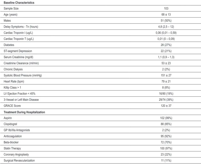
One hundred and three patients were studied, aged 68 ± 13 years, 50% males. As entry criteria, 59% were diagnosed as non-ST-segment elevation myocardial infarction and the remaining as unstable angina. In average, patients had intermediate risk for recurrent ischemic events, with a GRACE Score of 120 ± 37. In addition, ejection fraction < 45% was observed in 19% and severe coronary artery disease (triple-vessel or left main stenosis) in 39% of those who underwent angiography. Severe renal dysfunction (creatinine clearance < 30 ml/min) was present in 13%, but only two patients were on chronic dialysis. Baseline characteristics are shown in Table 1. During a median hospitalization of 8 days (interquartile range = 5 – 11), the incidence of cardiovascular events was 10% (5 deaths, 3 non-fatal myocardial infarctions and 2 non-fatal refractory anginas).
Median time delay between symptoms onset and blood draw to measure cardiac troponin was 4.8 hours (interquartile range = 2.5 - 12). At admission, the median level of hs-cTnI was 0.060 µg/L (0.010 – 0.590 µg/L), while median cTnT was 0.010 µg/L (0 – 0.090 µg/L).
Detectable levels of troponin I (≥ 0.012 µg/L) were more often observed than detectable troponin T (≥ 0.010
µg/L), respectively 77% vs. 52% (p < 0.001). Equally, the prevalence of hs-cTnI above the 99th percentile with 10%
CV (0.034 µg/L) was 58%, compared to as little as 36% of troponin T > 0.030 µg/L (p < 0.001). Thus, according to the universal definition of positive troponin (99th percentile),
hs-cTnI was often higher elevated than cTnT.
Prognostic Value of hs-cTnI versus cTnT
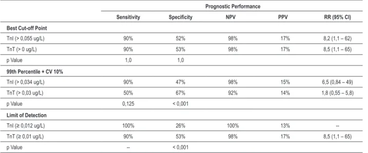
of positive cardiac marker that provided best the prognostic accuracy was hs-cTnI > 0.055 µg/L and cTnT > 0 µg/L.
Based on these definitions, hs-cTnI presented sensitivity of 90%, specificity of 52%, with negative predictive value of 98% and positive predictive value of 17% for cardiovascular events during hospitalization. Accordingly, hs-cTnI was associated with 17% incidence of events, compared with 2% in patients with negative hs-cTnI (relative risk = 8.2; 95% CI = 1.1 – 62; p = 0.02). Positive cTnT yielded equal numbers regarding accuracy and incidence of events – Table 2 and Figure 2. Therefore, both C-statistics and dichotomization analysis suggest that hs-cTnI and cTnT have similar prognostic accuracy.
Agreement between the two assays in defining positive troponin was 82% (Kappa = 0.63; 95% CI = 0.47 – 0.79; p < 0.001): both assays were positive in 44 patients, 10 patients had only hs-cTnI positive, 9 patients had only cTnT positive, and both methods were negative in 40 patients. Incidence of events was 22%, 0%, 0% and 3% in each of these four groups, respectively (p = 0.02) – Figure 2.
The GRACE score presented the same accuracy regardless whether hs-cTnI or cTnT was utilized in defining positive cardiac marker, based on the best cutoff points. When hs-cTnI was considered, the GRACE Score had a C-statistics of 0.755 (95% CI = 0.575 – 0.935; p = 0.008), almost identical to the GRACE Score calculated by cTnT (0.758; 95% CI = 0.575 – 0.940; p = 0.008) – p = 0.86 for comparison between the curves.
Prognostic Accuracy of Previously Deined Cutoff Points
When the universal definition of infarction (99th percentile
and CV < 10%) was utilized, hs-cTnI > 0.034 µg/L had higher sensitivity compared to standard cTnT > 0.030 µg/L (90% vs. 50%). Conversely, hs-cTnI had lower specificity than cTnT (47% vs. 67%). High-sensitivity troponin I > 0.034 µg/L was associated with 15% of events, compared with 2% in the other patients (p = 0.04), but cTnT > 0.030 µg/L was not significantly associated with events (14% vs. 8%; p = 0.49). Therefore, by the universal definition of infarction, hs-cTnI performed better than cTnT – Table 2.
Table 1 - Baseline characteristics
Baseline Characteristics
Sample Size 103
Age (years) 68 ± 13
Males 51 (50%)
Delay Symptoms - Tn (hours) 4,8 (2,5 – 12)
Cardiac Troponin I (ug/L) 0,06 (0,01 – 0,59)
Cardiac Troponin T (ug/L) 0,01 (0 – 0,09)
Diabetes 28 (27%)
ST-segment Depression 22 (21%)
Serum Creatinine (mg/dl) 1,1 (0,9 – 1,3)
Creatinine Clearance (ml/min) 53 ± 23
Chronic Dialysis 2 (2%)
Systolic Blood Pressure (mmHg) 151 ± 27
Heart Rate (bpm) 79 ± 21
Killip Class > 1 8 (8%)
LV Ejection Fraction < 45% 16/90 (19%)
3-Vessel or Left Main Disease 29/74 (39%)
GRACE Score 120 ± 37
Treatment During Hospitalization
Aspirin 102 (99%)
Clopidogrel 88 (85%)
GP IIb/IIIa Antagonists 2 (2%)
Anticoagulation 95 (92%)
Beta-blocker 72 (70%)
Statin Therapy 100 (97%)
Coronary Angioplasty 23 (22%)
When any detectable troponin level was taken as the
cutoff point, hs-cTnI ≥ 0.012 µg/L had a similar sensitivity to cTnT ≥ 0.010 µg/L (100% vs. 90%), but a worse specificity (24% vs. 53%). Individuals with hs-cTnI ≥ 0.012 µg/L had
13% of events, compared with 0% in the other patients
(p = 0.11), while cTnT ≥ 0.010 µg/L was associated with
significantly increased incidence of events (17% vs. 2%; p = 0.02). Therefore, by the criteria of any detectable level, cTnT performed better due to its higher specificity.
In summary, predefined cutoff points implies higher sensitivity and lower specificity for hs-cTnI compared to cTnT. As the cutoff point increases, hs-cTnI tends to perform better than cTnT, due to improved specificity. As the cutoff point decreases, hs-cTnI tends to perform worse than standard cTnT, due to impaired specificity.
Discussion
This study demonstrates a similar prognostic performance of hs-cTnI and cTnT in patients with non-ST elevation acute coronary syndromes, as indicated by the comparison of the areas under the ROC curves. The relevance of this result relies on the concern that a highly sensitive assay may suffer from lack of specificity. Recently, this concern was partially resolved by the demonstration that high-sensitivity cardiac troponins
provide better diagnostic accuracy in the setting of acute chest pain compared to standard assays5,6. However, the prognostic
accuracy of hs-cTn in patients with acute coronary syndromes had not been established.
In addition to the comparison of ROC curves, sensitivity and specificity were separately evaluated according to different cutoff points. When the Universal Definition of Infarction was utilized to define the cutoff points, the two methods performed differently: hs-cTnI had a better sensitivity, detecting more individuals with myocardial infarction compared to cTnT. On the other hand, the superior sensitivity took place at the expense of a reduced specificity. According to the prevention paradigm, prognostic markers are primarily focused on sensitivity, while specificity becomes a secondary aim. Based on this, one may conclude that hs-cTnI is the best choice. However, the Universal Definition of Infarction is not based on prognostic data. It is just an arbitrary definition of disease based on the analytical properties of the test. Thus, specific cutoff points should be defined for prognostic purposes. When the cutoff points were adjusted according to the best performance by the ROC curve analysis, the prognostic values of the two tests did not differ. Finally, when the limit of detection was utilized as the cutoff points, hs-cTnI became useless as a prognostic marker, with nearly null specificity. This demonstrates that lower cutoff points are not adequate for Figure 1 - ROC curves for high-sensitivity troponin I and conventional troponin T for prediction of cardiovascular events during hospitalization. High-sensitivity troponin
I signiicantly predicted cardiovascular events with a C-statistics of 0.73 (95% CI = 0.59 – 0.87; p = 0.02), similarly to cTnT (0.70; 95% CI = 0.55 – 0.84; p = 0.04) - p = 0.75 for comparison between the curves.
S
en
si
tiv
ity
100 - Speciicity
High-Sensitivity TnI
Table 2 - Prognostic performance of troponins assays according to different cut-off points
Prognostic Performance
Sensitivity Speciicity NPV PPV RR (95% CI)
Best Cut-off Point
TnI (> 0,055 ug/L) 90% 52% 98% 17% 8,2 (1,1 – 62)
TnT (> 0 ug/L) 90% 53% 98% 17% 8,5 (1,1 – 65)
p Value 1,0 1,0
99th Percentile + CV 10%
TnI (> 0,034 ug/L) 90% 47% 98% 15% 6,5 (0,84 – 49)
TnT (> 0,03 ug/L) 50% 67% 92% 14% 1,8 (0,55 – 5,8)
p Value 0,125 < 0,001
Limit of Detection
TnI (≥ 0,012 ug/L) 100% 26% 100% 13%
--TnT (≥ 0,01 ug/L) 90% 53% 98% 17% 8,5 (1,1 – 65)
p Value -- < 0,001
TnI - high-sensitivity cardiac troponin I; TnT - cardiac troponin T; NPV - negativa predictive value; PPV - positive predictive value.
high-sensitivity assays, while higher cutoff points derived from ROC analysis tend to improve accuracy. Conversely, the best cutoff point of cTnT was equal to the lower limit of detection, indicating that lower cutoff points improve accuracy of cTnT. Therefore, the present analysis emphasizes that high-sensitivity and conventional cardiac troponins behave differently and in order to maximize accuracy, cutoff points should be defined by the actual prognostic performance of each method.
A prognostic marker should be analyzed along with clinical predictive variables, as performed by risk scores which combine clinical, electrocardiographic, laboratory variables and markers of necrosis. Therefore, we compared the accuracy of the GRACE score considering hs-cTnI or cTnT, both defined by the best cutoff points. The C-statistics of the two GRACE scores were identical. This represents another evidence that hs-cTnI performs well enough as a prognostic marker in patients with ACS.
In
-H
os
pi
ta
l E
ve
nts
Figure 2 - Cardiovascular events during hospitalization according to high-sensitivity troponin I and conventional troponin T.
Both cTn Negative
(N = 40)
cTnT Positive
cTnI Negative
(N = 9)
cTnT Negative
cTnI Positive
(N = 10)
Both cTn Positive
(N = 44)
In-H
os
pit
al E
ve
nt
Previously, Bonaca et al and Apple et al evaluated the prognostic value of high-sensitivity troponin in the setting of chest pain, but did not compare with conventional troponins7,8. This comparison is essential to evaluate whether
the new assay should replace the old assay. Keller et al5
and Reichlin et al6 compared high-sensitivity troponins and
conventional troponins, but evaluated primarily diagnostic properties, in a heterogeneous sample of chest pain patients.5, 6 This year, Aldous et al11 demonstrated in a follow-up of two
years that high-sensitivity troponins have a better predictive value compared to conventional troponins. Also this year, Ndrepepa et al12 suggested the same result in a follow-up of
four years. To the best of our knowledge, this study is the first to compare the assays with focus on the acute phase prognosis, in a well-defined population of acute coronary syndromes.
The main limitation of our study is the total sample size and small number of events, which provides insufficient statistical power to guarantee a tolerable type II error probability. Therefore, this is a hypothesis-generating study, which should be retested by larger sample sizes. Nevertheless, we should point out that the areas under the ROC curves were very similar between the methods, which contributes to reduce the probability of type II error. While we suggest the importance of defining cutoff points, our analysis is underpowered for validating reference values. Thus, further studies are needed to precisely validate which cutoff points should be used in the prognostic setting. In fact, prognostic cutoff points may be different from diagnostic cutoff points. Moreover, this study is
limited by a sole and early measurement of troponin. Since a first evaluation may lack sensitivity for troponin changes, our findings should not be extrapolated for serial measurements.
Herein, we presented evidence that a new assay of high-sensitivity troponin has prognostic value comparable to the conventional assay. However, we did not demonstrate superiority. Nevertheless, the present findings coupled with previous knowledge that the high-sensitivity troponin is superior in terms of diagnosis indicate that the new method may be the preferred approach for the assessment of myocardial necrosis in patients with ACS.
In conclusion, this present study suggests that hs-cTnI has a similar prognostic performance to cTnT in the setting of non-ST-elevation ACS.
Potential Conflict of Interest
No potential conflict of interest relevant to this article was reported.
Sources of Funding
There were no external funding sources for this study.
Study Association
This study is not associated with any post-graduation program.
References
1. Jaffe AS, Ordonez-Llanos J. High sensitivity troponin in chest pain and acute coronary syndromes. A step forward? Rev Esp Cardiol. 2010;63(7):763-9. 2. Christenson RH, Phillips D. Sensitive and high sensitivity next generation
cardiac troponin assays: more than just a name. Pathology. 2011;43(3):213-9. 3. Diamond GA, Kaul S. How would the Reverend Bayes interpret
high-sensitivity troponin? Circulation. 2010;121(10):1172-5.
4. Katus HA, Giannitsis E, Jaffe AS, Thygesen K. Higher sensitivity troponin assays: Quo vadis? Eur Heart J. 2009;30(2):127-8.
5. Keller T, Zeller T, Peetz D, Tzikas S, Roth A, Czyz E, et al. Sensitive troponin I assay in early diagnosis of acute myocardial infarction. N Engl J Med. 2009;361(9):868-77.
6. Reichlin T, Hochholzer W, Bassetti S, Steuer S, Stelzig C, Hartwiger S, et al. Early diagnosis of myocardial infarction with sensitive cardiac troponin assays. N Engl J Med. 2009;361(9):858-67.
7. Bonaca M, Scirica B, Sabatine M, Dalby A, Spinar J, Murphy SA, et al. Prospective evaluation of the prognostic implications of improved assay performance with a sensitive assay for cardiac troponin I. J Am Coll Cardiol. 2010;55(19):2118-24.
8. Apple FS, Pearce LA, Smith SW, Kaczmarek JM, Murakami MM. Role of monitoring changes in sensitive cardiac troponin I assay results for early diagnosis of myocardial infarction and prediction of risk of adverse events. Clin Chem. 2009;55(5):930-7.
9. Hanley JA, McNeil BJ. A method of comparing the areas under receiver operating characteristic curves derived from the same cases. Radiology. 1983;148(3):839-43.
10. Granger CB, Goldberg RJ, Dabbous O, Pieper KS, Eagle KA, Cannon CP, et al. Predictors of hospital mortality in the global registry of acute coronary events. Arch Intern Med. 2003;163(19):2345-53.
11. Aldous SJ, Florkowski CM, Crozier IG, George P, Mackay R, Than M. High sensitivity troponin outperforms contemporary assays in predicting major adverse cardiac events up to two years in patients with chest pain. Ann Clin Biochem. 2011;48(Pt 3):249-55.