A New Tissue Doppler Index in Predicting Future Atrial Fibrillation in
Patients with Heart Failure
Cristian Mornos, Lucian Petrescu, Dragos Cozma, Adina Ionac, Sorin Pescariu, Stefan Iosif Dragulescu
Institute of Cardiovascular Diseases, Timisoara, RomâniaAbstract
Background: Onset of atrial fibrillation (AF) in patients with heart failure (HF) is usually associated with a high occurrence of cardiovascular complications. E/(E’×S’) ratio (E=early diastolic transmitral velocity, E’=early mitral annular diastolic velocity and S’=systolic mitral annulus velocity) has been shown to reflect left ventricular filling pressure.
Objetive: We investigate whether E/(E’×S’) could be a predictor of new-onset AF in patients with HF.
Methods: We analyzed 113 consecutive hospitalized patients with HF, in sinus rhythm, after appropriate medical treatment. Patients with histories of AF, inadequate echocardiographic images, congenital heart disease, paced rhythm, significant primary valvular disease, acute coronary syndrome, coronary revascularization during follow-up, severe pulmonary disease or renal failure were not included. E/(E’×S’) was determined using the average of septal and lateral mitral annular velocities. The primary study end-point was the new-onset AF.
Results: During the follow-up period (35.7 ± 11.2 months), 33 patients (29.2%) developed AF. Mean E/(E’×S’) was 3.09 ± 1.12 in these patients, while it was 1.7 2 ± 1.34 in the other patients (p < 0.001). The optimal E/(E’×S’) cut-off to predict new-onset AF was 2.2 (88% sensitivity, 77% specificity). There were 64 patients (56.6%) with E/(E’×S’) ≤ 2.2 and 49 (43.4%) with E/(E’×S’) > 2.2. New-onset AF was higher in patients with E/(E’×S’) > 2.2 than in patients with E/(E’×S’) ≤ 2.2 [29 (59.1%) versus 4 (6.2%), p < 0.001]. On multivariate Cox analysis including the variables that predicted AF on univariate analysis, E/(E’×S’) was the only independent predictor of new-onset AF (hazard ratio = 2.26, 95% confidence interval = 1.25-4.09, p = 0.007).
Conclusions: In patients with HF, E/(E’×S’) seems to be a good predictor of new-onset AF. (Arq Bras Cardiol 2011;97(6):468-477)
Keywords: Tissue doppler imaging, prognostic, heart failure, atrial fibrillation.
Mailing Address: Cristian Mornos •
Str G Adam 13 A – Timis – 300310 – Timisoara E-mail: mornoscristi@yahoo.com
Manuscript received January 30, 2011, revised manusript received June 15, 2011, accepted June 22, 2011.
development of AF11-17. Tissue Doppler Imaging (TDI), a new
echocardiographic method, can predict AF15-17. The early
diastolic transmitral velocity/early diastolic mitral annular velocity (E/E’) ratio, reflecting left ventricular (LV) filling pressures, was proposed as a single Doppler parameter in prediction of new-onset AF15-17.These studies support
the idea that increased intraventricular filling pressure correlates with the future development of AF. We have recently proposed a novel tissue Doppler index, E/(E’×S’), for the non-invasive assessment of LV end-diastolic pressure in a heterogeneous population of cardiac patients19. In this
study, E/(E’×S’) was the best predictor of LV end-diastolic pressure in sinus rhythm patients, and it was superior to E/E’, E’, S’ or E, regardless of LV ejection fraction (LVEF), particularly in those with E/E’ between 8 and 15 and in those with regional dysfunction. E/(E’×S’) associates an index of diastolic function (E/E’) and a parameter that explores LV systolic performance (S’) and therefore could provide supplementary information compared to each component alone. We hypothesized that this novel TDI index, E/(E’×S’), may be more sensitive than traditional echocardiographic methods to predict future development of new-onset AF in patients with HF.
Introduction
Atrial fibrillation (AF) is the most common arrhythmia in the general population1,2 and its incidence increases
with the severity of heart failure (HF)3.Mortality and
morbidity after the onset of AF remains high despite recent progress in the management of this condition, even after adjustment for multiple variables including age, hypertension, ischemic heart disease and congestive HF2,4-6. Accordingly, practice guidelines for HF and AF have
shifted their emphasis from treatment to prevention1,7.
The identification of individuals at risk for AF remains a challenge, though. Therefore, risk stratification based on clinical, biomarkers and echocardiographic parameters to define patients who are at risk for AF has been studied extensively8-18. With its ability to identify or
Methods
Population studied
We analyzed 158 consecutive hospitalized patients with HF in sinus rhythm, diagnosed according to the guidelines7,20. Patients were excluded from the study if
any of the following was present: history of AF, inadequate echocardiographic images, congenital heart disease, paced rhythm, significant primary valvular heart disease, acute coronary syndrome at inclusion, coronary artery bypass grafting during follow-up, severe pulmonary disease or renal failure. The other 113 patients formed our study group. The study was approved by the local research ethics committee.
Clinical variables recorded
The following clinical variables were recorded and included in the prognostic model: age, sex, body mass index, mean arterial pressure, heart rate, etiology of HF, New York Heart Association (NYHA) functional class, N-terminal pro-brain natriuretic peptide (NTproBNP) levels (determined simultaneously with echocardiography).
Echocardiography
Echocardiography was performed after appropriate medical treatment, within 24 hours of hospital discharge using a Vivid 7 ultrasonographic system (General Electric, Milwaukee, WI). Two-dimensional and M-mode measurements were performed according to the recommendations of the American Society of Echocardiography, working together with the European Association of Echocardiography21. The
mechanism of mitral regurgitation (MR) was identified by transtoracic and/or transesophageal echocardiography22.The
severity of MR was assessed from the apical views using the proximal convergence method; severe MR was considered if
the regurgitant orifice area was ≥ 40 mm2 and the regurgitant volume was ≥ 60 ml/beat23. Transmitral flow patterns were
recorded from apical four-chamber windows with 3 to 5 mm pulsed-sample Doppler volume placed between the mitral valve tips. Maximal velocities of E and late diastolic transmitral flow (A) waves were measured during end-expiratory apnoea and E/A ratio calculated24; the velocities were recorded for five
consecutive cardiac cycles, and the results were averaged. The global myocardial index (GMI) was determined using Doppler time intervals measured from mitral inflow and LV outflow Doppler tracings, as the sum of isovolumic contraction time and isovolumic relaxation time divided by ejection time25.
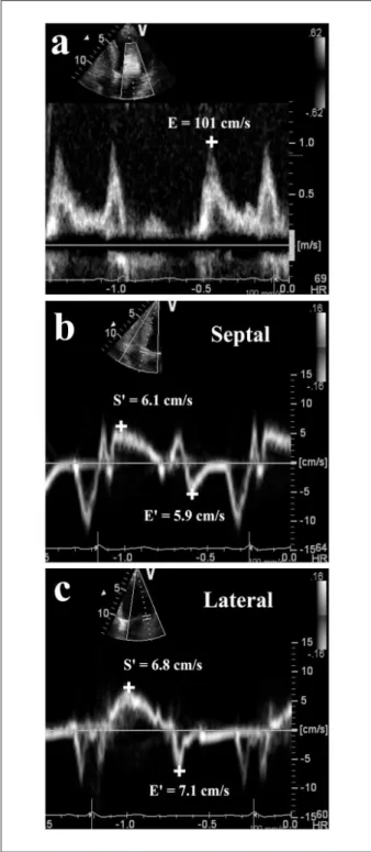
The tissue Doppler program was set in pulsed-wave Doppler mode. Motion of mitral annulus was recorded in the apical four-chamber view at a frame rate of 80 to 140 frames per second. A 4 to 5 mm sample volume was positioned sequentially at the lateral and septal corners of the mitral annulus. The peak E’ and S’ were recorded for five consecutive cardiac cycles during end-expiratory apnoea, and the results were averaged. E/E’ and E/(E’×S’) were calculatedusing the average of septal and lateral mitral annular velocities (Figure 1)19,24. The restrictive LV filling pattern was defined accordingly
to current guidelines24.All measurements were performed by
an experienced echocardiographer.
Clinical outcome
The primary event consisted of new-onset AF. We established the occurrence of AF only when a physician confirmed the diagnosis by reviewing an electrocardiogram. We did not assess the duration of AF and made no distinction between paroxysmal, persistent, or permanent AF.
Statistical analysis
Data are presented as the mean ± standard deviation (SD) for continuous variables and proportions for categorical variables. The mean values of continuous variables were compared by 2-independent sample t-tests, and differences in the prevalence between groups were compared via chi-square analyses. Receiver-operating characteristic (ROC) analysis was used to determine optimal cutoff values of continuous variables for the prediction of new-onset AF. Time-to-event analysis was performed using the Kaplan–Meier method. The relationship of parameters to the development of new AF was assessed with a Cox proportional hazards model. To test the independent predictor of new-onset AF, E/(E’×S’) was entered into a multivariable Cox model that also included as covariates all significant variables by univariate analysis associated with new-onset AF. All analyses were performed with SPSS statistical software (version 15.0, SPSS Inc., Chicago, Illinois) and a value of p <0.05 was considered for statistic significance. This work was supported by CNCSIS–UEFISCU, project number PN II/ RU code PD 526/2010.
Results
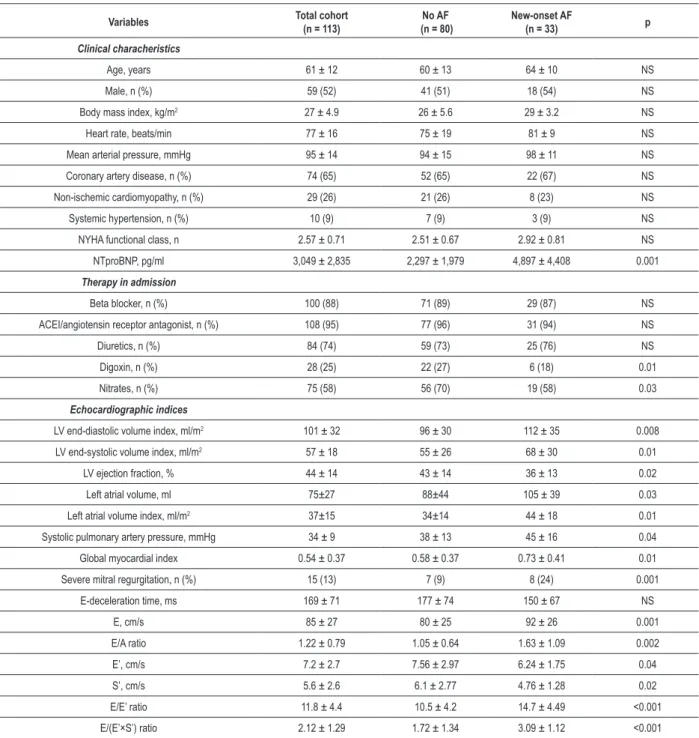
This study included 113 consecutive hospitalized patients (the mean age was 61 ± 12 years; 54 women), with HF in sinus rhythm. Clinical variables were available for all patients. The mean LVEF was 41±15%; 77 patients had HF with reduced LVEF (68.1%) and 36 patients presented HF with normal LVEF (31.9%). Baseline clinical and echocardiographic data are summarized in Table 1. Mitral annular velocities from TDI were recordable at both sites in all 113 patients. After a mean follow-up of 35.7±11.2 months, new-onset AF developed in 33 patients (29.2%). As compared with patients who did not develop new AF, patients who developed new-onset AF had significantly higher NTproBNP levels and pulmonary artery systolic pressures, larger left atrial (LA) and LV volumes, lower LVEF, higher values for E, E/A, E/E’ and GMI, lower E’ and S’ velocities, higher incidence of severe MR. In addition, there was no difference with regard to the distribution of age, gender, etiology, heart rate, mean arterial pressure, body mass index, NYHA class, medication (regarding beta blocker, angiotensin converting enzyme inhibitor/angiotensin receptor antagonist and diuretics) and E-deceleration time.
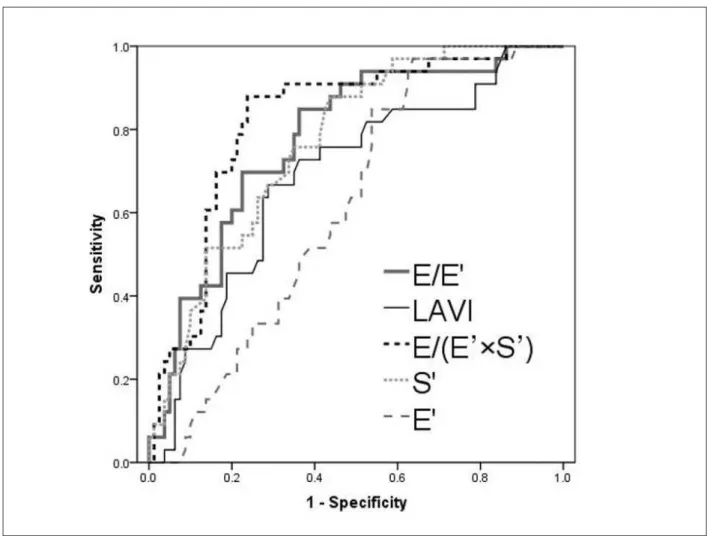
the E’ wave alone was an insignificant predictor (AUC = 0.61, 95%CI= 0.51–0.71, p = 0.056). A statistical comparison of the ROC curves demonstrates significant differences between E/(E’×S’) and E/E’ (p = 0.005), between E/(E’×S’)
and S’ (p = 0.002), and between E/(E’×S’) and LAVI (p < 0.001), respectively. The optimal cut-off value for E/(E’×S’) ratio to predict new-onset AF was 2.2 with 88% sensitivity and 77% specificity.
There were 64 patients (56.6%) with E/(E’×S’)≤2.2
and 49 (43.4%) with E/(E’×S’)>2.2. Mean E/(E’×S’) was 3.34±0.98 in the group of patients with E/(E’×S’)>2.2, while it was 1.19±0.47 in the others (p < 0.001). Patients with E/ (E’×S’)>2.2 presented significantly higher plasmatic NTproBNP levels (5379±4910 vs. 1265±1009 pg/ml, p < 0.001), larger LA and end–diastolic LV volumes (108±50 vs. 79±33 ml, p = 0.001, and 116±36 vs. 91±29 ml/m2, p < 0.001, respectively),
lower LVEF (34.8±13.2 vs. 46.2±13.3, p = 0.002), and higher incidence of severe MR [9 (18.3%) vs. 6 (9.3%), p = 0.01]. The incidence of new-onset AF was significantly higher in the group of patients with E/(E’×S’)>2.2 than in the group with E/
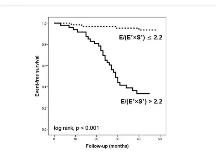
(E’×S’)≤2.2 [29 (59.1%) versus 4 (6.2%), p < 0.001]. Figure 3 shows the Kaplan-MeierAF event-free curves for patients
with E/(E’×S’)≤2.2 and >2.2. During the follow-up period,
cardiac death occurred in 18 patients (16%); new-onset AF was reported before the cardiac death in 14 of these patients. In the other group of 4 patients, cardiac death occurred between 28 and 41 months after the basal echocardiography. Non-cardiac death was not significantly different in the group of patients without AF compared to the group with new-onset AF [3 (3.75%) vs. 1 (3.03%), p = 0.16]. These patients were censored when we did new-onset AF analysis.
Table 2 shows the variables that predicted the new-onset AF on univariate Cox regression analysis. NTproBNP levels, severe MR, LVEF, LAV, LAVI, E/A, S’, E’, E/E’, E/(E’×S’),
LVEF≤40% combined with E/E’>15, and restrictive pattern emerged as predictors of AF in the patients studied. Age, sex, NYHA class, heart rate, mean arterial pressure, coronary artery disease, LV end-diastolic and end-systolic volume index, systolic pulmonary artery pressure, GMI, E and E-deceleration time, beta blocker, angiotensin converting enzyme inhibitor/angiotensin receptor antagonist or diuretics, were not significantly associated with new-onset AF in the univariate analysis.
Subsequently, all the variables that predicted the new-onset AF in the univariate analysis were entered into a forward multivariate Cox regression analysis. This analysis identified E/(E’×S’) ratio as the only independent predictor of new-onset AF (HR= 2.26, 95% CI= 1.25-4.09, p =0.007) in the study population.
The additional benefit of E/(E’×S’) to predict new-onset AF is shown in Figure 4. With regard to the incremental value, S’ offers an additional benefit (p =0.003) over conventional parameters (LVEF, LAVI, E/A and E/E’). However, the addition of E/(E’×S’) markedly improved the prognostic utility of the model containing LVEF, LAVI, E/A, E/E’ and S’ (p =0.001). We included in this model only the traditional echocardiographic parameters instead oft all of the variables that predicted the new-onset AF on univariate analysis.
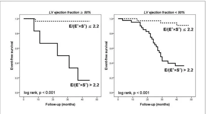
To determine whether this effect was due to abnormal LV systolic function, we analyzed patients with preserved
(LVEF≥50%) and those with reduced (LVEF<50%) LV Figure 1 –Bedside measurements of spectral Doppler peak early
systolic function separately. In patients with LVEF<50%, the subgroup with E/(E’×S’)≤2.2 (n =35) compared with the subgroup with E/(E’×S’)>2.2 (n =42) had a significantly better event-free survival rate (91.4% vs. 42.8%, p <0.001). The benefit was more prominent in patients
with LVEF≥50%. The patients with E/(E’×S’)≤2.2 (n =29) and the patients with E/(E’×S’)>2.2 (n =7) demonstrated a 96.5% and 28.5% event-free survival rate, respectively (p <0.001). Kaplan-Meier curves for AF event-free status in the two groups are shown in Figure 5.
Table 1 - Baseline characteristics of the study group
Variables Total cohort
(n = 113)
No AF (n = 80)
New-onset AF
(n = 33) p
Clinical characheristics
Age, years 61 ± 12 60 ± 13 64 ± 10 NS
Male, n (%) 59 (52) 41 (51) 18 (54) NS
Body mass index, kg/m2 27 ± 4.9 26 ± 5.6 29 ± 3.2 NS
Heart rate, beats/min 77 ± 16 75 ± 19 81 ± 9 NS
Mean arterial pressure, mmHg 95 ± 14 94 ± 15 98 ± 11 NS
Coronary artery disease, n (%) 74 (65) 52 (65) 22 (67) NS
Non-ischemic cardiomyopathy, n (%) 29 (26) 21 (26) 8 (23) NS
Systemic hypertension, n (%) 10 (9) 7 (9) 3 (9) NS
NYHA functional class, n 2.57 ± 0.71 2.51 ± 0.67 2.92 ± 0.81 NS
NTproBNP, pg/ml 3,049 ± 2,835 2,297 ± 1,979 4,897 ± 4,408 0.001
Therapy in admission
Beta blocker, n (%) 100 (88) 71 (89) 29 (87) NS
ACEI/angiotensin receptor antagonist, n (%) 108 (95) 77 (96) 31 (94) NS
Diuretics, n (%) 84 (74) 59 (73) 25 (76) NS
Digoxin, n (%) 28 (25) 22 (27) 6 (18) 0.01
Nitrates, n (%) 75 (58) 56 (70) 19 (58) 0.03
Echocardiographic indices
LV end-diastolic volume index, ml/m2 101 ± 32 96 ± 30 112 ± 35 0.008
LV end-systolic volume index, ml/m2 57 ± 18 55 ± 26 68 ± 30 0.01
LV ejection fraction, % 44 ± 14 43 ± 14 36 ± 13 0.02
Left atrial volume, ml 75±27 88±44 105 ± 39 0.03
Left atrial volume index, ml/m2 37±15 34±14 44 ± 18 0.01
Systolic pulmonary artery pressure, mmHg 34 ± 9 38 ± 13 45 ± 16 0.04
Global myocardial index 0.54 ± 0.37 0.58 ± 0.37 0.73 ± 0.41 0.01
Severe mitral regurgitation, n (%) 15 (13) 7 (9) 8 (24) 0.001
E-deceleration time, ms 169 ± 71 177 ± 74 150 ± 67 NS
E, cm/s 85 ± 27 80 ± 25 92 ± 26 0.001
E/A ratio 1.22 ± 0.79 1.05 ± 0.64 1.63 ± 1.09 0.002
E’, cm/s 7.2 ± 2.7 7.56 ± 2.97 6.24 ± 1.75 0.04
S’, cm/s 5.6 ± 2.6 6.1 ± 2.77 4.76 ± 1.28 0.02
E/E’ ratio 11.8 ± 4.4 10.5 ± 4.2 14.7 ± 4.49 <0.001
E/(E’×S’) ratio 2.12 ± 1.29 1.72 ± 1.34 3.09 ± 1.12 <0.001
Discussion
To the best of our knowledge, this is the first study investigating the value of a new tissue Doppler-derived index, E/(E’×S’), in predicting future development of AF. This parameter is useful to predict new-onset AF in patients with HF, in sinus rhythm, regardless of LVEF. The E/(E’×S’) ratio was the strongest predictor of new-onset AF compared to several other echocardiographic parameters (conventional and TDI parameters), clinical variables and plasmatic NTproBNP levels.
AF is the most common arrhythmia in the general population1,2. Onset of AF in HF patients is usually
associated with a high occurrence of cardiovascular complications2,4-7. In a large cohort of patients from the
Framingham Heart Study, at first diagnosis of HF, 20% of patients later developed new AF after about 4 years2. In
our study, the incidence of new-onset AF in HF was still high, even during optimal medical therapy, and occurred in 29.2% of the patients after a mean follow-up of 35.7±11.2 months. To enable the prevention of AF, risk stratification
on the basis of large observational studies has shown that several parameters are associated with AF8-18.
Conventional cardiovascular risk factors predict incident AF with reasonable accuracy18. A recent substudy of the
AFFIRM trial demonstrated a statistically significant difference in NYHA functional class among HF patients who were able to maintain sinus rhythm throughout the trial vs. those who did not maintain it5. The incidence of AF is increased with
the severity of HF3. In our study, NYHA functional class was
a significant predictor of AF in the univariate analysis but it was eliminated in the multivariate analysis.
Coronary artery disease was highly prevalent in this series and one cannot rule out the occurrence of ischemic events contributing to the new-onset AF. When coronary disease causes regional hibernation of the myocardium, the E’ velocity drops17,26
and it has been shown to rise again after percutaneous coronary intervention26. In these patients, the E/E’ ratio increase and S’
decrease due to regional changes in the myocardium are often caused by subclinical coronary disease17,26. In our study, the
Figure 3 –Kaplan-Meier curves of atrial ibrillation event-free survival in the 113 patients with heart failure according to E/(E’×S’) ratio below and above 2.2. E - peak
early diastolic transmitral velocity; E’ - peak early diastolic mitral annular velocity; S’ - peak systolic mitral annular velocity.
Table 2 - Clinical, laboratory, and echocardiographic variables associated with new-onset atrial fibrillation in Cox univariate and multivariate analysis
Variables Univariate
HR (95% CI) p -value
Multivariate HR (95%
CI) p -value
NTproBNP levels 1.04 (1.01 - 1.07) 0.001 1.01 (0.97 - 1.05) 0.43
Severe mitral regurgitation 1.03 (1.01 - 1.05) 0.003 0.97 (0.86 - 1.08) 0.48
LVEF 0.97 (0.94 - 1.00) 0.018 0.99 (0.95 - 1.04) 0.84
Left atrial volume 1.01 (1.00 - 1.02) 0.03 0.95 (0.91 - 0.99) 0.70
Left atrial volume index 1.04 (1.00 - 1.08) 0.02 0.98 (0.92 - 1.05) 0.61
E/A ratio 1.78 (1.31 - 2.42) 0.001 1.27 (0.75 - 2.13) 0.36
S’ velocity 0.58 (0.45 - 0.76) 0.008 1.01 (0.97 - 1.04) 0.66
E’ velocity 0.77 (0.64 - 0.93) 0.01 0.96 (0.71 - 1.29) 0.30
Restrictive pattern 2.06 (1.08 - 4.05) 0.029 0.80 (0.30 - 2.08) 0.65
E/E’ ratio 1.26 (1.13 - 1.29) <0.001 1.09 (0.91 - 1.29) 0.35
E/(E’×S’) ratio 2.46 (1.87 - 3.23) <0.001 2.26 (1.25 - 4.09) 0.007
LVEF≤40% and E/E’>15 2.21 (1.13 - 5.43) 0.009 0.30 (0.08 - 1.12) 0.08
presence of coronary artery disease was not an independent predictor of new-onset AF in the univariate analysis.
It has been suggested that biomarkers reflecting common pathophysiological processes may perform a better risk stratification7,9,10,18.A high NT-proBNP at baseline was associated
with future development of AF, as recently reported9.Smith et
al18 demonstrated that natriuretic peptides, rather than other
biomarkers, improve discrimination for new-onset AF. High natruretic peptides appear to identify patients at increased risk of paroxysmal AF in hypertensive patients15 or after isolated
coronary artery bypass grafting10. Univariate analysis of our data
supports the observation that NTproBNP has prognostic value but differently from what is observed in the literature, NTproBNP was not a predictor in the multivariate Cox regression.
Several previous studies with echocardiographic imaging have suggested that a larger LA volume is associated with a higher risk of AF in patients with abnormal LV relaxation8, in
elderly patients11,13 or in unselected patients14. Enlarged atria
are best correlated with increased wall tension because of chronic elevation of ventricular filling pressures and reflect the remodeling process, representing a quantifiable surrogate of the arrhythmogenic substrate. Chronic myocyte stretch increases the intercellular matrix, collagen production, and fibrosis, mediated through the renin-angiotensin-aldosterone system27. Some authors demonstrated the incremental value
of diastolic function (assessed with E-deceleration time, E/A,
and LA volume) to clinical risk factors alone as predictor of AF11,12. These parameters are influenced by the volemic status,
LA pressure, age and myocardial relaxation, and are associated with well recognized limitations24. In our study, LVEF, E/A, LA
volume, LAVI, restrictive pattern, severe MR, predictors of outcome in the univariate analysis, were eliminated in the multivariate analysis.
TDI is a relatively new technique available on echocardiographic equipment of various manufacturers, which can detect subclinical longitudinal LV dysfunction28.
Increased LV filling pressure is related to the enlargement of LA and to the future development of AF27. Reliable estimation
of LV filling pressure is the most useful information from the echocardiographic assessment of diastole. Conceptually speaking, it is very difficult to separate relaxation from contraction, and it is better to consider them together as part of a continuous cycle, where systolic and diastolic abnormalities have a variable contribution to the failing LV29. Some authors
consider that systolic function is in fact one of the most important determinants of diastolic function28-30. E/E’ ratio has
been proposed as the best single Doppler parameter in the prediction of AF15-17. Asymptomatic ventricular dysfunction
often precedes HF or AF18. Hirata et al31 showed that a
parameter combining the diastolic index (E/E’) with LVEF (a parameter that explores the systolic function) predicts outcomes
in patients with HF (LVEF≤40% and E/E’>15). In a previous Figure 4 –Prognostic value of echocardiographic parameters. Incremental prognostic value of the risk factors [left ventricular ejection fraction (LVEF), left atrial volume
study, we demonstrated that a new index, E/(E’×S’), is useful to assess the LV filling pressure, regardless of LVEF19. In this study,
E/(E’×S’) was the best predictor of LV end-diastolic pressure in a heterogeneous population of cardiac patients, and more closely related to LV filling pressure compared to E/E’, E’, S’ or E.In terms of new-onset AF, E/(E’×S’) was the only independent predictor in the multivariate analysis in this study. This novel parameter associates an index of diastolic function (E/E’) and a marker that explores LV systolic performance (S’) and therefore may provide supplementary information compared to each component considered separately. The superiority of E/(E’×S’) over the combined index of Hirata can be attributed to the capacity of reduced S’ to identify LV dysfunction in individuals with normal LVEF28. TDI does not require tracing of endocardial
contours, unlike LV volumes and LVEF. Regarding the future development of AF, the complex E/(E’×S’) index offers an additional benefit to more traditional echocardiographic parameters (LVEF, LAVI, E/A, S’ and E/E’).
Our results should be considered in the context of several limitations. The number of patients in this study was relatively small; however, we were able to reach several significant observations. We deliberately did not use more sophisticated Doppler parameters, such as pulmonary venous curves, the time interval between the onset of mitral inflow and early diastolic annular velocity by (TE’-E) and mitral inflow during a Valsalva
maneuver; these Doppler parameters are difficult to record and, thus, are not suitable for daily practice. We have limited TDI measurements at two sites (septal and lateral mitral annulus) and we did not examine anterior and posterior velocities that
could have provided additional information. Our study is a single center study and its reproduction in other centers or by multicenter studies would argue for its validity.
In conclusion, in this group of patients with HF in sinus rhythm, the novel tissue Doppler-derived index, E/(E’×S’), seems to be a good independent long-term predictor of new-onset AF. The E/(E’×S’) ratio >2.2 can be a simple, effective tool for assessing high risk patients for future development of new-onset AF, regardless of LVEF.
Acknowledgements
This work was supported by CNCSIS-UEFISCU, project number PN II/RU code PD 526/2010.
Potential Conflict of Interest
No potential conflict of interest relevant to this article was reported.
Sources of Funding
This study was funded by CNCSIS - UEFISCU project number PN II/ RU code PD 526/2010.
Study Association
This article is part of a postdoctoral grant submitted by Cristian Mornos, from The Institute of Cardiovascular Diseases and “Victor Babes” University of Medicine an Pharmacy, Timisoara, România.
References
1. Benjamin EJ, Chen PS, Bild DE, Mascette AM, Albert CM, Alonso A, et al. Prevention of atrial fibrillation: report from a National Heart, Lung, and Blood Institute workshop. Circulation. 2009;119(4):606-18.
2. Wang TJ, Larson MG, Levy D, Vasan RS, Leip EP, Wolf PA, et al. Temporal relations of atrial fibrillation and congestive heart failure and their joint influence on mortality: the Framingham Heart Study. Circulation. 2003;107(23):2920-5.
3. Kannel WB, Belanger AJ. Epidemiology of heart failure. Am Heart J. 1991;121(3 Pt 1):951-7.
4. Swedberg K, Olsson LG, Charlesworth A, Cleland J, Hanrath P, Komajda M, et al. Prognostic relevance of atrial fibrillation in patients with chronic heart failure on long-term treatment with beta-blockers: results from COMET. Eur Heart J. 2005;26(13):1303-8.
5. Chung MK, Shemanski L, Sherman DG, Greene HL, Hogan DB, Kellen JC, et al. Functional status in rate- versus rhythm-control strategies for atrial fibrillation: results of the Atrial Fibrillation Follow-Up Investigation of Rhythm Management (AFFIRM) Functional Status Substudy. J Am Coll Cardiol. 2005;46(10):1891-9.
6. Parkash R, Maisel WH, Toca FM, Stevenson WG. Atrial fibrillation in heart failure: High mortality risk even if ventricular function is preserved. Am Heart J. 2005;150(4):701-6.
7. Hunt SA, Abraham WT, Chin MH, Feldman AM, Francis GS, Ganiats TG, et al. 2009 focused update incorporated into the ACC/AHA 2005 guidelines for the diagnosis and management of heart failure in adults: a report of the American College of Cardiology Foundation/American Heart Association Task Force on Practice Guidelines. J Am Coll Cardiol. 2009;53(15):e1-90.
8. Tsang TS, Barnes ME, Gersh BJ, Bailey KR, Seward JB. Risks for atrial fibrillation and congestive heart failure in patients >/= 65 years of age with abnormal left ventricular diastolic relaxation. Am J Cardiol. 2004;93(1):54-8.
9. Asselbergs FW, van den Berg MP, Bakker SJ, Signorovitch JE, Hillige HL, van Gilst WH, et al. N-terminal pro-B-type natriuretic peptide levels predict newly detected atrial fibrillation in a population-based cohort. Neth Heart J. 2008;16(3):73-8.
10. Gibson PH, Croal BL, Cuthbertson BH, Rae D, McNeilly JD, Gibson G, et al. Use of preoperative natriuretic peptides and echocardiographic parameters in predicting new-onset atrial fibrillation after coronary artery bypass grafting: a prospective comparative study. Am Heart J. 2009;158(2):244-51.
11. Tsang TS, Gersh BJ, Appleton CP, Tajik AJ, Barnes ME, Bailey KR, et al. Left ventricular diastolic dysfunction as a predictor of the first diagnosed nonvalvular atrial fibrillation in 840 elderly men and women. J Am Coll Cardiol. 2002;40(9):1636-44.
12. Jons C, Joergensen RM, Hassager C, Gang UJ, Dixen U, Johannesen A, et al. Diastolic dysfunction predicts new-onset atrial fibrillation and cardiovascular events in patients with acute myocardial infarction and depressed left ventricular systolic function: a CARISMA substudy. Eur J Echocardiogr. 2010;11(7):602-7.
13. Tsang TS, Barnes ME, Bailey KR, Leibson CL, Montgomery SC, Takemoto Y, et al. Left atrial volume: important risk marker of incident atrial fibrillation in 1655 older men and women. Mayo Clin Proc. 2001;76(5):467-75.
14. Leung DY, Chi C, Allman C, Boyd A, Ng AC, Kadappu KK, et al. Prognostic implications of left atrial volume index in patients in sinus rhythm. Am J Cardiol. 2010;105(11):1635-9.
15. Badran HM, Eid MA, Michael A. Doppler-derived indexes and B-type natriuretic peptide in prediction of paroxysmal atrial fibrillation in essential hypertension: a prospective study. Echocardiography. 2007;24(9)
16. Li C, Ding X, Zhang J, Zhou C, Chen Y, Rao L. Does the E/e’ index predict the maintenance of sinus rhythm after catheter ablation of atrial fibrillation? Echocardiography. 2010;27(6):630-6.
17. Sharp AS, Tapp RJ, Thom SA, Francis DP, Hughes AD, Stanton AV, et al. Tissue Doppler E/E’ ratio is a powerful predictor of primary cardiac events in a hypertensive population: an ASCOT substudy. Eur Heart J. 2010;31(6):747-52.
18. Smith JG, Newton-Cheh C, Almgren P, Struck J, Morgenthaler NG, Bergmann A, et al. Assessment of conventional cardiovascular risk factors and multiple biomarkers for the prediction of incident heart failure and atrial fibrillation. J Am Coll Cardiol. 2010;56(21):1712-9.
19. Mornos C, Cozma D, Rusinaru D, Ionac A, Maximov D, Petrescu L, et al. A novel index combining diastolic and systolic Tissue Doppler parameters for the non-invasive assessment of left ventricular end-diastolic pressure. Int J Cardiol. 2009;136(2):120-9.
20. Paulus WJ, Tschöpe C, Sanderson JE, Rusconi C, Flachskampf FA, Rademakers FE, et al. How to diagnose diastolic heart failure: a consensus statement on the diagnosis of heart failure with normal left ventricular ejection fraction by the Heart Failure and Echocardiography Associations of the European Society of Cardiology. Eur Heart J. 2007;28(20):2539-50.
21. Lang RM, Bierig M, Devereux RB, Flachskampf FA, Foster E, Pellikka PA, et al. Recommendations for chamber quantification. Eur J Echocardiogr. 2006;7(2):79-108.
22. Otto CM. Valvular regurgitation: diagnosis, quantitation and clinical approach. In: Otto CM, ed. Textbook of clinical echocardiography. Philadelphia: W.B. Saunders; 2000. p. 265-300.
23. Zoghbi WA, Enriquez-Sarano M, Foster E, Grayburn PA, Kraft CD, Levine RA, et al. Recommendations for evaluation of the severity of native valvular regurgitation with two-dimensional and Doppler echocardiography. J Am Soc Echocardiogr. 2003;16(7):777-802.
24. Nagueh SF, Appleton CP, Gillebert TC, Marino PN, Oh JK, Smiseth OA, et al. Recommendations for the evaluation of left ventricular diastolic function by echocardiography. Eur J Echocardiogr. 2009;10(2):165-93.
25. Tei C, Ling LH, Hodge DO, Bailey KR, Oh JK, Rodeheffer RJ, et al. New index of combined systolic and diastolic myocardial performance: a simple and reproducible measure of cardiac function: a study in normals and dilated cardiomyopathy. J Cardiol. 1995;26(6):357-66.
26. Diller GP, Wasan BS, Thom SA, Foale RA, Hughes AD, Francis DP, et al. Evidence of improved regional myocardial function in patients with chronic stable angina and apparent normal ventricular function—a tissue Doppler study before and after percutaneous coronary intervention. J Am Soc Echocardiogr. 2009;22(2):177-82.
27. Boixel C, Fontaine V, Rücker-Martin C, Milliez P, Louedec L, Michel JB, et al. Fibrosis of the left atria during progression of heart failure is associated with increased matrix metalloproteinases in the rat. J Am Coll Cardiol. 2003;42(2):336-44.
28. Vinereanu D, Nicolaides E, Tweddel AC, Fraser AG. “Pure” diastolic dysfunction is associated with long-axis systolic dysfunction. Implications for the diagnosis and classification of heart failure. Eur J Heart Fail. 2005;7(5):820-8.
29. Yip G, Wang M, Zhang Y, Fung JWH, Ho PY, Sanderson JE. Left ventricular long axis function in diastolic heart failure is reduced in both diastole and systole: time for a redefinition? Heart.2002;87(2):121-5.
30. Yu CM, Lin H, Yang H, Kong SL, Zhang Q, Lee SWL. Progression of systolic abnormalities in patients with “isolated” diastolic heart failure and diastolic dysfunction. Circulation. 2002;105(10):1195-201.