Heart Failure with Normal Ejection Fraction – New Diagnostic Criteria
and Pathophysiological Advances
Evandro Tinoco Mesquita
1,2e Antonio José Lagoeiro Jorge
2Hospital Pró-Cardíaco1, Rio de Janeiro, RJ; Universidade Federal Fluminense2, Niterói, RJ - Brasil
Mailing Address: Evandro Tinoco Mesquita
Rua General Polidoro, 192 - Botafogo, 22.280-000, Rio de Janeiro, RJ, Brazil. E-mail: [email protected]
Manuscript received February 29, 2008; revised manuscript received May 09, 2008; accepted May 15, 2008.
Summary
Heart failure (HF) is a highly prevalent complex cardiovascular syndrome, and its clinical presentation is usually associated with ventricular dilatation, decreased contractility and reduced left ventricular ejection fraction (EF). However, in the past two decades studies have demonstrated that many patients with signs and symptoms of HF have normal EF (higher than 50%). The great challenge for doctors lies in the identification of patients presenting heart failure with normal ejection fraction (HFNEF) and this challenge seems to be mainly related to the high complexity of the syndrome and to the lack of a standardized method to confirm or exclude the diagnosis that could be used in the daily clinical practice. Unlike in heart failure with reduced ejection fraction (HFREF) in which one single parameter – EF lower than 50%, is sufficient to confirm the diagnosis of the syndrome, in HFNEF different diastolic indexes have been used to characterize the presence or absence of diastolic dysfunction (DD). The purpose of this review is to show new concepts related to the diastolic function that will help understand the cardiovascular pathophysiology of HFNEF, and to discuss the new guideline of the European Society of Cardiology for the diagnosis and exclusion of HFNEF based on cardiac function indices obtained using tissue Doppler imaging (TDI) and natriuretic peptide determination.
Introduction
Assessment of the overall cardiac performance using left ventricular (LV) ejection fraction measurement has raised heated debates and controversies regarding nomenclature, definition and diagnosis of HFNEF1.
HFNEF is frequently referred to as diastolic heart failure (DHF) because of the presence of diastolic dysfunction (DD) characterized by reduced relaxation and increased ventricular stiffness2-4. However, the utilization of the term DHF may not
be appropriate since the diastolic dysfunction does not occur only in these patients, but also in those with HF with systolic dysfunction. Thus, in the absence of a differentiated role for diastolic dysfunction, patients presenting with HF without EF
reduction would be better defined by the term HFNEF than by DHF5.
The differentiation between HFNEF and HFREF is based on the EF measurement by Doppler echocardiography, and this gives the impression that patients with HFNEF have only diastolic function changes with preserved systolic function. However, new techniques to evaluate cardiac function with measurement of the long axis velocity using tissue Doppler imaging have proven to be a more sensitive index for the assessment of systolic function than EF4.Thus, HFNEF would
be the result of the systolic dysfunction of the ventricular muscular pump in the presence of a preserved performance of the hemodynamic pump6, that is, when the EF is analyzed
separately from the left ventricle, the identification of myocardial contractility abnormalities may be missed4,6.
Despite its unfavorable prognosis, HFNEF is currently a poorly valued clinical syndrome in comparison with other non-cardiac conditions such as cancer and diabetes, and heart diseases such as myocardial infarction.1 The little importance
given to the diagnosis of HFNEF may be mainly related to the high complexity of the syndrome, poor approval by the medical establishment due to the difficulty in identifying a standardized method to quantify the diagnosis that could be used in the clinical practice, and also due to controversies involving the definition of diastolic dysfunction, as well as of criteria for the diagnosis of HFNEF1.
Recently, the European Society of Cardiology published a new guideline on how to diagnose HFNEF using two algorithms to exclude or confirm the syndrome with emphasis on the findings of TDI and natriuretic peptides5.
Epidemiology
Different authors have demonstrated that HFNEF is currently the most common form of presentation of HF, with a prognosis similar to that of HFREF7,8. Epidemiological studies
show that the prevalence of HFNEF is higher than 50% among patients with HF7,8.In a recent article published in Arquivos Brasileiros de Cardiologia, Moutinho et al observed a HFNEF prevalence of 64.2% in a population of patients seen in the
Programa Médico de Família (Family Medical Program) in the city of Niterói, State of Rio de Janeiro, with signs and symptoms of HF9.
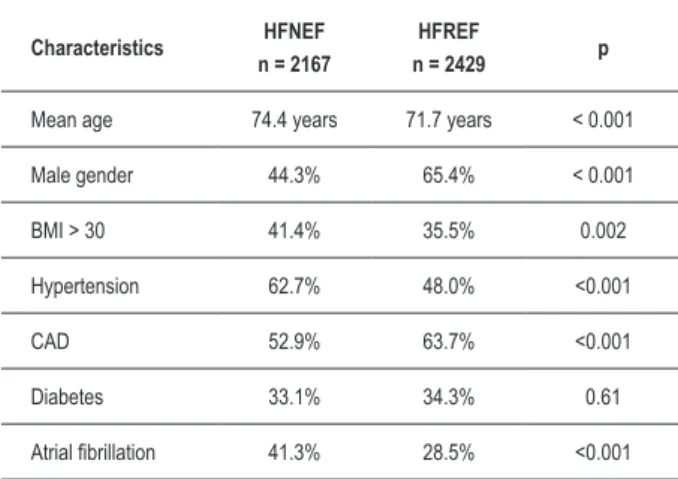
It is important to point out that the demographics and comorbidities of patients with HF vary according to EF (Table 1)8. When patients with HFNEF are compared with
those with HFREF, we can observe the first ones are older, more obese and most of them are females. Patients with HFNEF have a history of hypertension, diabetes and atrial fibrilation8.
Key Words
Table 1 – Epidemiology of heart failure stratiied by EF
Characteristics HFNEF n = 2167
HFREF
n = 2429 p
Mean age 74.4 years 71.7 years < 0.001
Male gender 44.3% 65.4% < 0.001
BMI > 30 41.4% 35.5% 0.002
Hypertension 62.7% 48.0% <0.001
CAD 52.9% 63.7% <0.001
Diabetes 33.1% 34.3% 0.61
Atrial ibrillation 41.3% 28.5% <0.001
BMI – Body Mass Index; CAD – Coronary Artery Disease; EF – ejection fraction; Adapted from Owan et al8
Key elements of the pathophysiology of
cardiac dysfunction
The normal diastolic function allows the heart to have adequate filling both at rest and during exercise, without the occurrence of increased diastolic pressures. However, changes in cardiac relaxation, the presence of myocardial hypertrophy, and remodeling are key defects that change the ventricular stiffness and filling pressures, thus leading to exercise intolerance, which would be the first symptom of HFNEF and the first determinant of reduced quality of life14.
In the heart with diastolic dysfunction, EF remains normal for quite a while. Maintenance of the cardiac performance is due to a compensatory period composed of two phases – the systolic activation phase, and the systolic dysfunction phase of the muscular pump1.
The systolic activation phase (Figure 1) is characterized by increased pressure, volume and ventricular flow, which is reversible and reflects the activation of all the adaptive mechanisms of the heart as a muscular and hemodynamic pump1.
If cardiac stress is maintained, the systolic activation may progress to the systolic dysfunction phase (Figure 2)1.
Therefore, the complete understanding of the systolic dysfunction of the muscular pump in the presence of a preserved performance of the hemodynamic pump, that is, normal EF, would undoubtedly be a key pathway to the understanding of the initial stage of the HF syndrome, which we call HFNEF1,4.
Systolic Activation - Early relaxation delay
V P
EDV
ESV
Modulation of duration of systole
Modulation of relaxation velocity - secondary to changes in duration of systole - sensitive to heart rate, neurohormones, load changes - Heterometric autoregulation - pressure or volume, hypertrophy I - Homeometric autoregulation - neurohormones, heart rate - Endothelial autoregulation - NO, BNP, cytokines
time
Figure 1 – Early relaxation delay (or prolonged contraction is – together with
peak and increased contraction velocity – the typical characteristic of the Systolic Activation Phase of the muscular heart pump. Changes in relaxation velocity are merely an effect secondary to the delayed modulation of systole. Usually early relaxation delay does not lead to increased end-diastolic pressure/volume. P – LV pressure; V – LV volume; EDV – end diastolic volume; ESV – end systolic volume; NO – nitric oxide; BNP – B-type natriuretic peptide (reproduction authorized by Dirk L. Brutsaert 1).
Although patients with HFNEF have a better prognosis than those with HFREF, they present significant morbidity and mortality, and the prognosis after hospitalization for HF is poor7,8,10, with a mortality rate of approximately 20% in one
year9. Although strategies based on recent evidences for the
treatment of HF have favorably modified the outcomes for patients with HFREF, studies have shown increased prevalence rates of HFNEF without change in mortality over the past 20 years8.
HFNEF and HFREF – a single syndrome or not?
There is no consensus as to whether HF should be considered a single syndrome or whether HFNEF and HFREF are two different clinical forms11.
If HF were characterized as a single syndrome12, it would
be defined by a progressive decline in the systolic performance that can be better evaluated by the analysis of velocities or measurements involving the long axis shortening using TDI than by using the EF measurement alone13.
The theory that HF is not a single syndrome but rather two diseases is supported by structural, functional and molecular changes associated with the diastolic function as well as by clinical studies with pharmacological intervention showing that patients with HFNEF do not have the same response as patients with HFREF, thus suggesting the existence of different pathophysiological mechanisms11.
We emphasize that it is totally artificial to split the two phases of the cardiac cycle (systole and diastole), but some authors11 have argued that in HFNEF the systolic function is
completely normal, and that this clinical condition is due to DD alone. Hence, HFNEF and HFREF should be considered different clinical entities11. These studies are based on global
measurements derived from the LV volume pressure curve that do not take into consideration regional changes of the long axis function4, which are compensated by an increase in
Abnormal relaxation
Abnormal relaxation (DD) is a functional alteration of the heart and one of the three mandatory criteria for the diagnosis of HFNEF, and should not be mistaken for or used as synonymous with HFNEF15.
The best way to demonstrate that patients with HFNEF have abnormal relaxation is to show whether the end-diastolic pressure/volume ratio (EDPVR) is higher when compared to its normal value. Certain conditions such as hypertrophy (hypertensive heart disease, hypertrophic cardiomyopathy and/or aortic stenosis) and myocardial ischemia affect relaxation making it slower and incomplete, thus leading to increased EDV15. The P/EDV-R allows for the demonstration
that not only is the intraventricular diastolic pressure elevated but also that this elevation is seen in ventricles presenting decreased filling volume11.
Demonstration of increased EDPVR is important because in the absence of this alteration, the EDV may be increased because of an increase in preload, such as can be observed in patients with aortic and mitral regurgitation with no contractility impairment and no significant change in the relaxation properties, andbecause a surgical correction of the alteration also corrects the overload16.
Cardiac hypertrophy
Ventricular hypertrophy is considered an adaptive mechanism of the heart in face of an increased load. Through intracellular mechanisms this overload may elicit different
responses that can or not be associated with functional myocardial impairment17.
The concept of hypertrophy is based on the identification of increased heart weight which is mainly determined by the increase in cardiomyocyte size.17 We should not forget
that cardiac muscle cells comprise the least percentage of all myocardial cells; however, given that they are the largest cells, variation in their size will determine a significant impact on the final heart weight17.
The hypertrophic response may be triggered by natural overload mechanisms such as those determined by growth, pregnancy, and those induced by physical activity, or also by pathological mechanisms such as hypertension, heart valve stenosis and regurgitation, cardiomyopathy, and myocardial infarction.
Pathological hypertrophy is accompanied by loss of contractility. The analysis of the fibers of the LV long axis on TDI shows decreased contraction in hypertrophic hearts4 due
to hypertension in comparison with normal hearts or athletes’ hearts with physiological hypertrophy17.
Ventricular remodeling
The main pathophysiological difference between HFNEF and HFREF is the increased ventricular volume and the change in the ventricular shape because of the remodeling process2.
Myocardial infarction is a powerful stimulus to the remodeling process, leading to enlargement of the LV and reduction of EF, whereas in the hypertensive heart disease the remodeling is a slow process, with LV hypertrophy leading to LV systolic and diastolic dysfunction, in which the compensatory increase in radial contraction tends to normalize the EF. However, in late stages, remodeling will occur with LV volume increase and the patient will go from HFNEF to HFREF1.
Currently, the use of medications that act on remodeling have proven to be efficient in the treatment of HF, and signs of reverse remodeling are a powerful predictor of clinical improvement2.
Diagnosing HFNEF in the clinical practice
The first step in the assessment of HF is to establish its diagnosis by the presence of signs and symptoms and, when available, with BNP determination. Next, a heart imaging method should be used to objectively assess LV function and determine the main etiology and its mechanisms (HFNEF, HFREF, pericardial diseases and heart valve diseases). The third step is to determine whether remodeling is already present (increased ventricular volume), and finally, to look for the presence of additional deleterious factors such as dyssynchrony, arrhythmias, and metabolic and electrolyte changes2.
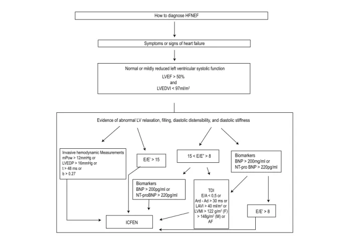
The ESC guideline5 presents an algorithm model (Figures 3
and 4) on how to diagnose and how to exclude HFNEF using the new concepts of cardiac function and TDI measurements. It establishes three mandatory stages to diagnose HFNEF, which are: presence of signs or symptoms for the diagnosis of HF, presence of greater than 50% EF, and evidences of diastolic dysfunction (relaxation, filling and stiffness)5.
Figure 2 – The early systolic dysfunction stage is characterized by worsened
relaxation, which is slow or incomplete, with progressive loss of capacity to modulate the early relaxation. Worsened relaxation may result in increased end diastolic volume/pressure. The grey vertical bar right before peak P and peak ESV shows the transition between intracellular contraction and the relaxation process. P – LV pressure; V – LV volume; EDV – end diastolic volume; EDV – end diastolic volume (reproduction authorized by Dirk L. Brutsaert 1).
Systolic dysfunction - Relaxation worsening
V P
EDV
ESV
Etiology
Mechanisms
Failure of the triple control
- inadequate load - inadequate inactivation
- load non-uniformities and inadequate inactivation Stages II and III hypertrophy
Mild ischemia
Signs and symptoms of HF
The signs and symptoms of HF include peripheral edema, hepatomegaly, lung crepitations, pulmonary edema, fatigue and breathlessness. Breathlessness is the most common symptom and also leads to a greater difficulty in the differential diagnosis5. This difficulty is more frequently seen in elderly and
obese patients, who represent the largest proportion of patients with HFNEF7. Unlike hospitalized patients who are admitted
with a classical presentation of HF which usually leaves no doubt as to the diagnosis, outpatients frequently complain of breathlessness with no detectable signs of congestion, which leads to the need for tests to confirm the diagnosis5.
Normal or mildly abnormal systolic left ventricular function
The choice of a cut-off point to the EF value to discriminate HFNEF from HFREF remains arbitrary5. The ESC guideline
established the value of EF≥50% as necessary for the
assessment according to recent recommendations of the American Society of Echocardiography and of the European Association of Echocardiography5.
Criteria for the presence of normal EF need to be implemented with measures of ventricular volume. Thus EDV, which must be indexed to body surface, should not exceed 97 ml/m2 for the diagnosis of HFNEF5.
Evidences of abnormal LV relaxation
The first question is whether it is necessary to evaluate relaxation changes in all patients with suspected HFNEF5.
Considering this possibility, Zile et al18 tested the hypothesis
that measurements of diastolic function are not always necessary to establish the diagnosis of HFNEF. The authors evaluated patients with HF who had EF >50% and evidences of concentric remodeling and demonstrated that 92% of these patients had high end-diastolic pressure and all had at least one abnormal relaxation index, abnormal filling or stiffness. In this group of patients, the DD parameters obtained did not add data for the diagnosis, but only confirmed DD18. We should keep in mind
that this study evaluated patients with history of established HF and, therefore, these data cannot be extrapolated to patients presenting only symptoms of breathlessness19.
An important finding of this study is that the evidence of concentric remodeling has implications for the diagnosis of HFNEF and is a potential substitute for the characterization of diastolic dysfunction18,20. The study shows that a LV wall
mass index > 122g/m2 for women and > 149 g/m2 for men
is sufficient evidence for the diagnosis of HFNEF when TDI is not conclusive or when plasma BNP is elevated18.
Assessment of the left ventricular function using TDI
TDI has a fundamental role in the assessment of this process since the clinical presentation of HFNEF is indistinguishable from that of HFREF and the EF measurement is certainly not relevant. Thus, assessments of systolic and diastolic dysfunction using measurements of the long axis and strain velocity in early diastole (E’) with TDI are more important5,14,21.
The development of new TDI techniques13 has provided
greater accuracy in the assessment of the ventricular function. In a recent study, Wang et al20 showed that the measurement of
strain velocity in early diastole (E’) is a strong predictor of mortality in comparison with clinical data and standard echocardiography. This measurement is easy to be taken and adds significant value to the clinical management of patients with HF20,22.
E’ has proven to be a strong predictor of the prognosis of HF, since this measurement reflects both the LV systolic and diastolic function. Additionally, the subendocardial fibers, which are responsible for contraction of the long axis, may be more susceptible to the effect of fibrosis, hypertrophy and ischemia due to their position, and this would explain why E’ is a good marker of the disease5.
Another frequently used echocardiographic parameter is the measurement of the early mitral valve flow velocity (E), which can be obtained either at the septal or at the lateral side of the mitral annulus5.
TDI has, therefore, permited the quantitative assessment of the LV systolic and diastolic function12. Recent studies22-24
investigated the prognostic role of TDI in several heart diseases such as HF, acute myocardial infarction and hypertension.
E/E’ ratio
The E/E’ ratio correlates closely with LV filling pressures. E depends on the left atrial pressure, LV relaxation, and age, whereas E’ depends mostly on the LV relaxation and age13.
Therefore, in the E/E’ ratio, effects of LV relaxation and age are eliminated and the ratio becomes a measurement of the left atrial pressure or LV filling pressure13.The E/E’ ratio can
thus be conceptualized as the amount of blood entering the LV during the fast filling phase, where E represents the gradient necessary to make this blood enter the LV. Thus, an increase in E/E’ means a high gradient to a low shift in volume13.
When E/E’ is higher than 15, LV filling pressures are elevated and when the ratio is lower than 8 these pressures are low5.
The E/E’ ratio is therefore considered diagnostic evidence of the presence of HFNEF if greater than 15, and evidence of absence of HFNEF if lower than 85,25. E/E’ values between 8 and
15 are considered suggestive, but not diagnostic and needs to be evaluated with other non-invasive data to confirm HFNEF5.
Left atrial volume index
Increased left ventricular diastolic pressures lead to left atrial remodeling with increased LA volume. Therefore, the left atrial volume index (LAV-I) may be seen as a morphologic expression of the left ventricular diastolic dysfunction21.
Increased LAV-I (>29 mL/m2) would be a load-independent
marker of LV filling pressure in patients with suspected HFNEF14. LAV-I could then be considered the barometer of
the heart21.
When E/E’ is not conclusive (15 < E/E’ > 8) for the diagnosis of HFNEF, the presence of LAV-I > 40 ml/m2 provides sufficient
evidence to confirm the diagnosis5,25. Similarly, a LAV-I <
26 ml/m2 is proposed as a prerequisite for the exclusion of
HFNEF5.
Natriuretic peptides
results in natriuresis, vasodilatation, and improved ventricular relaxation21. In patients with HFNEF, BNP values correlate
with indices that evaluate the early and late LV diastolic relaxation. High values have been observed in patients with late or abnormal relaxation1.
Since BNP levels may be influenced by different conditions (sepsis, liver failure, kidney failure, COPD, obesity), high BNP values do not provide sufficient evidence for the diagnosis of diastolic dysfunction, requiring additional tests5. For the
diagnosis of HFNEF, a high positive predictive value was determined when the BNP cut-off point was chosen (200 pg/ml). For the exclusion of HFNEF, a high negative predictive value was determined for the choice of the BNP cut-off point (<100 pg/ml)5.
Therefore, natriuretic peptides are mainly recommended for exclusion, and not for diagnosis, of HFNEF. Since BNP alone does not provide evidence for the diagnosis of HFNEF, it should always be used with other non-invasive tests5,25.
How to conirm and exclude HFNEF
Diagnosing HFNEF (Figure 3) is as important as excluding this diagnosis in patients with breathlessness and no signs of
Figure 3 – How to diagnose HFNEF – lowchart; mPCW – mean pulmonary capillary wedge pressure; t – time constant of LV relaxation; b – constant of LV chamber
stiffness; E – early mitral valve low velocity; E’ – Early TD lengthening velocity; NT-proBNP – N-terminal proBNP; E/A – ratio of early to late mitral valve low velocity; TDI – tissue Doppler imaging; LAVI – left atrial volume index; Avd – duration of reverse pulmonary vein atrial systole low; Ad – durarion of mitral valve atrial wave low; European Heart Journal– How to diagnose diasolic heart failure: a consensus statement on the diagnosis of heart failure with normal left ventricular ejection fraction by the Heart Failure and Echocardiography Associations of the European Society of Cardiology (5)
Evidence of abnormal LV relaxation, filling, diastolic distensibility, and diastolic stiffness
E/E’ > 15
ICFEN Biomarkers BNP > 200pg/ml or NT-proBNP > 220pg/ml
15 < E/E” > 8 Biomarkers
BNP > 200mg/ml or NT-pro BNP > 220pg/ml
TDI E/A < 0,5 or Ard - Ad > 30 ms or LAVI > 40 ml/m2 or LVMI > 122 g/m2 (F) > 148g/m2 (M) or
AF
E/E’ > 8 How to diagnose HFNEF
Symptoms or signs of heart failure
Normal or mildly reduced left ventricular systolic function
LVEF > 50% and LVEDVI < 97ml/m2
Invasive hemodynamic Measurements mPow > 12mmHg or
LVEDP > 16mmHg or t > 48 ms or b > 0.27
congestion (Figure 4), since the differential diagnosis of HFNEF is very difficult, especially in these patients5.
We can exclude HFNEF in patients with breathlessness who present lower than 100 pg/ml BNP values because of its high negative predictive value5,25.
If echocardiography rules out the presence of heart valve or pericardial disease, if the EF is higher than 50% and the EDV index is lower than 76 ml/m2, if there is no atrial
fibrillation, atrial dilatation or left ventricular hypertrophy, and if the E/E’ ratio is lower than 8, the diagnosis of HFNEF can be ruled out5.
Conclusion
The ECS guideline which established criteria for diagnostic confirmation and exclusion of HFNEF should be increasingly more frequently used by cardiologists and echocardiographers in the assessment of patients with suspected HFNEF.
Figure 4 – How to exclude HFNEF; E – early mitral valve low velocity; E’ – early TD lengthening velocity; BNP – N-terminal proBNP; LVEF – left ventricular ejection
fraction; LAVI – left atrial volume index; EDVI – end diastolic volume index; TDI – tissue Doppler imaging; S – myocardial of mitral annulus shortening velocity; AF – atrial ibrillation; HFREF – heart failure with reduced ejection fraction; HFREF – heart failure with normal ejection fraction; CAD – coronary artery disease; European Heart Journal – How to diagnose diastolic heart failure: a consensus statement on the diagnosis of heart failure with normal left ventricular ejection fraction by the Failure and Echocardiography Associations of the European Society of Cardiology (5)
How to exclude HFNEF
Breathlessness without signs of fluid overload
BNP > 100 pg/ml
Evidence of pulmonary disease
Consider pulmonary disease
Consider valvular or pericardial disease
Consider HFREF
Consider high output state
Consider HFNEF
Consider CAD TDI
LVEV > 50%
LVEDVI < 97 ml/m2
LAVI < 29 ml/m2 - and no AF
E/E’ < 8 or S > 6.5 cm/s
No HFNEF Evidence of valvular or pericardial disease
yes
yes
yes yes
yes
yes
no
no
no
no no
Although uncommonly used in the clinical practice, TDI is currently the best tool for the diagnosis of HFNEF, and its utilization will gradually replace the conventional transmitral echocardiographic criteria in the assessment of diastolic dysfunction.
Acknowledgements
We would like to thank Professor Dirk L. Brutsaert, from Middelheim Hospital, University of Antwerp, Belgium, for the information provided to the authors, and also for authorizing the use of the figures in this review.
Potential Conflict of Interest
No potential conflict of interest relevant to this article was reported.
Sources of Funding
There were no external funding sources for this study.
Study Association
This article is part of the thesis of Master submitted by Antônio Lagoeiro e Evandro Tinoco Mesquita, from Universidade Federal Fluminense.
1. Brutsaert DL, Cardiac dysfunction in heart failure: the cardiologist’s love affair with time. Prog Cardiovasc Dis. 2006; 49: 157-81.
2. Sanderson JE. Heart failure with a normal ejection fraction Heart. 2007; 93: 155-8.
3. Zile MR, Baicu CF, Gaasch WH. Diastolic heart failure—abnormalities in active relaxation and passive stiffness of the left ventricle. N Engl J Med. 2004; 350: 1953-9.
4. Yip G, Wang M, Zhang Y, Fung JWH, Ho PY, Sanderson JE. Left ventricular long axis function in diastolic heart failure is reduced in both diastole and systole:
References
time for a redefinition?Heart. 2002; 87: 121-5.
5. Paulus WJ, Tschöpe C, Sanderson JE, Rusconi C, Flachskampf FA, Rademakers FE, et al. How to diagnose diastolic heart failure: a consensus statement on the diagnosis of heart failure with normal left ventricular ejection fraction by the Heart Failure and Echocardiography Associations of the European Society of Cardiology. Eur Heart J. 2007; 28: 2539-50.
6. Brutsaert DL; De Keulenaer GW. Diastolic heart failure: a myth. Curr Opin Cardiol. 2006; 21 (3): 240-8.
heart failure in subjects with normal versus reduced left ventricular ejection fraction: prevalence and mortality in a population-based cohort. J Am Coll Cardiol. 1999; 33: 1948-55.
8. Owan TE, Hodge DO, Herges RM, Jacobsen SJ, Roger VL, Redfield MM. Trends in prevalence and outcome of heart failure with preserved ejection fraction N Engl J Med. 2006; 355: 251-9.
9. Moutinho MAE, Colucci FA, Alcoforado V, Tavares LR, Rachid MBF, et al. Insuficiência cardíaca com fração de ejeção preservada e com disfunção sistólica na comunidade. Arq Bras Cardiol. 2008; 90 (2): 145-50.
10. Mesquita ET, Sócrates J, Rassi S, Villacorta H, Mady C. Insuficiência cardíaca com função sistólica preservada. Arq Bras Cardiol. 2004; 82: 494-500.
11. Baicu CF, Zile MR, Aurigemma GP, Gaasch WH. Left ventricular systolic performance, function, and contractility in patients with diastolic heart failure. Circulation. 2005; 111: 2306-12.
12. De Keulenaer GW, Brutsaert DL. Systolic and diastolic heart failure: different phenotypes of the same disease? Eur J Heart Fail. 2007; 9: 136-43.
13. Yu CM, Sanderson JE, Marwick TH, Oh JK. Tissue Doppler imaging: a new prognosticator for cardiovascular diseases. J Am Coll Cardiol. 2007; 49: 1903-14.
14. Westermann D, Kasner M, Steendijk P, Spillmann F, Riad A, Weitmann K, et al. Role of left ventricular stiffness in heart failure with normal ejection fraction. Circulation. 2008; 117 (16): 2051-60.2008; 117 (16): 2051-60.
15. Boo JG. Entendiendo la insuficiencia cardíaca. Arch Cardiol Mex. 2006; 76 (4): 431-47.
16. Burkhoff D, Maurer MS, Packer M. Heart failure with a normal ejection fraction: is it really a disorder of diastolic function? Circulation. 2003; 107:Circulation. 2003; 107: 656-8.
17. Escudero EM, Pinilla AO. Paradigmas y paradojas de la hipertrofia ventricular izquierda: desde el laboratorio de investigación a la consulta clínica. ArchArch
Cardiol Mex. 2007; 77 (3): 237-48.
18. Zile MR, Gaasch WH, Carroll JD, Feldman MD, Aurigemma GP, Schaer GL, et al. Heart failure with a normal ejection fraction: is measurement of diastolic function necessary to make the diagnosis of diastolic heart failure? Circulation. 2001; 104: 779-82.
19. Kawaguchi M, Hay I, Fetics B, Kass DA. Combined ventricular systolic and arterial stiffening in patients with heart failure and preserved ejection fraction. Circulation. 2003; 107: 714-20.
20. Wang M, Yip GW, Wang AY, Zhang Y, Ho PY, Tse MK, et al. Peak early diastolic mitral annulus velocity by tissue Doppler imaging adds independent and incremental prognostic value. J Am Coll Cardiol. 2003; 41: 820-6.
21. Lester SJ, Tajik AJ, Nishimura RA, Oh JK, Khandheria BK, Seward JB. Unlocking the mysteries of diastolic function deciphering the rosetta stone 10 years later. J Am Coll Cardiol. 2008; 51: 679-89.
22. Yamamoto T, Oki T, Yamada H, Tanaka H, Ishimoto T, Wakatsuki T, et al. Prognostic value of the atrial systolic mitral annular motion velocity in patients with left ventricular systolic dysfunction. J Am Soc Echocardiogr. 2003; 16: 333-9.
23. Richartz BM, Werner GS, Ferrari M, Figulla HR. Comparison of left ventricular systolic and diastolic function in patients with idiopathic dilated cardiomyopathy and mild heart failure versus those with severe heart failure. Am J Cardiol. 2002; 90: 390-4.
24. Wang M, Yip GW, Wang AY, Zhang Y, Ho PY, Tse MK, et al. Tissue Doppler imaging provides incremental prognostic value in patients with systemic hypertension and left ventricular hypertrophy. J Hypertens. 2005; 23: 183-91.