Case Report
Key Words
Myocardial infarction; thoracic injuries; heart injuries; wounds, non penetrating.
We report the case of a 29-year-old man, victim of a car accident, who suffered a severe blunt chest trauma, with evolving congestive heart failure. He had previously had a good overall health status, with no symptoms of cardiovascular disease. At the initial assessment, the electrocardiogram showed Q waves in the precordial leads and the echocardiogram disclosed severe left ventricular dysfunction. Coronary angiogram showed a proximal left anterior descending coronary artery lesion, with anterior wall akinesis on contrast-enhanced ventriculography. A Thallium-201 single photon emission computed tomography (SPECT) showed no viability. He remained on medical treatment with good evolution.
Myocardial Infarction Caused by Coronary Artery Injury after a Blunt
Chest Trauma
Márcio Silva Miguel Lima
1, Jeane Mike Tsutsui
1, Victor Sarli Issa
2Laboratório de Ecocardiografia - Instituto do Coração, Faculdade de Medicina da Universidade de São Paulo1; Unidade de Transplante Cardíaco e
Insuficiência Cardíaca - Instituto do Coração, Faculdade de Medicina da Universidade de São Paulo2, São Paulo - Brazil
Mailing address: Márcio Silva Miguel Lima•
Departamento de Ecocardiografia, Instituto do Coração do Hospital das Clínicas (InCor HCFMUSP),
Av. Enéias de Carvalho Aguiar 44, Cerqueira César, 05.403-000, São Paulo, SP - Brazil
E-mail: [email protected]
Manuscript received February 20, 2008; revised manuscript received March 26, 2008; accepted April 02, 2008.
Case report
A 29-year-old man from the state of Minas Gerais was referred to the Heart Failure Clinic of a tertiary university hospital in September 2005, because of shortness of breath during the previous six months. At that time, the patient received the diagnosis of congestive heart failure.
This patient had been in his usual state of healthuntil February 2005, when he was victim of a car accident, suffering blunt chest trauma, with rib fractures and a pneumothorax. He was hospitalized for 20 days and treated with pleural drainage and analgesics due to chest pain. No cardiovascular abnormalities were observed during that period. He resumed his regular activities after hospital discharge.
One month later, he observed progressive shortness of breath that prevented him from performing ordinary activities; lower limbs edema and abdominal enlargement ensued. He did not have chest pain at that time.
This patient had been in previous good health, with no symptoms suggestive of cardiovascular disease and had an excellent exercise tolerance so far. He was a current cigarette smoker of a package per day, with moderate alcohol consumption, and had no history of hypertension, diabetes, dyslipidemia or illicit drug use.His family history was negative for coronary artery disease, cardiomyopathy or thrombotic disease.
On physical examination, he was in good overall condition, eupneic, with normal-colored mucosa. His body weight was 81 Kg; height was 1.73 m, heart rate was 120 beats per minute, blood pressure was 130/70 mmHg, and the respiratory rate was 22 breaths per minute. The examination of the neck revealed normal carotid pulses with no bruits. The chest examination disclosed a normal apical impulse, normal heart sounds, and reduced breath sounds over the middle-lower portion of the right hemithorax, consistent with pleural effusion. Peripheral pulses were symmetric with normal amplitudes, and there was no peripheral edema.
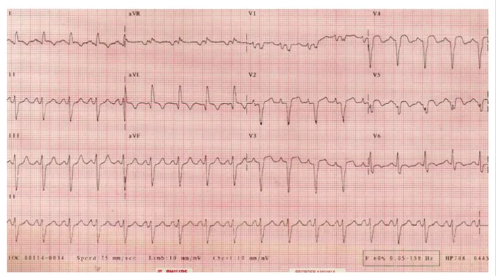
An electrocardiogram showed ST-segment elevation in leads V2 to V5, with Q waves from V1 to V6 and in DI and aVL, and signs of left atrial and ventricular enlargement (Fig 1). The chest roentgenogram showed mildly enlarged cardiac silhouette and evidence of pulmonary venous congestion. Serologic test for detection of Trypanosoma cruzi infection by complement fixation test (Machado-Guerreiro test) was negative. The echocardiogram disclosed a moderately dilated left ventricle (LVDed 6,0cm, LVDsys 5,0cm) with normal wall thickness (septum 0.7cm, posterior wall 0.8 cm) and severe global hypokinesis (estimated ejection fraction of 0.35). The left atrium was mildly dilated (4,5cm). The right ventricle was also dilated and hypokinetic, with a systolic pressure in the pulmonary artery estimated as 22 mmHg.
The patient was submitted to coronary angiography. There was a proximal left anterior descending coronary artery lesion with estimated 99% obstruction, estimated TIMI 2 flow, with the aspect of recanalization. The left-main artery, the circumflex and the right coronary artery were completely normal (Fig 2). A ventriculography was performed and disclosed diffuse akinesis, more severe in the anterior wall. A Thallium-201 single photon emissioncomputed tomography (SPECT), using the rest reinjection technique, was performed and showed no viability of the anterior wall, septum and apex. The gated blood pool imaging estimated a left ventricular ejection fraction of 0.22.
Case Report
Lima et al Coronary artery injury after a blunt chest trauma
Arq Bras Cardiol 2009;93(1):e1-e3
Figure. 1 -- Eletrocardiogram.
Figure 2 -Coronary angiograms showing a proximal left anterior descending coronary artery lesion with estimated 99% obstruction (Panel A and B) and a normal right coronary artery (Panel C).
Case Report
Lima et al
Coronary artery injury after a blunt chest trauma
Arq Bras Cardiol 2009;93(1):e1-e3
He remained on medical treatment with carvedilol, enalapril, aspirin, and furosemide, with doses titrated until the therapeutic goals recommended for congestive heart failure were attained. His symptoms gradually improved.
Discussion
Motor vehicle accidents are directly related to blunt chest trauma. Injuries to the heart and great vessels are the common presentation1. Coronary artery injury due to
blunt chest trauma occurs in about 2% of the cases, carrying a high risk of morbidity and mortality2. Such injuries are
often associated with sternal and rib fractures3. Myocardial
infarction following a blunt chest trauma is extremely uncommon with just a few cases having been reported to date2,4-8. Injury to the coronary vessel can lead to myocardial
infarction either immediately or after some hours. Although the pathogenesis is not clearly understood, some of the mechanisms proposed are intimal tear, subintimal hematoma, coronary artery wall dissection, thrombus formation, focal spasm, vessel rupture and coronary embolism2,9. In this case, there was clear evidence of the
causal relation of the chest trauma, the coronary lesion due to the trauma and the evolving heart failure. Despite being a current cigarette smoker, this young patient had no symptoms related to ischemic heart disease at exercise practice and no sign of atherosclerosis in the other normal arteries (right coronary artery and circumflex artery).
It is often difficult to diagnose cardiac contusion with myocardial infarction in the emergency department after a thoracic trauma, as the pain may be attributed to the chest
trauma itself and to thoracic wall lesions. Electrocardiography, measurements of creatine phosphokinase-MB isoenzyme levels and echocardiography may be helpful and should be carried out for every patient with suspected cardiac injury, as these tests can provide information about the existence and extension of the cardiac damage. Although the condition is difficult to diagnose, it is crucial to attain an early identification and treatment. Reperfusion therapies present clear benefits to treat a complete vessel occlusion after a coronary injury. Modalities of treatment are the percutaneous coronary intervention (PCI) and coronary artery bypass graft (CABG). Because the cardiac infarction might often be the result of an intimal tear or dissection, thrombolytic therapy might worsen the situation and acute PCI or CABG must be considered preferable10. Without
specific treatment, a coronary artery occlusion can lead to ventricular impairment and clinical heart failure, and a worse prognosis4,6.
Potential Conflict of Interest
No potential conflict of interest relevant to this article was reported.
Sources of Funding
There were no external funding sources for this study.
Study Association
This study is not associated with any post-graduation program.
References
1. Pêtre R, Chilcott M. Blunt trauma to the heart and great vessels. N Engl J Med. 1997; 336: 626-32.
2. Mattox KL, Estrera AL, Wall MJ Jr. Traumatic heart disease. In: Zipes DP, Libby P, Bonow RO, Braunwald E (eds). Braunwald’s heart disease: a textbook of cardiovascular medicine. 7th ed. Philadelphia: Elsevier Saunders; 2005. p. 1781-8.
3. Lai CH, Ma T, Chang TC, Chang MH, Chou P, Jong GP. A case of blunt chest trauma induced acute myocardial infarction involving two vessels. Int Heart J. 2006; 47: 639-43.
4. Wei T, Wang L, Chen L, Wang C, Zeng C. Acute myocardial infarction and congestive heart failure following a blunt chest trauma. Heart Vessels. 2002; 17: 77-9.
5. Banzo I, Montero A, Uriarte I, Vallina N, Hernández A, Guede C, et al. Coronary artery occlusion and myocardial infarction: a seldom encountered complication of blunt chest trauma. Clin Nucl Med. 1999;
24: 94-6.
6. Tsai TN, Yang SP, Tsao TP, Huang KA, Cheng SM. Delayed diagnosis of post-traumatic acute myocardial infarction complicated by congestive heart failure. J Emerg Med. 2005; 29: 429-31.
7. Fang BR, Li CT. Acute myocardial infarction following blunt chest trauma. Eur Heart J. 1994; 15: 705-7.
8. Goktekin O, Unalir A, Gorenek B, Kudaiberdieva G, Cavusoglu Y, Melek M, et al. Traumatic total occlusion of left main coronary artery caused by blunt chest trauma. J Invasive Cardiol. 2002; 14: 463-5.
9. Neiman J, Hui WKK. Posteromedial papillary muscle rupture as a result of right coronary occlusion after blunt chest trauma treated medically: a case report and review. Am Heart J. 1992; 123: 1694-9.
10. Christensen MD, Nielsen PE, Sleight P. Prior blunt chest trauma may be a cause of single vessel coronary disease; hypothesis and review. Int J Cardiol. 2006; 108: 1-5.