Initial circulatory response to active standing
in Parkinson
’
s disease without typical
orthostatic hypotension
Resposta circulatória inicial após ortostatismo ativo na doença de Parkinson sem
hipotensão ortostática típica
Guillermo Delgado1, Bruno Estañol1, Mayela Rodríguez-Violante2, Jesús Antonio González-Hermosillo3, Óscar Infante-Vázquez4
ABSTRACT
While the circulatory response to orthostatic stress has been already evaluated in Parkinson’s disease patients without typical orthostatic hypotension (PD-TOH), there is an initial response to the upright position which is uniquely associated with active standing (AS). We sought to assess this response and to compare it to that seen in young healthy controls (YHC).Method:In 10 PD-TOH patients (8 males, 60±7 years, Hoehn and Yahr#3) the changes in systolic blood pressure (SBP) and heart rate that occur in the first 30 seconds (sec) of standing were examined. Both parameters were non-invasively and continuously monitored using the volume-clamp method by Peñáz and the Physiocal criteria by Wesseling. The choice of sample points was prompted by the results of previous studies. These sample points were compared to those of 10 YHC (8 males, 32±8 years).Results:The main finding of the present investigation was an increased time between the AS onset and SBP overshoot in PD-TOH group (24±4 vs. 19±3 sec; p,0.05).Conclusion:This delay might reflect a prolonged latency in the baroreflex-mediated vascular resistance response, but more studies are needed to confirm this preliminary hypothesis.
Keywords:Parkinson’s disease, autonomic nervous system, orthostatic hypotension, hemodynamics, cardiovascular physiological Phenomena.
RESUMO
Apesar da resposta circulatória ao estresse ortostático já foi estudada em pacientes com doença de Parkinson sem hipotensão ortostática típica (PD-TOH), não há uma resposta inicial que é exclusivamente associada com o ortostase ativa (AS). Portanto, buscou-se avaliar esta resposta e compará-la à observada em jovens saudáveis (YHC).Método:Em 10 PD-TOH pacientes (8 homens, 60±7 anos, Hoehn e Yahr#3) as mudanças na pressão arterial sistólica (PAS) e da frequência cardíaca que ocorrem nos primeiros 30 segundos (seg) de pé foram examinados. Ambos parâmetros foram monitorizados continuamente através do método Penõáz e os critérios de Wesseling. Os pontos de amostragem foram escolhidos com base em estudos anteriores. Estes pontos foram comparados com os de 10 YHC (32±8 anos).
Resultados:O principal achado deste estudo foi o aumento do tempo entre o início de AS e rebote sistólica no grupo PD-TOH (24±4 vs 19±3 seg, p,0,05). Conclusão: Este atraso pode refletir uma latência prolongada na resposta da resistência vascular mediado pelo barorreflexo, mas outros estudos são necessários para confirmar esta hipótese preliminar.
Palavras-chave:doença de Parkinson, sistema nervoso autônomo, hipotensão ortostática, hemodinâmica, fenômenos fisiológicos cardiovasculares.
There is an initial circulatory or hemodynamic response ( first 30 seconds) to the upright position which is solely associated with active standing (AS)1. This response has
been described in both young and elderly healthy subjects1-4.
When orthostatic hypotension occurs within 15 seconds (sec) of standing, it is termed initial orthostatic hypotension
1Laboratory of Clinical Neurophysiology, Department of Neurology and Psychiatry, National Institute of Medical Sciences and Nutrition Salvador Zubirán; 2Movement Disorders Clinic, National Institute of Neurology and Neurosurgery Manuel Velasco Suárez;
3Department of Electrocardiography and Electrophysiology, National Institute of Cardiology Ignacio Chávez; 4Department of Electromechanical Instrumentation, National Institute of Cardiology Ignacio Chávez.
Correspondence:Guillermo Delgado; Laboratory of Clinical Neurophysiology, Department of Neurology and Psychiatry, National Institute of Medical Sciences and Nutrition Salvador Zubirán (INNSZ); Vasco de Quiroga 15, Tlalpan; C.P. 14000; México D.F. - México; E-mail: [email protected], [email protected]
Conflict of interest:There is no conflict of interest to declare.
Received 01 April 2013; Received in final form 14 October 2013; Accepted 11 November 2013.
DOI:10.1590/0004-282X20130247
(IOH) and is attributed to a mismatch between cardiac out-put and peripheral vascular resistance (PVR)1. The aforesaid
mismatch is due to a reduction in PVR and moreover, this decrease is clearly associated with the depth of the blood pressure (BP) drop1. Symptoms of cerebral hypoperfusion
are likewise present during this drop1.
Orthostatic symptoms (OS) occur when cerebral per-fusion is sufficiently impaired and, in turn, hypoperper-fusion develops when cerebrovascular autoregulation (CVA) fails to cope with the BP reduction5,6. This is why, in typical
orthostatic hypotension (TOH), these symptoms takes place more readily in patients with severe cerebrovascular autore-gulatory failure5. Nevertheless, autonomic dysfunction in
Parkinson’s disease (PD) does not seem to impair the CVA nor its response to orthostatic stress6,7. These CVA
differ-ences could account for the reduced reporting of OS in PD6.
Autonomic dysfunction in PD is associated with a gener-alized sympathetic denervation8. Recently, it has been
reported that PD patients with TOH (PD+TOH) have a lower basal leg vascular resistance, which might suggest that sym-pathetic denervation is more pronounced in PD+TOH than in PD patients without TOH (PD-TOH)8. Yet, Oka et al.
docu-mented that latent cardiac and vasomotor sympathetic dys-function occurs in PD-TOH patients9.
While the hemodynamic response to orthostatic stress has been already evaluated in PD-TOH8, IOH can be
docu-mented only by continuous beat-to-beat BP monitoring dur-ing AS1. Consequently, in this study, we assessed
beat-to-beat the initial hemodynamic response to AS in PD-TOH and compared this response to that seen in young healthy controls (YHC).
METHOD
Study population
We enrolled 20 non-institutionalized patients with mild to moderate PD (16 males, mean age 60±7 years, mean dura-tion of disease 3.5±2 years, Hoehn and Yahr stage 2[1-3])10.
All of them fulfilled the UK Brain Bank Clinical Criteria for PD11. PD+TOH patients were excluded from the present
study; the foregoing condition was defined as a fall in BP of at least 20mmHg systolic (SBP) and 10mmHg diastolic (DBP) within 3 minutes (min) in the upright position12,13.
BP measurement was performed using standard sphygmo-manometry. These patients were all investigated after 12 hours off medication. We also included ten YHC (8 males, mean age 32±8 years) as control group, since the transient hypotension seen during AS does not increase with age1.
Their health status was determined by a thoughtful medical history and a directed physical examination. This study was
approved by the Ethical Committee of the National Institute of Medical Sciences and Nutrition and an informed written consent was obtained from all participants.
Protocol
All evaluations were performed in the morning. Participants in both groups were instructed to avoid alcohol, caffeinated beverages and over-the-counter medications after 22:00 on the night before the evaluation. Finger arterial pressure (FAP) was non-invasively and continuously (beat-to-beat) monitored using the volume-clamp method by Penõáz and the Physiocal (physiological calibration) criteria by Wesseling14. SBP, DBP and mean BP (MBP) were then
reconstructed from FAP; and heart rate (HR) was meanwhile computed as the inverse of the interbeat interval (Finometer1
PRO, Finapres Medical Systems, Amsterdam)14. Measurements were made at supine rest
before AS (5 min), during AS, and at the upright position after AS (5 min). Participants were not specifically instructed to stand up quickly. Hemodynamic parameters were extracted with BeatScope for Windows1
(v.1.1a, Finapres Medical Systems).
Data analysis and statistics
This investigation was focused on SBP and HR changes in the initial hemodynamic response to AS and was carried out utilizing BeatScope software. Supine values of SBP and HR were calculated as the corresponding arithmetic means of the 5 min supine rest, and afterwards SBP response was determined at time (t)=-10 sec, -5 sec, 0 sec, time at peak
SBPa (tSBPa), time at valley SBPb (tSBPb), time at SBP
over-shoot (tSBPos) and +30 sec. HR response was concurrently
measured at t=-10 sec, -5 sec, 0 sec, time at peak HRa
(tHRa), time at valley HRb(tHRb), time at peak HRc(tHRc), time
at valley HRd(tHRd) and +30 sec.t0was defined as the start of
AS. The selection of sample points was prompted by the results of previous studies2,3,15,16. Unless otherwise stated,
hemodynamic data are reported as absolute values and as changes between two different sample points (D). When possible, vasoconstrictor (VCI) and baroreflex sensitivity (BRI) indices, as advanced by Yamaguchi et al., were addi-tionally calculated16
. Baroreflex sensitivity (BRS) was also obtained using a slightly modified version of the method already employed by Imholz et al.15Statistical analysis was
performed with STATISTICA for Windows1
RESULTS
All data were visually inspected prior to analysis. Five patients were excluded either because of tremor-induced artifact (n=2) or asymptomatic TOH (n=3). Besides, the char-acteristic hemodynamic pattern seen during AS was almost completely lost in five patients1-3,17
, thus hampering attempts to locate the sample points and further comparison between groups. Data from these patients were hence eliminated from study analysis (n=5).
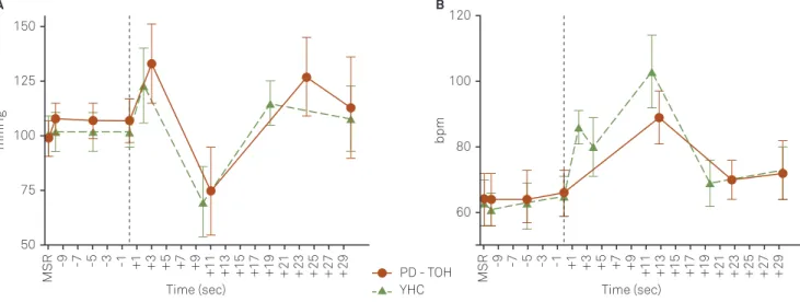
Initial hemodynamic response(Tables 1 and 2). In the ten remaining patients (8 males, mean age 60±7 years), basal SBP and HR were, respectively, 99±8 millimetres of mercury (mmHg) and 64±8 beats per min (bpm). On standing SBP increased suddenly from 107±10 (t0) to 133±18mmHg
(SBPa) and then decreased from that value to 75±20mmHg
(SBPb). An overshoot was evident in seven patients during
the recovery phase of SBP (SBPos, 127±18 mmHg). These
changes reached its maximum (SBPa) after 3±1 sec (tSBPa)
and its minimum (SBPb) after 11±2 sec (tSBPb). For its part,
in the seven aforementioned patients, SBPos was reached after 24±4 sec (tSBPos). Thereafter, at the end of the initial
hemodynamic response (t+30), SBP was 113±23mmHg
(Figure 1).
Standing was accompanied by an absence of a true HRa
in all patients and, as a consequence, also by an absence of HRb. The awaited transition from the HR surge to an
increscent further rise was likewise not precisely definable. In this manner, HR increased gradually from 66±7 (t0) to
89±8bpm (HRc) upon standing and subsequently dropped
from that value to 70±6 (HRd). These changes reached its
maximum (HRc) after 13±4 sec (tHRc) and its minimum
(HRd) after 23±5 sec (tHRd). HR was lastly 72±8bpm at t+30
(Figure 1).
Patients were slower to stand up than YHC (11.7 [8.8-14] vs. 4.9 [4.2-5] sec; p,0.001). SBPoswas evident in nine YHC
during the recovery phase of SBP (115±10mmHg). While its value in mmHg was not significantly different between groups, its timing (tSBPos) was delayed in the PD-TOH
group (24±4 vs. 19±3 sec; p,0.05). The remaining SBP parameters did not significantly differ from those found in YHC (Figures 1 and 2). HRa(or its corresponding
aforede-scribed transition) was present in nine YHC (86±5bpm) at 2±1 sec (tHRa); HRbwas afterwards identified in only seven
YHC (80±9bpm) at 4±1 sec (tHRb). HRc was smaller in the
PD-TOH group than in YHC (89±8 vs. 103±11bpm; p,0.05), howbeit its timing (tHRc) did not significantly differ
between groups. No significant differences were found between groups for the rest of the HR parameters (Figures 1 and 2).
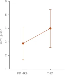
Baroreflex and vasoconstriction(Table 3). BRI and VCI were determined in seven patients and in almost all YHC (n=9). BRI was lower in PD-TOH than in YHC. VCI was also decreased, although not significantly (p=0.09), in the former group (Figure 3). BRS was measured in all the participants and did not significantly differ between groups.
BP abnormalities. Three patients and six YHC met the criteria for asymptomatic IOH1. In one patient, SBP recovery
phase was noticeably prolonged (28 sec) but nevertheless it does not fulfilled the TOH criteria, since BP after standing for 3 min was not altered1. This matter will be addressed
in detail in a subsequent communication.
Table 1.Initial HR response to active standing.
PD-TOH (n=10) YHC (n=10)
MHRSR(bpm) 64±8 63±7
HR-10 64±8 61±5
HR-5 64±9 63±6
HR0 66±7 65±6
HRa – 86±5†
HRb – 80±9‡
HRc 89±8* 103±11
HRd 70±6 69±7
HR+30 72±8 73±9
tHRa(sec) – 2±1†
tHRb – 4±1‡
tHRc 13±4 12±3
tHRd 23±5 20±2
All results are expressed as mean±standard deviation. PD-TOH: Parkinson’s disease without typical orthostatic hypotension; YHC: young healthy controls; MHRSR: mean heart rate (HR) during a 5-min supine rest;
bpm: Beats per minute; HR-10: HR at 10 seconds (sec) before active
standing (AS); HR-5: HR at 5 sec before AS; HR0: HR at the start of AS;
HRa: HR at first peak; HRb: HR at first valley; HRc: HR at second peak or
maximum; HRd: HR at second valley; HR+30: HR at 30 sec after AS;tHRa:
Time at HRa;tHRb: Time at HRb;tHRc: Time at HRc;tHRd: Time at HRd.
*p,0.05 PD-TOH vs. YHC.†n=9.‡n=7.
Table 2.Initial SBP response to active standing.
PD-TOH (n=10) YHC (n=10)
MSBPSR(mmHg) 99±8 100±9
SBP-10 108±7 102±9
SBP-5 107±8 102±9
SBP0 107±10 102±7
SBPa 133±18 123±17
SBPb 75±20 70±16
SBPOS 127±18† 115±104
SBP+30 113±23 108±15
DSBP0-SBPb(mmHg) 31±17 31±13
tSBPa(sec) 3±1 2±0.8
tSBPb 11±2 10±1
tSBPos 24±4†* 19±34 All results are expressed as mean±standard deviation. PD-TOH: Parkinson’s disease without typical orthostatic hypotension; YHC: Young healthy controls; MSBPSR: Mean systolic blood pressure (SBP) during a
5-min supine rest; mmHg: Millimetres of mercury; SBP-10: SBP at 10
seconds (sec) before active standing (AS); SBP-5: SBP at 5 sec before AS;
SBP0: SBP at the start of AS; SBPa: SBP at first peak; SBPb: SBP at valley;
SBPOS: SBP at overshoot; HR+30: SBP at 30 sec after AS;DSBP0-SBPb:
Absolute change from SBP0to SBPb;tSBPa: Time at SBPa;tSBPb: Time at
SBPb;tSBPos: Time at SBPos.
DISCUSSION
The main finding of the present investigation was an increased time (tSBPos) between the start of AS (t0) and the
overshoot (SBPos) in PD-TOH group. When compared
between groups, SBP (in mmHg) was not significantly differ-ent attSBPos; notwithstanding it could not be determined in
three patients. Peripheral sympathetic vasoconstriction, in part baroreflex-mediated, is responsible for the rebound of BP from SBPb to SBPos following AS1,18. Local reflexes are
involved as well in this PVR increment8,19,20. Except for the
venoarteriolar response (VAR) in those aged over 75 years, both central and local responses are independent of age in healthy subjects1,19,21. Thus, we suggest that an increased tSBPos may be caused either by an impaired VAR or by an
abnormal baroreflex response. Indeed, these mechanisms are not mutually exclusive22.
With respect to the first possibility, Andersen et al. found that, in PD patients without symptoms or signs of auto-nomic dysfunction, local reflexes did not differ from those
150
A 120
100
80
60 125
100
mmHg bpm
Time (sec) Time (sec)
SBP HR
MSR
-9 -7 -5 -3 -1 +1 +3 +5 +7 +9 +11 +13 +15 +17 +19 +21 +23 +25 +27 +29
75
50
150
B 120
100
80
60 125
100
mmHg bpm
MSR
-9 -7 -5 -3 -1 +1 +3 +5 +7 +9 +11 +13 +15 +17 +19 +21 +23 +25 +27 +29
75
50
Figure 1.Initial hemodynamic response in PD-TOH. The vertical dashed line indicatest0, i.e., the AS onset (A). Initial hemodynamic
response in YHC (B); mmHg: millimetres of mercury; bpm: beats per minute; MSR: mean value (SBP or HR) during a 5-min supine rest; sec: seconds; SBP: systolic blood pressure; HR: heart rate; PD-TOH: Parkinson’s disease without typical orthostatic hypotension; AS: active standing; YHC: young healthy controls.
A
mmHg
Time (sec)
MSR
-9 -7 -5 -3 -1 +1 +3 +5 +7 +9 +11 +13 +15 +17 +19 +21 +23 +25 +27 +29
50 75 100 125 150
bpm
B
Time (sec)
MSR
-9 -7 -5 -3 -1 +1 +3 +5 +7 +9 +11 +13 +15 +17 +19 +21 +23 +25 +27 +29
60 80 100 120
PD - TOH YHC
Figure 2.Comparison of the initial SBP response between PD-TOH (solid line) and YHC (dashed line). The vertical dashed line indicatest0, i.e., the AS onset (A). Comparison of the initial HR response between PD-TOH (solid line) and YHC (dashed line). Note
that the bimodal pattern of this response is entirely lost in PD-TOH (B); mmHg: millimetres of mercury; bpm: beats per minute; MSR: mean value (SBP or HR) during a 5-min supine rest; sec: seconds; PD-TOH: Parkinson’s disease without typical orthostatic hypotension; YHC: young healthy controls; SBP: systolic blood pressure; AS: active standing; HR: heart rate.
Table 3.Baroreflex and vasoconstriction.
PD-TOH (n=10) YHC (n=10)
VCI (mmHg/sec) 2.9+1.2† 4.0+1.44
BRI (bpm/mmHg) 0.6+0.5†* 2.2+1.04
BRS (msec/mmHg) 9.1 (5.4-10.8) 9.9 (8.5-16.6) All results are expressed as mean ± standard deviation or median (interquartile range). PD-TOH: Parkinson disease without typical ortho-static hypotension; YHC: Young healthy controls; VCI: Vasoconstrictor index; BRI: Baroreflex sensitivity index as calculated by Yamaguchi et al.14; BRS: Baroreflex sensitivity as computed by Imholz et al.13; mmHg: Millimetres of mercury; sec: Second; bpm: Beats per minute; msec: Millisecond.
in age-matched healthy controls19. This finding was
corro-borated by Fusina et al.23 and more recently by Groothuis
et al.8 Regarding the second possibility, it is noteworthy
that baroreflex function assessment has concentrated on baroreceptor-HR reflex, even though PVR control is of greater importance during orthostatic stress. In the same manner, this attention has focused on the sensitivity of the baroreflex response and not in its latency. Gulli et al. reported, in connection with the foregoing, a prolonged delay in the bar-oreflex-mediated PVR response, after baroreceptor unloading (simulation of BP drop), in patients with poor orthostatic tol-erance24. Additionally, it must be noted that the temporal
response to baroreceptor unloading is similar in young and older healthy subjects25. We therefore deemed the latter
pos-sibility as more likely in the present case.
VCI was proposed as an indicator of PVR function16,26,
and in our study, it was found decreased in PD-TOH com-pared to YHC (although not significantly). The lack of significant difference may be due to the way in which it is calculated (i.e., from the ratio between MBP recovery and its corresponding time)26, inasmuch as this calculus
takes into account both the magnitude (mmHg) and the duration (sec) of the recovery and, as in the case of SBP, the former did not significantly differ between groups (data not shown). Even PD-TOH patients have baroreflex abnor-malities, albeit more subtle than those experienced by PD +TOH patients27. In our study, baroreceptor-HR reflex
func-tion was assessed using two different procedures. While it is true that BRS can be determined more easily than BRI in PD-TOH, its results were inconsistent with those obtained by BRI, since it did not detect blunted baroreceptor-HR reflex function.
Getting out of the bed is often difficult for PD patients28.
This situation was patent when our patients assumed the upright posture from the supine and were comparatively slower than YHC. We also found that the bimodal pattern of the initial HR response to AS was entirely lost in all patients, this absence could be attributed to the aforesaid motor slowness2. For its part, HR
cattenuation in PD-TOH
group might be explained by the age effect4.
In conclusion, the present study demonstrates that, in PD-TOH patients, there is a delay between the AS onset and SBP overshoot. This delay possibly reflects a prolonged latency in the baroreflex-mediated PVR response, but more studies are needed to confirm this preliminary hypothesis.
References
1. Wieling W, Krediet CT, van Dijk N, Linzer M, Tschakovsky ME. Initial orthostatic hypotension: review of a forgotten condition. Clin Sci (Lond) 2007;112:157-165.
2. Borst C, Wieling W, van Brederode JF, Hond A, de Rijk LG, Dunning AJ. Mechanisms of initial heart rate response to postural change. Am J Physiol 1982;243:676-681.
3. Borst C, van Brederode JF, Wieling W, van Montfrans GA, Dunning AJ. Mechanisms of initial blood pressure response to postural change. Clin Sci (Lond) 1984;67:321-327.
4. Wieling W, Veerman DP, Dambrink JH, Imholz BP. Disparities in circulatory adjustment to standing between young and elderly subjects explained by pulse contour analysis. Clin Sci (Lond) 1992;83:149-155.
5. Novak V, Novak P, Spies JM, Low PA. Autoregulation of cerebral blood flow in orthostatic hypotension. Stroke 1998;29:104-111.
6. Jamnadas-Khoda J, Koshy S, Mathias CJ, Muthane UB, Ragothaman M, Dodaballapur SK. Are current recommendations to diagnose orthostatic hypotension in Parkinson’s disease satisfactory? Mov Disord 2009;24:1747-1751.
7. Niehaus L, Böckeler GC, Kupsch A, Meyer BU. Normal cerebral hemodynamic response to orthostasis in Parkinson’s disease. Parkinsonism Relat Disord 2002;8:255-259.
8. Groothuis JT, Esselink RA, Seeger JP, van Aalst MJ, Hopman MT, Bloem BR. Lower vascular tone and larger plasma volume in Parkinson’s disease with orthostatic hypotension. J Appl Physiol 2011;111:443-448.
9. Oka H, Toyoda C, Yogo M, Mochio S. Cardiovascular dysautonomia in de novo Parkinson’s disease without orthostatic hypotension. Eur J Neurol 2011;18:286-292.
10. Goetz CG, Poewe W, Rascol O, et al. Movement Disorder Society Task Force report on the Hoehn and Yahr staging scale: status and recommendations. Mov Disord 2004;19:1020-1028.
11. Hughes AJ, Daniel SE, Kilford L, Lees AJ. Accuracy of clinical diagnosis of idiopathic Parkinson’s disease: a clinico-pathological study of 100 cases. J Neurol Neurosurg Psychiatr 1992;55:181-184.
12. Lahrmann H, Cortelli P, Hilz M, Mathias CJ, Struhal W, Tassinari M. EFNS guidelines on the diagnosis and management of orthostatic hypotension. Eur J Neurol 2006;13:930-936.
6
5
4
3
2
1
PD -TOH
mmHg
/sec
YHC
13. Freeman R, Wieling W, Axelrod FB, et al. Consensus statement on the definition of orthostatic hypotension, neurally mediated syncope and the postural tachycardia syndrome. Auton Neurosci 2011;161:46-48.
14. Guelen I, Westerhof BE, Van Der Sar GL, et al. Finometer, finger pressure measurements with the possibility to reconstruct brachial pressure. Blood Press Monit 2003;8:27-30.
15. Imholz BP, Settels JJ, van der Meiracker AH, Wesseling KH, Wieling W. Non-invasive continuous finger blood pressure measurement during orthostatic stress compared to intra-arterial pressure. Cardiovasc Res 1990;24:214-221.
16. Yamaguchi H, Tanaka H, Adachi K, Mino M. Beat-to-beat blood pressure and heart rate responses to active standing in Japanese children. Acta Paediatr 1996;85:577-583.
17. Wieling W, Dambrink JH, Borst C. Cardiovascular effects of arising suddenly. N Engl J Med 1984;310:1189.
18. Gerhardt U, Schäfer M, Hohage H. Arterial blood pressure oscillation after active standing up in kidney transplant recipients. J Auton Nerv Syst 2000;80:93-100.
19. Andersen EB, Boesen F. Sympathetic vasoconstrictor reflexes in Parkinson’s disease with autonomic dysfunction. Clin Auton Res 1997;7:5-11.
20. Crandall CG, Shibasaki M, Yen TC. Evidence that the human cutaneous venoarteriolar response is not mediated by adrenergic mechanisms. J Physiol 2002;538:599-605.
21. Gniadecka M, Gniadecki R, Serup J, Søndergaard J. Impairment of the postural venoarteriolar reflex in aged individuals. Acta Derm Venereol 1994;74:194-196.
22. Henriksen O. Sympathetic reflex control of blood flow in human peripheral tissues. Acta Physiol Scand 1991;603(Suppl):S33-S39.
23. Fusina S, Bongiovanni LG, Cacciatori V, Gemma L, Nardelli, Rizzuto N. Venoarteriolar response in hemi Parkinson’s disease. Auton Neurosci 2001;87:66.
24. Gulli G, Cooper VL, Claydon VE, Hainsworth R. Prolonged latency in the baroreflex mediated vascular resistance response in subjects with postural related syncope. Clin Auton Res 2005;15:207-212.
25. Fisher JP, Kim A, Young CN, et al. Influence of ageing on carotid baroreflex peak response latency in humans. J Physiol 2009;587:5427-5439.
26. Tanaka H, Thulesius O, Yamaguchi H, Mino M. Circulatory responses in children with unexplained syncope evaluated by continuous non-invasive finger blood pressure monitoring. Acta Paediatr 1994;83:754-761.
27. Jain S, Goldstein DS. Cardiovascular dysautonomia in Parkinson’s disease: from pathophysiology to pathogenesis. Neurobiol Dis 2012;46:572-580.