O
RIGINALA
RTICLE Revista Brasileira de FisioterapiaAutonomic control of heart rate in patients
with chronic cardiorespiratory disease and
in healthy participants at rest and during a
respiratory sinus arrhythmia maneuver
Controle autonômico da frequência cardíaca de pacientes com doenças
cardiorrespiratórias crônicas e indivíduos saudáveis em repouso e durante a
manobra de acentuação da arritmia sinusal respiratória
Michel S. Reis, Ana P. Deus, Rodrigo P. Simões, Isabela A. V. Aniceto, Aparecida M. Catai, Audrey Borghi-Silva
Abstract
Objective: To evaluate the autonomic modulation of heart rate (HR) at rest in the supine position and during a respiratory sinus arrhythmia maneuver (M-RSA) among participants with chronic obstructive pulmonary disease (COPD) or with chronic heart failure (CHF). Methods: Twenty-eight men were divided into three groups: ten with COPD, aged 69±9 years; nine with CHF, aged 62±8 years; and nine healthy participants aged 64±5 years (controls). At rest, the R-R interval of the electrocardiographic signal was obtained in the following situations: 1) 15 min in the supine position; and 2) 4 min during M-RSA in the supine position. The data were analyzed in the time domain (RMSSD and SDNN indices) and the frequency domain (LFab and HFab). During M-RSA, the expiratory/inspiratory ratio (E/I) and the inspiratory/expiratory difference (∆IE) were calculated. Results: The main findings showed that the CHF patients presented lower RMSSD (12.2±2.6 vs. 20.4±6.5), LFab (99.2±72.7 vs. 305.3±208.9) and HFab (53.4±29.9 vs. 178.9±113.1), compared with the controls. The LFab band was significantly lower in the COPD group than in the controls (133.8±145.5 vs. 305.3±208.9). Additionally, both CHF patients and COPD patients showed lower E/I ratios (1.1±0.06 vs. 1.2±0.1 and 1.1±0.03 vs. 1.2±0.1) and ∆IE values (7.0±3.5 vs. 12.7±0.1 and 4.9±1.6 vs. 12.7±0.1), respectively, compared with the controls during M-RSA.
Conclusion: The results from this study suggest that both COPD and CHF have a negative impact on the autonomic control of heart rate. Article registered on the Australian New Zealand Clinical Trials Registry (ANZCTR) under the number: ACTRN12609000467235
Key words: chronic obstructive pulmonary disease; chronic heart failure; autonomic control of heart rate; respiratory sinus arrhythmia.
Resumo
Objetivo: Avaliar a modulação autonômica da frequência cardíaca (FC) em repouso, na postura supina e durante a manobra de acentuação da arritmia sinusal respiratória (M-ASR) de pacientes com doença pulmonar obstrutiva crônica (DPOC) ou com insuficiência cardíaca crônica (ICC). Métodos: Vinte e oito homens foram subdivididos em três grupos: 10 com DPOC (GD) e 69±9 anos; 9 com ICC (GI) e 62±8 anos; e 9 saudáveis (GC) com 64±5 anos. Em repouso, os intervalos R-R a partir do sinal eletrocardiográfico foram obtidos nas seguintes situações: 1) 15 minutos na posição supina e 2) 4 minutos durante M-ASR na posição supina. Os dados foram analisados nos domínios do tempo (índices RMSSD e SDNN) e da frequência. Durante M-ASR, foram calculadas a razão expiração/inspiração (E/I) e a diferença inspiração/expiração (∆IE). Resultados: Os principais achados mostraram que os pacientes com ICC apresentaram menores valores de RMSSD (12,2±2,6 vs 20,4±6,5), BFab (99,2±72,7 vs 305,3±208,9) e AFun (53,4±29,9 vs 178,9±113,1) quando comparados ao controle. Além disso, a banda de BFab foi significantemente reduzida no grupo DPOC quando comparado ao controle (133,8±145,5 vs 305,3±208,9). Adicionalmente, pacientes com ICC e DPOC mostraram menor razão E/I (1,1±0,06 vs 1,2±0,1 e 1,1±0,03 vs 1,2±0,1) e ∆IE (7,0±3,5 vs 12,7±0,1 e 4,9±1,6 vs 12,7±0,1), respectivamente, comparados ao GC durante a M-ASR. Conclusão: Os resultados deste estudo sugerem que tanto a DPOC como a ICC produzem impacto negativo sobre o controle autonômico da FC.
Artigo registrado no Australian New Zealand Clinical Trials Registry (ANZCTR) sob o número: ACTRN12609000467235
Palavras-chaves:doença pulmonar obstrutiva crônica; insuficiência cardíaca crônica; controle autonômico da frequência cardíaca; arritmia sinusal respiratória.
Received: 02/09/2008 – Revised: 09/02/2009 – Accepted: 26/05/2009
1 Laboratory of Cardiovascular Physical Therapy, Center of Research in Physical Exercise, Universidade Federal de São Carlos (UFSCar), São Carlos, SP, Brazil.
Correspondence to: Audrey Borghi Silva, Laboratório de Fisioterapia Cardiorrespiratória, Núcleo de Pesquisa em Exercício Físico, Departamento de Fisioterapia, Universidade Federal de São Carlos (UFSCar), Rodovia Washington Luis, Km 235, Bairro Monjolinho, CEP 13565-905, São Carlos (SP), Brazil, e-mail: [email protected]
Introduction
It has been reported that chronic cardiopulmonary diseases cause imbalance of the sympathovagal balance of heart rate (HR) control1-3. In chronic obstructive pulmonary disease (COPD), the coexistence of expiratory flow limita-tion and loss of elastic recoil of the lung causes progressive changes in the respiratory pattern and in lung volume and capacity. Accordingly, a series of hemodynamic adjustments occurs, especially in the autonomic control of HR to main-tain homeostasis4.
In patients with COPD, there is inconsistency in the changes in autonomic modulation of HR. By observing the indices of the time domain, some authors found that these individuals have a reduced heart rate variability (HRV), however the val-ues of spectral analysis showed a predominance of vagal action (relecting the vagal hyperactivity observed in the airway)5-8. Other authors9,10 found an increase in sympathetic activity in HR control, although they agree that there is a reduction in HRV in patients with COPD.
With regard to chronic heart failure (CHF), the reduced ejection fraction has a signiicant impact on the respiratory system, leading to congestive phenomena. hus, through the sympathetic hyperactivity and reduced vagal action, there is an increase in HR and vasoconstriction of capillaries and veins with low redistribution11. Musialik-Lydka, Sreidniawa and Pasyk12, Tulppo and Huikuri13 and Rosen et al.14 suggest the pre-dominance of sympathetic activity in the sinus node. In con-trast, van de Borne et al.15, Ponikowski et al.16 and Mortara et al.17 observed the sympathovagal imbalance of the autonomic control of HR without evidence of sympathetic hyperactivity.
he autonomic control of HR is also modulated by re-spiratory cycles. During spontaneous respiration, there is a synchronous interaction between the inspiratory phase and the increase in HR, due to vagal withdrawal, and between the expiratory phase and the reduction in HR, due to the resump-tion of vagal activity in the sinus node18. his phenomenon is called respiratory sinus arrhythmia (RSA), which is char-acterized by physiological HR oscillations in synchrony with respiration19,20. Under experimental conditions, the RSA can be enhanced through controlled breathing maneuvers (M-RSA). Based on the processing of the HR values and RR inter-vals (RRI), it is posible to extract the M-RSA indices, which relect the modulatory efect of respiration on the sinus node, caused primarily by the parasympathetic eferent21. Given that the analysis of HRV has been very useful in the prognosis of several chronic cardiopulmonary diseases, the analysis of the interaction between respiratory cycles and periodic HR oscil-lations through this maneuver can also provide an important prognostic index. In this sense, it would be reasonable to
107 assume that certain disorders that cause changes in
respira-tory cycles may indirectly produce autonomic changes in HR. Given these considerations, the study aimed to evaluate the autonomic modulation of HR at rest in the supine position and during the M-RSA of patients with COPD or CHF and in healthy individuals.
Methods
Observational and cross-sectional study with three groups.
Participants
Participants with the following proiles were recruited: 1) COPD group (DG) - moderate to severe COPD and forced expiratory volume in 1s (FEV1) <60% of the predicted value (GOLD Scientiic Committee, 2001), clinically stable and without exacerbations of disease, ex-smokers, non-alcoholics, not involved in a regular exercise program over the last six months, and with symptoms of dyspnea on low or moder-ate exertion; CHF group (FG) - documented CHF in the past six months, with echocardiography showing ejection frac-tion (EF) of the left ventricle <50% and classiicafrac-tion of the disease from I to III according to the New York Heart Asso-ciation (NYHA)22, no hospitalization in the last month, non-smokers, non-alcoholics, without COPD (FEV1/FVC>70% and FEV1>70% of the predicted value), unstable angina at rest or a history of myocardial infarct within the last six months; and inally 3) control group (CG) - apparently healthy individuals who served as controls and who were screened with a clinical evaluation.
All participants underwent the following evaluations: a clinical evaluation ( full medical history, family history, lifestyle, physical examinations) with the cardiologist, pul-monologist or general physician; a physical therapy evalu-ation (postural evaluevalu-ation and muscle tests); a dyspnea evaluation using the scale developed by the British Council of Modified Medical Research (Medical Research Coun-cil - MRC)23 for patients with COPD and using the NYHA classification22 for CHF; laboratory tests ( full blood test, triglycerides, total cholesterol and fractions, urinalysis); spirometry; 12-lead electrocardiogram (ECG) and maximal or symptom-limited incremental exercise test. Individuals were excluded if they were not clinically stable, had elec-trocardiographic changes that prevented HRV collection, refused to participate in the study or did not fulfill the abovementioned inclusion criteria.
108
informed consent form to take part in the research. he study was approved by the Research Ethics Committee of Universi-dade Federal de São Carlos under approval number 238/2005.
Medication
he participants with COPD routinely used short-acting (n=10) and long-acting (n=6) bronchodilators. In contrast, all CHF participants made use of beta-blockers (n=9), furosemide (n=3), digoxin (n=5), nitrates (n=2), angiotensin-converting en-zyme inhibitors (n=6) and acetylsalicylic acid (n=2).
Spirometry
he pulmonary function tests were performed using a hand-held spirometer (Vitalograph Spirometer 2120, Ennis, Ireland). Slow vital capacity (SVC) and forced vital capacity (FVC) ma-neuvers were carried out to determine the FEV1 and the FEV1/ FVC ratio. he technical procedures and the acceptability and reproducibility criteria followed the standards recommended by the American horacic Society24.
Experimental procedure
he research was conducted in a laboratory with controlled temperature between 22°C and 24°C and relative air humidity between 50% and 60% at the same time of day (between 8 a.m. and 12 p.m.). he participants were familiarized with the experimental environment and with the examiners. he par-ticipants were instructed to avoid stimulating beverages on the day before the test and on the test day and to avoid exercise 24 hours before the tests. Light meals and adequate sleep (at least eight hours) were also recommended.
Initially the participants remained at rest in the supine position for about 10 minutes for the HR to reach baseline values. Next, the ECG and instantaneous HR were collected for 15 minutes in this position. The collection was then repeated during the performance of M-RSA in the same position in the following order: 1 minute at rest with spon-taneous respiration, 4 minutes performing M-RSA, and 1 final minute at rest. During M-RSA, the participants were instructed to perform a series of deep and slow inspirations and expirations, varying the lung volume from total lung capacity (maximal inspiration) to residual volume (maximal expiration). Therefore, each respiratory cycle was performed in 10 seconds (5 seconds of inspiration and 5 seconds of expiration) with a total of five to six respiratory cycles per minute, during which maximum RSA was expected to be reached21. The participants were also instructed to monitor their respiratory rate by the time displayed on an analog
clock, while the examiner used the ECG signals on the com-puter monitor to give positive feedback if the cycle matched the previous cycle. At the end of the maneuver, the partici-pants were instructed to breathe spontaneously, remaining at rest for a period of 1 minute. During all experimental pro-cedures, the participants were monitored using the bipolar MC5 lead. The ECG signal was recorded on a one-channel heart monitor (TC 500, Ecafix, São Paulo, Brazil) and pro-cessed by a Lab-PC+ multifunction I/O board (National Instruments Co., Austin, TX, USA), which is an interface between the cardiac monitor and a personal computer (Pentium III). The HR was obtained and calculated from the intervals between the RRIs that were recorded at a sampling frequency of 500 Hz and stored by a specific software25.
Data analysis
HRV was analyzed in the time and frequency domains using a specific routine developed in MatLab 6.1 (Release 12.1). The selection of the analysis interval of the supine rest conditions was done through visual inspection of RRI distribution (ms) within a 15-minute period. We selected the most stable signal period that produced a frequency sampling of, at least, 256 points as recommended by the Task Force1. The noise caused by ectopic beats, heart rhythm disturbances or interference in the signal was manually removed because it could interfere with the analysis and interpretation of the data.
The analysis of the time domain was based on the RMSSD (ms) and SDNN (ms) indices1,26. In contrast, the analysis of the frequency domain consisted in the application of the fast Fourier transform (FFT) with Hanning windowing of the RRI of the time series and cubic spline interpolation. By applying this model, the total spectral density (TSD) and its three frequency bands were identified: very low frequency (VLF), low frequency (LF) and high frequency (HF)1,26. These components were determined in absolute values (ms2; LFab and HFab) and normalized units (nu; LFnu and HFnu), ob-tained by dividing the spectral component in question by the TSD, subtracting the VLF component and multiplying by 100. Additionally, the components were expressed as the ratio of low to high frequency bands (LF/HF ratio), reflect-ing the sympathovagal balance.
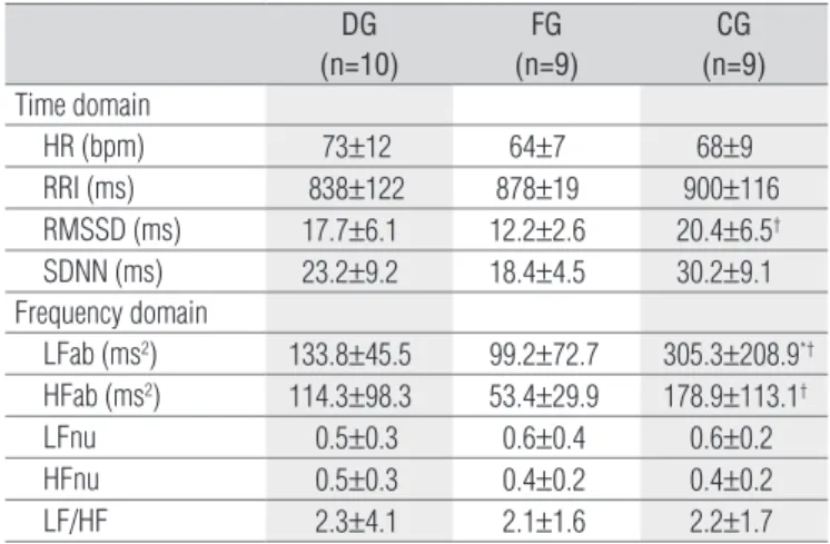
DG (n=10)
FG (n=9)
CG (n=9) Time domain
HR (bpm) 73±12 64±7 68±9
RRI (ms) 838±122 878±19 900±116
RMSSD (ms) 17.7±6.1 12.2±2.6 20.4±6.5†
SDNN (ms) 23.2±9.2 18.4±4.5 30.2±9.1
Frequency domain
LFab (ms2) 133.8±45.5 99.2±72.7 305.3±208.9*†
HFab (ms2) 114.3±98.3 53.4±29.9 178.9±113.1†
LFnu 0.5±0.3 0.6±0.4 0.6±0.2
HFnu 0.5±0.3 0.4±0.2 0.4±0.2
LF/HF 2.3±4.1 2.1±1.6 2.2±1.7
Table 2. Comparison of heart rate variability at rest in the time and
frequency domains for all groups.
Values are means±SD. DG=COPD group; FG=CHF group; CG=control group; HR=heart rate; RRI=RR intervals in the ECG; RMSSD=root mean square of successive differences between adjacent RRI; SDNN= standard deviation of normal to normal RRI; LFab=low frequency in absolute values; HFab=high frequency in absolute va-lues; LFnu=low frequency in normalized units; HFnu=high frequency in normalized units;† p<0.05=FG vs. CG and * p<0.05=DG vs CG (one-way ANOVA with Tukey’s
post-hoc test).
109 M-RSA); inspiration-expiration diference (ΔIE; diference
be-tween the mean of the highest HR values obtained during the inspiratory phase and the mean of the lower HR values in the expiratory phase of the M-RSA).
Statistical analysis
For the statistical tests, the data were transformed into decimal logarithms. Parametric statistical tests were used because the data showed normal distribution (Shapiro-Wilk test) and homogeneity of variances (Levene’s test). One-way ANOVA with Tukey’s post-hoc test was used in the intergroup comparisons (DG vs. FG vs. CG). he analyses were performed in SPSS Release 10.0.1 (1999) with the signiicance level set at p<0.05.
Results
Twenty-eight participants were studied: 10 patients with COPD (DG), 9 patients with CHF (FG) and 9 control partici-pants (CG). Table 1 shows the demographic, anthropometric and clinical data of the participants at rest. here were no signiicant diferences between groups for the anthropometric measures, however the DG values were signiicantly lower than the CG values for body mass, even though the CG individuals were classiied as healthy28.
The DG showed a moderate stage of disease (Stage IIb)29. In addition, the FEV1 and FEV1/FVC ratio for this group
were significantly lower than the CG (p<0.05). The func-tional capacity of the DG by the MRC scale showed that the participants belonged to classes I (N=1), II (N=3) and III (N=6). The FG was composed of participants with systolic dysfunction of idiopathic etiology (N=5) due to myocardial infarction (N=4) and with functional class I (N=2), II (N=4) and III (N=3) by the NYHA22. According to the MRC and NYHA scales for functional class, the DG and FG showed similarities in the degree of dyspnea, i.e. limitation in the performance of activities of daily living that require light or moderate effort. Additionally, the SpO2 was lower in the DG than in the CG (p<0.05), but the DG and the FG did not dif-fer (Table 1).
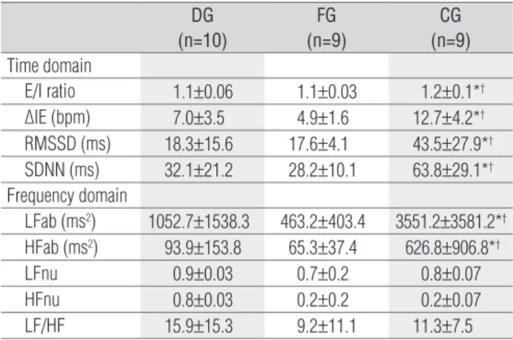
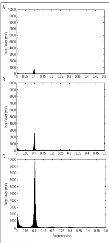
Regarding the HRV indices at rest (Table 2), it was ob-served that the DG showed lower LFab when compared to the CG (p<0.05). In contrast, the FG showed lower values of RMSSD, LFab and HFab than the CG (p<0.05). No signifi-cant differences were observed between the DG and the FG. In the comparisons during the M-RSA (Table 3), the DG and FG showed lower values for the E/I ratio and ΔIE when compared to the CG (p<0.05). For the HRV indices in the M-RSA in the time domain, the participants in the DG and FG showed significantly lower values for RMSSD and SDNN than the CG. Similarly, in the frequency domain, the DG and FG showed lower LFab and HFab values com-pared to the CG (p<0.05). Additionally, Figure 1 illustrates the spectral behaviour that represents each group. The DG and FG showed lower values of total spectral density com-pared to the CG.
Variables DG
(N=10)
FG (N=9)
CG (N=9) Demographics / Anthropometrics
Age (years) 69±9 62±7 64±5
Height (m) 1.67±0.08 1.68±0.06 1.71±0.05
Body mass (kg) 64±9.0 69±8.5 75±6.3*
BMI (kg/m²) 23±3.3 25±3.1 25±1.2
Echocardiogram
EF (%) --- 41±6
---Spirometry
FEV1 (% of predicted value) 41±11 80±9 91±20*
FEV1/FVC 59±12 82±11 101±7*
Clinical characteristics
SpO2 (%) 92±3 96±2 96±1*
RF (rpm) 15±4 14±4 12±3
Table 1. Demographic, anthropometric and clinical characteristics for
all groups.
Values are means±SD. DG=COPD group; FG=CHF group; CG=control group; BMI=body mass index; EF=ejection fraction of the left ventricle; FEV1=forced expiratory volume in the first second; FEV1/FVC=FEV1 to forced vital capacity ratio; SpO2=peripheral oxygen saturation; RF=respiratory frequency in respirations per min. * p<0.05=DG vs. CG;
DG (n=10)
FG (n=9)
CG (n=9) Time domain
E/I ratio 1.1±0.06 1.1±0.03 1.2±0.1*†
∆IE (bpm) 7.0±3.5 4.9±1.6 12.7±4.2*†
RMSSD (ms) 18.3±15.6 17.6±4.1 43.5±27.9*†
SDNN (ms) 32.1±21.2 28.2±10.1 63.8±29.1*†
Frequency domain
LFab (ms2) 1052.7±1538.3 463.2±403.4 3551.2±3581.2*†
HFab (ms2) 93.9±153.8 65.3±37.4 626.8±906.8*†
LFnu 0.9±0.03 0.7±0.2 0.8±0.07
HFnu 0.8±0.03 0.2±0.2 0.2±0.07
LF/HF 15.9±15.3 9.2±11.1 11.3±7.5
Values are means±SD. DG=COPD group; FG=CHF group; CG=control group; E/I ratio=inspiratory/expiratory ratio; ∆IE=inspiratory-expiratory differences in beats per min; root mean square of successive differences between adjacent RRI; SDNN= standard deviation of normal to normal RRI; LFab=low frequency in absolutes values; HFab= high frequency in absolutes values; LFnu=low frequency in normalized units; HFnu=high fre-quency in normalized units;* p<0.05=DG vs CG; † p<0.05=FG vs. CG (one-way ANOVA
with Tukey’s post-hoc test).
Table 3. I/E ratio, IE-differences and heart rate variability during
respiratory sinus arrhythmia maneuver of the groups studied.
110
Discussion
he main indings of the present study showed that both participants with COPD and participants with CHF showed changes in autonomic modulation of HR at rest and during the M-RSA when compared to apparently healthy individuals matched for age.
Impact of the cardiorespiratory diseases on
HRV at rest
In the present study, the COPD participants showed re-duced sympathetic activity when compared to the controls. Some authors8,30 have observed that both sympathetic and parasympathetic activity are reduced in individuals with COPD. In contrast, Volterrani et al.7 observed that patients with COPD showed higher values of the HF band (nu), re-flecting an increase in parasympathetic activity over the control of HR. These authors consider that these findings can be related to bronchoconstriction and, consequently, to the reduction in FEV1 showed by these patients. How-ever, in the study by Volterrani et al.7, some methodologi-cal features that differ from the present study may have influenced the results. In the study by these authors7, the COPD group had a wide age-range (31 to 68 years), with a large number of young individuals diagnosed with asthma who may have already had an exacerbated vagal response. Our results are similar to those reported by Chen, Chen and Kuo9, who showed a reduction in sympathetic activity in patients with COPD.
In the present study, the participants with CHF also ex-perienced a significant reduction in HRV when compared to the control group, with reduced sympathetic and parasym-pathetic activity. These findings corroborate those of other authors who also observed a reduction in time domain indi-ces in patients with CHF31. In accordance with our findings, Saul et al.32 observed that patients with congestive CHF had a reduction in all frequency bands compared to the healthy group with similar anthropometric characteristics and age. Guzetti et al.31 observed that patients with CHF showed a reduction in the time domain indices and lower values of LF band when compared to healthy participants, resulting in lower sympathetic activity.
In contrast, other authors15-17 reported reduced HRV in CHF patients with a predominance of sympathetic activity. hese authors suggest that sympathetic hyperactivity in HR control is a relection of compensatory changes in the au-tonomic system caused by the evolution of the disease and aimed at ensuring homeostasis with an appropriate increase in cardiac output. In the present study, we found a reduction in both sympathetic and parasympathetic action on the sinus node, possibly due to regulatory changes in the autonomic centers12, sensitivity of chemoreceptors13 or the respiratory pattern marked by periodic oscillations in these patients14.
However, it is important to note that our indings were observed in the absolute values of the LF band, which is in-luenced by the VLF bands, combined with the systems of long-term autonomic control. Additionally, the CHF patients involved in the present study continued the drug treatment. he action of beta-blockers33 on the sinus node receptors, the action of digitalis drugs34 on the sodium and potassium pumps of the myocardial and on the angiotensin-converting enzyme inhibitors promote the autonomic regulation of the heart, attenuating sympathetic hyperactivity and possibly inluenc-ing the present results.
Impact of cardiorespiratory diseases on HRV
indices during the M-RSA
Regarding the indices of HR and RRI obtained with the M-RSA, it was observed that both the COPD and CHF partici-pants showed reduced parasympathetic response compared to the control participants. he results corroborate other indings in the literature that reported a signiicant reduction in RSA in patients with DPOC35,36 and CHF32 compared to healthy par-ticipants. However, no previous study has compared the HRV indices and RSA in both pathologies.
111
A
B
C
Total Power (ms
2)
Total Power (ms
2)
Total Power (ms
2)
10000
9000
8000
7000
6000
5000
4000
3000
2000
1000
0.05 0.1 0.15 0.2 0.25 0.3 0.35 0.4 0.45 0.5
0 0
10000
9000
8000
7000
6000
5000
4000
3000
2000
1000
0.05 0.1 0.15 0.2 0.25 0.3 0.35 0.4 0.45 0.5
0 0
10000
9000
8000
7000
6000
5000
4000
3000
2000
1000
0.05 0.1 0.15 0.2 0.25 0.3 0.35 0.4 0.45 0.5
0 0
Frequency (Hz)
Figure 1. Decomposition of the spectrum into single spectral
components - very low frequency (VLF), low frequency (LF), and high frequency (HF) - during respiratory sinus arrhythmia maneuver. (A) COPD patient, (B) CHF patient, and (C) control.
participants in the CG. RSA, a product of the interaction be-tween the respiratory and cardiovascular systems, is inluenced by respiratory frequency and tidal volume37. In the present study, in which the respiratory frequency was controlled during the M-RSA, it can be assumed that the lower values of the RSA indices are related to reduced tidal volume, further strengthening our speculation that the reduction in HRV observed in patients of both groups may be related to changes in lung compliance and the response of pulmonary stretch receptors.
Methodological considerations
he present study has some limitations. Regarding the participants, groups with more participants would have been ideal; however, the strict exclusion criteria did not allow a larger sample because both diseases can be associated with respira-tory and cardiovascular changes. Additionally, we considered that an echocardiogram of the participants with COPD would be important to exclude the coexistence of cor pulmonale or right ventricular failure, and we considered that arterial blood gas analysis was needed to classify the patients with COPD as hypoxemic or hypercapnic. Similarly, the measurement and control of tidal volume during controlled respiration, which were not performed in the present study, could contribute to the consolidation and interpretation of the results. Finally, the evaluation of complete lung function (static volumes) would be of particular relevance in the evaluation of patients, but such measurements involve expensive equipment.
Clinical relevance
Autonomic control is a major means of adjustment in response to postural, physical changes under stress or during physical exercise in conditions of health or coexisting pathol-ogies. In this sense, the knowledge of autonomic variations in HR in patients with cardiopulmonary diseases contributes to a more appropriate physical therapy evaluation and prescrip-tion of rehabilitaprescrip-tion programs and to a better understanding of the efects of diferent physical therapy interventions.
Conclusion
he results of the present study suggest that patients with COPD or CHF show modiication of the autonomic control of HR, with reduced sympathetic and/or parasympathetic activi-ties, when compared to healthy participants due to the integra-tion of both cardiorespiratory diseases. Moreover, both COPD and CHF showed attenuated responses to enhanced parasym-pathetic activity during the M-RSA.
Acknowledgments
112
References
1. Task force of european society of cardiology the North American society of pacing electrophysiology. Heart rate variability: standards of measurement, physiological interpretation, and clinical use. Circulation. 1996;93(5):1043-65.
2. Sztajzel J. Heart rate variability: a noninvasive electrocardiographic method to measure the autonomic nervous system. Swiss Med Wkly. 2004;134:514-22.
3. Castello V, Mendes RG, Simões RP, Reis MS, Catai AM, Borghi-Silva A. Atividade autonômica em uma adolescente com ventrículo único submetida à intervenção fisioterapêutica: relato de caso. Res Bras Fisioter. 2008;12(2):157-60.
4. De Burgh Daly M. Interactions between respiration and circulation. In: Fishman AP, Cherniack NS, Widdicombe JG, Geiger SR, editors. Handbook of physiology. The respiratory system, section 3. Bethesda: American Physiological Society; 1986. p. 529-94.
5. Borghi-Silva A, Reis MS, Mendes RG, Pantoni CB, Simões RP, Martins LE, et al. Noninvasive ventilation acutely modifies heart rate variability in chronic obstructive pulmonary disease patients. Respir Med. 2008;102(8):1117-23.
6. Pantoni CBF, Reis MS, Martins LEB, Catai AM, Costa D, Borghi-Silva A. Estudo da modulação autonômica da freqüência cardíaca em repouso de pacientes idosos com doença pulmonar obstrutiva crônica. Res Bras Fisioter. 2007;11(1):35-41.
7. Volterrani M, Scalvini S, Mazzuero G, Lanfranchi P, Colombo R, Clark AL, et al. Decreased heart rate variability in patients with chronic obstructive pulmonaty disease. Chest. 1994;106(5):1432-7.
8. Scalvini S, Porta R, Zanelli E, Volterrani M, Vitacca M, Pagani M, et al. Effects of oxygen on autonomic nervous system dysfunction in patients with chronic obstructive pulmonaty disease. Eur Respir J. 1999;13(1):119-24.
9. Chen WL, Chen GY, Kuoa CD. Hypoxemia and autonomic nervous dysfunction in patients with chronic obstructive pulmonaty disease. Respir Med. 2006;100(9):1547-53.
10. Stewart AG, Waterhouse JC, Howard P. The QTc interval, autonomic neuropathy and mortality in hypoxaemic COPD. Respir Med. 1995;89(2): 79-84.
11. Leung RS, Bradley TD. Respiratory modulation of heart rate and blood pressure during cheyne-stokes respiration. J Electrocardiol. 2003;36 Suppl:S213-7.
12. Musialik-Lydka AM, Sredniawa B, Pasyk S. Heart rate variability in heart failure. Kardiol Pol. 2003;58(1):10-6.
13. Tulppo M, Huikuri HV. Origin and significance of heart rate variability. J Am Coll Cardiol. 2004;43(12):2278-80.
14. Rosen SD, Murphy K, Leff AP, Cunningham V, Wise RJ, Adams L, et al. Is central nervous system processing altered in patients with heart failure? Eur Heart J. 2004;25(11):952-62.
15. van de Borne P, Montano N, Pagani M, Oren R, Somers VK. Absence of low-frequency variability of sympathetic nerve activity in severe heart failure. Circulation. 1997;95(6):1449-54.
16. Ponikowski P, Chua TP, Piepoli M, Ondusova D, Webb-Peploe K, Harrington D, et al. Augmented peripheral chemosensitivity as a potential input to baroreflex impairment and autonomic imbalance in chronic heart rate failure. Circulation. 1997;96(8):2586-94.
17. Mortara A, Sleight M, Pinna GD, Maestri R, PRPA A, La Rovere MT, et al. Abnormal awake respiratory patterns are common in chronic heart failure and may prevent evaluation of autonomic tone by measures of heart rate variability. Circulation. 1997;96(1):246-52.
18. Grossman P, Wilhelm FH, Spoerle M. Respiratory sinus arrhythmia, cardiac vagal control and daily activity. Am J Physiol Heart Circ Physiol. 2004;287(2):728-34.
19. Carrasco-Sosa S, Gaitán-González MJ, González-Camarena R, Yáñez-Suárez O. Baroreflex sensitivity assessment and heart rate variability: relation to maneuver and technique. Eur J Appl Physiol. 2005;95(4):265-75.
20. Katona PG, Jih F. Respiratory sinus arrhythmia: noninvasive measure of parasympathetic cardiac control. J Appl Physiol. 1975;39(5):801-5.
21. Hayano J, Mukai S, Sakakibara M, Okada A, Takata K, Fujinami T. Effects of respiratory interval on vagal modulation of heart rate. Am J Physiol. 1994;267(1 Pt 2):33-40.
22. The criteria committee of the New York Heart Association. Nomenclature and criteria for diagnosis of diseases of the heart and great vessels. 9ª ed. Boston: Little Brown & Co; 1994.
23. Ferrer M, Alonso J, Morera J, Marrandes RM, Khalaf A, Aquar MC, et al. Chronic obstructive pulmonary disease and healthy-related quality of life. The quality of life obstructive pulmonary disease study group. Ann Intern Med. 1997;127(12):1072-9.
24. Crapo MD, Hankison JL, Irvin C, Mancityre NL, Voter KZ, Wise RA. American Thoracic Society: Standardization of spirometry 1994 update. Am J Respir Crit Care Med. 1995;152(3):1107-36.
25. Silva E, Catai AM, Trevelin LC, Guimarães JO, Silva Jr. LP, Silva LMP, et al. Design of a computerized system to evaluate the cardiac function during dynamic exercise. Phys Med Biol. 1994;39:409.
26. Pyetan E, Toledo E, Zoran O, Akselrod S. Parametric description of cardiac vagal control. Auton Neurosci. 2003;109(1-2):42-52.
27. O’Brien IA, O’Hare P, Corrall RJ. Heart rate variability in healthy subjects: effect of age and the derivation of normal ranges for tests of autonomic function. Br Heart J. 1986;55(4):348-54.
28. World Health Organization. Diet nutrition and the prevention of chronic diseases (1990). Geneva: WHO Technical Report Series 797; 1989.
113
30. Stein PK, Nelson P, Rottman JN, Howard D, Ward SM, Kleiger RE, et al. Heart rate variability reflects severity of COPD in PiZ a1-antitrypsin deficiency. Chest. 1998;113(2):327-33.
31. Guzzetti S, Mezzetti S, Magatelli R, Porta A, De Angelis G, Rovelli G, et al. Linear and non-linear 24 h heart rate variability in chronic heart failure. Auton Neurosci. 2000;86(1-2):114-9.
32. Saul JP, Arai Y, Berger RD, Lilly LS, Colucci WS, Cohen RJ. Assessment of autonomic regulation in chronic congestive heart failure by heart rate spectral analysis. Am J Cardiol. 1988;31(15):1292-9.
33. Goldsmith RL, Bigger JT, Bloomfield DM, Krum H, Steinman RC, Sackner-Bernstein J, et al. Long-term carvedilol therapy increases parasympathetic nervous system activity in chronic congestive heart failure. Am J Cardiol. 1997;80(8):1101-4.
34. Flapan AD, Goodfield NE, Wright RA, Francis CM, Neilson JM. Effects of digoxin on time domain measures of heart rate variability in patients with stable chronic cardiac failure: withdrawal and comparison group studies. Int J Cardiol. 2007;59(1):29-36.
35. Pagani M, Lucini D, Pizzinelli P, Sergi M, Bosisio E, Mela GS, et al. Effects of aging and of chronic obstructive pulmonary disease on RR interval variability. J Auton Nerv Syst. 1996;59(3):125-32.
36. Giardino ND, Chan L, Borson S. Combined heart rate variability and pulse oximetry biofeedback for chronic obstructive pulmonary disease: preliminary findings. Appl Psychophysiol Biofeedback. 2004;29(2):121-33.