VIEW AND REVIEW
Depression and temporal lobe epilepsy represent
an epiphenomenon sharing similar neural
networks: clinical and brain structural evidences
Depressão e epilepsia de lobo temporal representam um epifenômeno compartilhando
redes neurais similares: evidências clínicas e de neuroimagem estrutural
Kette D. R. Valente1,2, Geraldo Busatto Filho2
Around 400 BC, Hippocrates wrote: “Melancholics ordinar-ily become epileptics, and epileptics, melancholics; what deter-mines the preference is the direction the malady takes; if it bears upon the body, epilepsy, if upon the intelligence, melancholy”.
he relationship between depression and epilepsy has been known since ancient times, however, to date, it is not fully understood.
he prevalence of psychiatric disorders (PD) in persons with epilepsy is high compared to general population. Mood disorders occur in 11 to 62%, psychosis in 7 to 10% and per-sonality disorders in 5 to 33%1-8.
Depression is the most frequent PD in persons with epi-lepsy9-11. It is assumed that the rate of depression ranges from
20 to 55% in patients with refractory epilepsy, especially con-sidering those with temporal lobe epilepsy caused by mesial temporal sclerosis (TLE-MTS). Furthermore, people with epi-lepsy have a lifetime prevalence of any type of mood disor-der higher than that observed in general population or peo-ple with other chronic medical disorders. Despite its high prevalence, depression remains underdiagnosed and under-treated in persons with epilepsy, with relevant personal and social costs. he impact of depression in morbidity can be
1Laboratório de Neuroisiologia Clínica, Instituto de Psiquiatria do Hospital das Clínicas da Faculdade de Medicina da Universidade de São Paulo (USP), São
Paulo SP, Brazil;
2Laboratório de Neuroimagem em Neurociências, Departamento de Psiquiatria da Faculdade de Medicina da USP, São Paulo SP, Brazil.
Correspondence: Kette D. R. Valente; Rua Doutor Ovidio Pires de Campos 785; 05403-010 São Paulo SP - Brasil; E-mail: [email protected]
Conflict of interest: There is no conlict of interest to declare.
Received 05 March 2012; Received in inal form 27 July 2012; Received in inal form 03 August 2012
ABSTRACT
The relationship between depression and epilepsy has been known since ancient times, however, to date, it is not fully understood. The prevalence of psychiatric disorders in persons with epilepsy is high compared to general population. It is assumed that the rate of depression ranges from 20 to 55% in patients with refractory epilepsy, especially considering those with temporal lobe epilepsy caused by mesial temporal sclerosis. Temporal lobe epilepsy is a good biological model to understand the common structural basis between depression and epilepsy. Interestingly, mesial temporal lobe epilepsy and depression share a similar neurocircuitry involving: temporal lobes with hippocampus, amygdala and entorhinal and neocortical cortex; the frontal lobes with cingulate gyrus; subcortical structures, such as basal ganglia and thalamus; and the connecting pathways. We provide clinical and brain structural evidences that depression and epilepsy represent an epiphenomenon sharing similar neural networks.
Key words: epilepsy, depression, neuroimaging, temporal lobe.
RESUMO
A relação entre depressão e epilepsia é conhecida desde a antiguidade; entretanto, até o momento, não é completamente compreendida. A prevalência de transtornos psiquiátricos nas pessoas com epilepsia é elevada quando comparada à população em geral. A taxa de de-pressão varia de 20 a 55% nos pacientes com epilepsia refratária, especialmente considerando-se aqueles com epilepsia do lobo temporal causada por esclerose mesial temporal. A epilepsia do lobo temporal é um bom modelo biológico para compreender as bases estruturais comuns entre a epilepsia e a depressão. É relevante ressaltar que a epilepsia do lobo mesial e a depressão apresentam circuitos similares envolvendo: os lobos temporais com o hipocampo, a amigdala, o córtex entorrinal e o neocortex; os lobos frontais com o giro cíngulo; estrutu-ras subcorticais, como os núcleos da base e o tálamo, e suas vias de conexão. Postulamos por meio de evidências clínicas e estruturais que a depressão e a epilepsia representam um epifenômeno com redes neuronais similares.
evaluated by some studies of quality of life in patients with refractory epilepsy (9.7 seizures/month; mean duration of epilepsy of 12.7 years)12,13. According to Gilliam et al.14,
qual-ity of life (QOLIE-89) summary score was signiicantly as-sociated with depression scales, but not with the type or the frequency of seizures, although patients averaged 0.3 to 51 seizures/month. In addition, in a recent population-based study15, depression was a risk factor for death in epilepsy.
Suicide remains as a major cause of death in epilepsy, since 32.5% of all deaths of persons with epilepsy are due to suicide and 13.5% of all registered suicides are committed by these persons16. herefore, although complete cessation of seizures
remains the paramount goal of therapy in epilepsy, these data clearly clamor for the need to better appreciate the im-portance of mood disorders in the overall treatment plan of these patients.
Although prevalence of depression in epilepsy, as well as its consequence in terms of mortality and morbidity, have been extensively debated and remains as a major concern for those in charge for these patients, the mechanisms underly-ing this association — epilepsy and depression — have not been properly explored. For this reason, many clinicians, pa-tients and relatives still believe that epilepsy and depression present a cause and consequence relationship. In this review article, we aimed to provide clinical and brain structural ev-idences that depression and epilepsy represent an epiphe-nomenon sharing similar neural networks. In particular, we give emphasis to in vivo neuroimaging studies that
investi-gated volumetric abnormalities in samples of individuals with epilepsy compared to control groups.
DEPRESSION AND EPILEPSY: CLINICAL EVIDENCE OF THESE CONDITIONS AS AN EPIPHENOMENA
he hypothesis that epilepsy, as a disabling and stigma-tizing disease, may lead to depression has been weakened by some clinical evidences that speaks against the concept of long-lasting epilepsy causing depression. In addition, it has a clinical impact since it keeps clinicians aware for the fact that depression may occur early in the course of epilepsy and even precede its onset.
Depression in children with epilepsy
he same scenario, described in adults, is observed in children with epilepsy that even without the long lasting ef-fect of epilepsy present a high prevalence of PD, especially de-pression17. he prevalence of depression ranges from 28% in
children with uncomplicated epilepsy to 58% in children with refractory epilepsy18. A study conducted in a Brazilian
tertia-ry care facility showed that depression was the most frequent PD in children with refractory epilepsy19. As adults, children
with epilepsy remain undertreated and underdiagnosed, es-pecially those with depression3,20. Suicide rates are high even
in children and adolescents with epilepsy21.
Bidirectional relationship
Some studies have shown that depression and suicide attempt are independent risk factors for the onset of sei-zures and epilepsy. he irst scientiic studies to show the bidirectional view of epilepsy and depression were carried out in the last decade. One study established that a histo-ry of depression preceding the onset of epilepsy was seven times more frequent among patients than among age- and sex-matched controls and that a signiicant number of pa-tients with new-onset epilepsy were already sufering from depression prior to their irst seizure22-24. Another study
found that people with epilepsy were 3.7 times more likely to have a history of depression preceding their initial sei-zure than the control group23, corroborating Hippocrates
clinical observations and suggesting a bidirectional rela-tionship between epilepsy and depression.
herefore, the high prevalence of the comorbidity of these two disorders in children and in patients with new-onset epi-lepsy associated with the occurrence of depression prior to seizure or epilepsy onset suggests that, although it is natural-ly assumed that mood disorders are a consequence of seizure disorders, as a normal reaction to the impact of epilepsy, we have data that depression and epilepsy may share common pathogenic mechanisms, manifesting with the involvement of the same neuroanatomic structures, as will be discussed25.
DEPRESSION IN TEMPORAL LOBE EPILEPSY
Temporal lobe epilepsy (TLE) is a good biological model to understand the common structural basis between depres-sion and epilepsy.
Temporal lobe epilepsy can be determined by neocortical lesions (lateral temporal lobe epilepsy) and by mesial tempo-ral sclerosis. Mesial tempotempo-ral sclerosis (MTS) is a condition characterized by hippocampal atrophy. Hippocampal scle-rosis is the most common histological abnormality observed in patients with mesial temporal lobe epilepsy26. It usually
presents marked cell loss in Sommer’s sector (CA1 and pro-subiculum) and endofolium (hilus and CA4). Between these regions, there was relative sparing in CA3 and CA227.
In patients with temporal lobe epilepsy caused by mesial temporal sclerosis (TLE-MTS), the frequency of depression is extremely high even when compared to other types of epi-lepsy (50–60%), including neocortical TLE26,27. Patients with
TLE-MTS show signiicantly higher depression scores than patients with neocortical temporal lesions, independent of the lateralization of the lesion28. he prevalence of suicide
persons with TLE-MTS. In this group, suicide and suicide attempts in persons with TLE-MTS may be 25 times higher than average population4,29, the highest rate observed in all
persons with epilepsy. Moreover, compared to TLE patients without hippocampal sclerosis, patients with TLE-MTS have greater frequency of cognitive side efects and mood distur-bances when treated with antiepileptic drugs (AEDs)30.
Despite the known clinical relationship between TLE-MTS and depression, only a few studies have investigated whether patients with this comorbidity display a characteris-tic pattern of clinical and imaging indings. Interestingly, neu-rocircuitry involved in both TLE-MTS31-34 and depression35
include the temporal lobes with hippocampus, amygdala and entorhinal and neocortical cortex; the frontal lobes with cingulate gyrus; subcortical structures, such as basal ganglia and thalamus; and the connecting pathways.
hese similarities between TLE-MTS and depression will be discussed.
TEMPORAL LOBE EPILEPSY AND DEPRESSION: COMMON BRAIN STRUCTURAL ABNORMALITIES
here are many lines of evidence to support the notion of biological underpinnings of depression and temporal lobe epilepsy as comorbid conditions25. he common brain
path-ways involved in emotional processing, encompassing the limbic system and its complex connections, constitute one of these pieces of evidence (Fig 1).
NEUROIMAGING STUDIES IN MTLE
In vivo neuroimaging methods had advanced
dramati-cally in past decades, with advances both in regard to novel techniques for data acquisition and new models for image processing and analysis. his has allowed increasingly more sophisticated investigations of structural and functional brain abnormalities associated with TLE-MTS.
In morphometric neuroimaging studies using mag-netic resonance imaging (MRI), measures of regional brain volumes can be obtained in groups of subjects with TLE in comparison to control groups of healthy individu-als, matched for demographic variables. The most widely employed method to perform such between-group com-parisons are region-of-interest (ROI) techniques. Such ROI-based methods involve the delineation of selected an-atomical structures in sequential brain slices, most often manually, in order to obtain quantitative volumetric indi-ces for such regions. Manual ROI-based methods are time-consuming and subject to observer bias, and the brain areas studied are often circumscribed using variable ana-tomical borders across different studies of TLE36-38. Such
inconsistencies may limit data comparisons between sep-arate studies and the conduction of meta-analyses of the results obtained. As an alternative to ROI-based measure-ments, recent morphometric MRI studies have employed automated methods that allow hypothesis-free voxelwise comparisons of regional brain volumes across the entire brain, without requiring the a priori selection of anatom-ical ROI borders and with perfect repeatability. This ap-proach is referred to as voxel-based morphometry (VBM)39.
The VBM technique initially involves spatial transforma-tions of the MRI scans of all subjects included in a study onto a common anatomical space, in order to remove in-ter-individual variations in brain size and shape. In such spatial normalization process, images are conformed by linear and non-linear transformations to a standard tem-plate (based on databanks of images normalized to a ste-reotactic atlas), or to a customized template created spe-cifically for the study (obtained from a pool of images from the population under investigation). Spatially normalized MRI datasets of each subject are then automatically seg-mented into gray matter, white matter and cerebral spinal fluid compartments, and smoothed with a Gaussian filter. Segmented images are subsequently compared statistical-ly between groups on a voxel-by-voxel basis, and statistical maps are produced in standardized brain space showing the location of voxel clusters where significant between-group volume differences are present, at a predefined sta-tistical level of inference. Because it is fully automated, rat-er-independent and capable of investigating the presence of morphometric abnormalities across the whole brain (rather than solely on selected brain regions), the VBM has been an important tool in TLE-MTS, substantiating the concept of a more widespread diseased, not restricted to the hippocampus.
Stimulus presentation
DPFC Dorsal AC Hippocampus
VLPFC Orbitofrontal cortex
Ventral AC
Amygdala Insula
Thalamus Ventral striatum Mesencephalon Emotional regulation
Affective expression
Evaluation
+/-On the left, the steps involved in the perception and evaluation of emotions are shown. On the right, the brain structures involved in the mediation of those emotional processing steps are displayed, including the hippocampus, other limbic structures, cortical regions and subcortical gray matter nuclei. DPFC: dorsal prefrontal cortex; VLPFC: ventrolateral prefrontal cortex; AC: anterior cingulate. Adapted from Phillips ML et al.72.
Keller and Roberts34 reviewed the applications and
results of VBM studies that have reported limbic and ex-tralimbic changes associated with TLE considering gray and white matter. A PubMed search yielded 18 applica-tions of VBM to study brain abnormalities in patients with TLE up to May 2007. Across studies, 26 brain regions were found to be significantly reduced in volume relative to healthy controls.
Limbic structures
here was a strong asymmetrical distribution of tempo-ral lobe abnormalities preferentially observed ipsilatetempo-ral to the seizure focus, particularly of the hippocampus (82.35% of all studies), parahippocampal gyrus (47.06%) and entorhinal (23.52%) cortex. he contralateral hippocampus was report-ed as abnormal in 17.65% of studies.
EXTRALIMBIC STRUCTURES
There was bilateral distribution of extratemporal lobe atrophy, preferentially affecting the thalamus (ipsi-lateral=61.11%, contralateral=50%) and parietal lobe (ip-silateral=47.06%, contralateral=52.94%). VBM generally reveals a distribution of brain abnormalities in patients with TLE consistent with the region-of-interest neuroim-aging and postmortem literature.
Careful manual and automated morphometrical stud-ies showed that patients with TLE-MTS show signiicant extrahippocampal atrophy that involves the temporal lobe and extratemporal brain structures40-44. Notably, the
distri-bution of brain atrophy in TLE-MTS preferentially afects a network of regions that are functionally and anatomically connected to the hippocampus31-34.
Clinical implication
Studies with VBM in TLE-MTS have helped to under-stand some of the distinct clinical proiles presented by these patients. For instance, Yasuda et al.45 showed that gray
matter (GM) atrophy with a bilateral and widespread pat-tern, encompassing the entire ipsilateral temporal lobe, as well as areas in the thalami, cerebellum, occipital, parietal and frontal lobes, was correlated with negative family his-tory for epilepsy.
Patients with a higher seizure frequency and longer period of active epilepsy have also been correlated with more intense gray and white matter reduction over time46.
Morphometrical MRI studies of patients with epilepsy dem-onstrated that extrahippocampal gray matter loss likely fol-lows a progressive course47. Cognitive deicits commonly
exhibited by patients with TLE-MTS, in particular memory impairment, are directly related to the degree of medial tem-poral and frontal lobe atrophy48,49.
NEUROIMAGING STUDIES IN DEPRESSION: LIMBIC AND EXTRALIMBIC STRUCTURES
Hippocampus
he most robust inding concerns volumetric changes in the hippocampus. High-resolution MRI volumetric studies of individuals with major depressive disorders (MDD) consis-tently showed decreased volume of hippocampus in adult, as well as in paediatric series50-52. In depression,
hippocam-pal volume is decreased bilaterally50-52 or in the left
hippo-campus only53. Antidepressants are thought to exert a
neu-roprotective function, particularly in MDD. Indeed, the work of Sheline et al.52, using ROI, showed that the hippocampal
volume loss was related with longer duration of untreated disease52-54. he volume reduction appears to be more
pro-nounced with longer illness duration and in the presence of a positive family history for MDD.
Amygdala
Amygdala, which plays a major role in the processing of fear and related emotions55, seems to undergo a two-staged
process with initial bilateral enlargement during acute and a subsequent shrinkage during chronic depression. here are several controversies as to amygdala volume, howev-er a recent meta-analysis phowev-erformed by Hamilton et al.56,
based on VBM indings, has shown that amygdala
enlarge-ment in MDD was associated primarily with antidepressant treatment, whereas unmedicated patients showed volume decrements in this region. Amygdala and parahippocam-pal gray matter volumes were also signiicantly reduced in VBM studies including patients with comorbid anxiety dis-orders, as well as in irst-episode/drug free samples57.
herefore, preservation of hippocampal and amygdala volume in MDD has been associated with antidepressant treatment52,56. hese indings may be partially explained by
the efect of antidepressant treatment that has a number of potentially important efects on multiple biological systems; at present, antidepressant-induced normalization of the se-rum levels of the neurotrophin brain-derived neurotrophic factor is considered a key modulator of the neurotrophic ef-fect of these drugs on brain structures58.
EXTRALIMBIC STRUCTURES – PREFRONTAL CORTEX AND CINGULATE GYRUS
Although hippocampus and amygdala have received the greatest interest, extralimbic structures, such as the prefron-tal cortex and the anterior portion of cingulate gyrus, are af-fected as well (Fig 2).
In depression, smaller volumes of frontal lobes have been found59. he work of Lavretsky et al.59 used distinct MRI image
and cortical parcellation methods) to measure the gray and white matter volumes in two prefrontal subregions: the an-terior cingulate and orbitofrontal cortex showed that the de-pressed group had smaller orbitofrontal gray matter volumes compared to the age-matched normal comparison group. he severity of apathy was associated with the decreased gray matter volume in the right anterior cingulate gray mat-ter volumes using partial correlation and regression analyses after controlling for age, sex and diagnosis.
Bora et al.57, in a systematic search of VBM studies
ap-plied in MDD, performed a metanalysis to data collated from a total of 23 studies comparing regional gray matter volumes of 986 MDD patients and 937 healthy controls. GM was sig-niicantly reduced in a conined cluster located in the rostral anterior cingulate cortex (ACC). here were also gray matter reductions in dorsolateral and dorsomedial prefrontal cor-tex, and decrease in the latter region was evident in patients with multiple-episodes. Gray matter reduction in rostral ACC was the most consistent inding in VBM studies of MDD.
he data reviewed above indicates that there is a substan-tial degree of similarities in regard to the brain regions that show morphometric abnormalities in TLE-MTS and MDD.
NEUROIMAGING STUDIES IN TLE-MTS AND DEPRESSION
In addition to all the similarities about the regions in-volved in TLE-MTS and depression, there are some direct ev-idences based on studies with patients with TLE and depres-sion compared to those TLE subjects without depresdepres-sion.
Limbic structures
MRI volumetric studies of individuals with TLE and de-pression have found decreased volumes of the hippocampus. Hippocampal volumes loss is expected in TLE-MTS, by dei-nition, especially related to the site of seizure origin – ictal onset zone60-62. However, there are evidences of a more
pro-nounced decrease, or bilateral decrease, in patients with TLE-MTS and depression61.
Next to the hippocampus, the temporal lobe structure that received greatest attention is the amygdala. Richardson et al.63 examined the relationship between self-reported
de-pression severity and both structural MRI volumetry and [(18)F]luorodeoxyglucose positron emission tomography (PET)-measured resting metabolism of the amygdala and hippocampus of 18 patients with TLE. Signiicant positive relationships were noted between right and left amygdala volumes and depression, indicating that both right and left amygdala volumes are associated with depression severity among persons with TLE.
Patients with TLE and depressive symptoms have in-creasing amygdala volumes measured by MRI63. his
inding conirmed earlier reports by Tebartz van Elst et al.64.
Moreover, the latter authors found a signiicant correlation between left amygdala volume and depression severity64,65.
hus, the amygdala is hyperactive in anxiety and mood dis-orders and may increase in size during acute depression in patients with TLE63,64. hese indings overlie those previously
demonstrated in patients with depression without epilepsy66,
transient amygdala enlargement may be secondary to en-hanced regional blood low and vascular volume as detect-ed by positron emission tomography (PET)67 or because of
dendritic remodeling with increased branching of amygda-loid neurons68. However, as previously mentioned in this text,
amygdala and hippocampal volumes decrease in recurrent and chronic untreated depression52,62. herefore, in this
con-text, there are common characteristics between these ind-ings and those observed in patients with depression without epilepsy.
Extralimbic structures
In TLE, for instance, smaller volumes of frontal lobes have been found62.
Woermann et al.69 studied patients with TLE and
inter-ictal episodes of aggression — intermittent explosive dis-order (IED) — that may represent one expression of inter-ictal dysphoric depression in patients with temporal lobe epilepsy. Interictal dysphoric depression, a term coined by
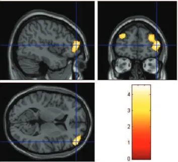
Example of a morphometric magnetic resonance imaging (MRI) study in which patients with severe major depression (MD) with psychotic features (n=20) were found to present reduced gray matter volume bilaterally in the dorsolateral prefrontal cortex compared to a group of healthy controls (n=94), after correction for group differences in demographic variables. Foci of reduced gray matter in MD patients relative to controls (p<0.001) were overlaid on brain slices spatially normalized into an approximation to the Talairach and Tournoux stereotactic atlas, and are in the Figure displayed on the sagittal, coronal and transaxial planes. Data extracted from Azevedo-Marques Périco et al.73.
Fig 2. Findings of extralimbic brain structural abnormalities in
Blumer et al.70, to describe the clinical manifestations of
de-pression in epilepsy is characterized by intermittent dys-thmia with recurrent episodes of dysphoria. In the work of Woermann et al.69, after automated segmentation of
cere-bral grey matter from T1 weighted MRI, the objective tech-nique of statistical parametric mapping (SPM) was applied to the analysis of 35 control subjects, 24 patients with TLE with a history of repeated, interictal episodes of aggression, and 24 patients with TLE without episodes of aggression. Patients with TLE with aggressive episodes had a decrease of grey matter, most markedly in the left frontal lobe, com-pared with the control group and with patients with TLE without aggressive episodes, suggesting that a reduction of frontal neocortical grey matter might underlies the patho-physiology of aggression in TLE.
Later on, the elegant study conducted by Salgado et al.71
with 96 health controls and 48 TLE-MTS (24 with major de-pression and 24 without major dede-pression) revealed a sig-niicant group efect regarding gray matter volume (GMV) in some brain regions. he number of areas of GMV loss was signiicantly higher in the group with MTLE with depres-sion, with a more widespread distribution of GMV loss in patients with depression.
CONCLUDING REMARKS
In temporal lobe epilepsy, VBM studies showed gray/white matter atrophy extending beyond the atrophic hippocam-pus. hese widespread abnormalities have been associated with higher seizure frequency, longer epilepsy duration and higher incidence of precipitating factors, cognitive impair-ment and worse surgical outcome. In addition, in this review, we observed that these patients with a more widespread dis-ease, demonstrated by VBM, also have more severe depres-sion. Even though hippocampal and extrahippocampal gray
matter atrophy are related to seizure control and cognitive performance, the mechanisms underlying brain damage in patients with MTLE remain largely unknown. Voxel-based morphometry may provide an important instrument to un-derstand the diferent clinical proiles presented by patients with the same pathology and perhaps, in the future, to iden-tify those patients with epilepsy who are at-risk for a worse outcome. his may represent an attempt to provide early di-agnosis and treatment for these patients. he morphometric MRI studies reviewed in this article, most notably those us-ing VBM, provide strong support to the notion that depres-sion and MTLE represent an epiphenomenon sharing similar neural networks involving several brain regions. It is reason-able to postulate that widespread brain abnormalities may point to a more diseased cortex and consequently to worse outcome in many senses — severity of epilepsy, worse cog-nition and higher prevalence of depression. It is important to stress that VBM methods were designed to perform mean group comparisons for research purposes and are not suit-able for diagnosis of individual cases. herefore, it is unlikely that VBM has any clinical utility given the lack of robustness for individual comparisons. However, VBM may help eluci-date some unresolved important research questions such as how recurrent temporal lobe seizures afect hippocampal and extrahippocampal morphology using serial imaging ac-quisitions. Furthermore, neuroimaging studies and, in this context, VBM have shown that several structures involved in mesial temporal sclerosis are equally involved in depression and, in a structural level, provide a piece of evidence to better understand this complex puzzle.
ACKNOWLEDGEMENTS
We thank Fabio Luiz de Souza Duran for assistance in the production of the illustrations.
1. Gibbs FA. Ictal and non-ictal psychiatric disorders in temporal lobe epilepsy. J Nervous Mental Dis 1951;113:522-528.
2. Ettinger A, Reed M, Cramer J, Epilepsy Impact Project Group. Depression and comorbidity in community-based patients with epilepsy or asthma. Neurology 2004;63:1008-1014.
3. Ettinger AB, Weisbrot DM, Nolan EE, et al. Symptoms of depression and anxiety in pediatric epilepsy patients. Epilepsia 1998;39:595-599. 4. Jones JE, Hermann BP, Barry JJ, Gilliam FG, Kanner AM, Meador
KJ. Rates and risk factors for suicide, suicidal ideation, and suicide attempts in chronic epilepsy. Epilep Behav 2003;4:S31-S38. 5. Kanner AM, Dunn DW. Diagnosis and management of depression and
psychosis in children and adolescents with epilepsy. J Child Neurol 2004;19:S65-S72.
6. de Araújo Filho GM, da Silva JM, Mazetto L, Marchetti RL, Yacubian EM. Psychoses of epilepsy: a study comparing the clinical features of patients with focal versus generalized epilepsies. Epilepsy Behav 2011;20:655-658.
7. de Araújo Filho GM, Pascalicchio TF, Sousa Pda S, Lin K, Ferreira Guilhoto LM, Yacubian EM. Psychiatric disorders in juvenile myoclonic epilepsy: a controlled study of 100 patients. Epilepsy Behav 2007;10:437-441.
8. Moschetta S, Fiore LA, Fuentes D, Gois J, Valente KD. Personality traits in patients with juvenile myoclonic epilepsy. Epilepsy Behav 2011;21:473-477.
9. Hermann BP, Seidenberg M, Bell B, Woodard A, Rutecki P, Sheth R. Comorbid psychiatric symptoms in temporal lobe epilepsy: association with chronicity of epilepsy and impact on quality of life. Epilepsy Behav 2000;1:184-190.
10. Kanner AM. Depression in Epilepsy is much more than a reactive process. Epilepsy Curr 2003;3:202-203.
11. Boylan KR, Bieling PJ, Marriott M, Begin H, Young LT, MacQueen GM. Impact of comorbid anxiety disorders on outcome in a cohort of patients with bipolar disorder. J Clin Psychiatry 2004;65:1106-1113.
12. Gilliam F. Optimizing health outcomes in active epilepsy. Neurology 2002;58:S9-S20.
13. Boylan LS, Flint LA, Labovitz DL, Jackson SC, Starner K, Devinsky O. Depression but not seizure frequency predicts quality of life in treatment-resistant epilepsy. Neurology 2004;62:258-261.
14. Gilliam F. Optimizing epilepsy management: seizure control, reduction, tolerability, and co-morbidities. Introduction. Neurology 2002;58:S1.
15. Ridsdale L, Charlton J, Ashworth M, Richardson MP, Gulliford MC. Epilepsy mortality and risk factors for death in epilepsy: a population-based study. Br J Gen Pract 2011;61:271-278.
16. Pompili M, Girardi P, Tatarelli R. Death from suicide versus mortality from epilepsy in the epilepsies: a meta-analysis. Epilepsy Behav 2006;9:641-648.
17. Hoare P. The development of psychiatric disorder among schoolchildren with epilepsy. Dev Med Child Neurol 1984;26:3-13. 18. Rutter ML. Psycho-social disorders in childhood, and their outcome in
adult life. J R Coll Physicians Lond 1970;4:211-218.
19. Thome-Souza S, Kuczynski E, Assumpção F Jr., et al. Which factors may play a pivotal role on determining the type of psychiatric disorder in children and adolescents with epilepsy? Epilepsy Behav 2004;5:988-994.
20. Ott D, Siddarth P, Gurbani S, et al. Behavioral disorders in pediatric epilepsy: unmet psychiatric need. Epilepsia 2003;44:591-597. 21. Baker GA. Depression and suicide in adolescents with epilepsy.
Neurology 2006;66:S5-S12.
22. Hesdorffer DC, Hauser WA, Olafsson E, Ludvigsson P, Kjartansson O. Depression and suicide attempt as risk factors for incident unprovoked seizures. Ann Neurol 2006;59:35-41.
23. Hesdorffer DC, Hauser WA, Annegers JF, Cascino G. Major depression is a risk factor for seizures in older adults. Ann Neurol 2000;47:246-249.
24. Jones JE, Hermann BP, Woodard JL, et al. Screening for major depression in epilepsy with common self-report depression inventories. Epilepsia 2005;46:731-735.
25. Kanner AM. Depression in epilepsy: a neurobiologic perspective. Epilepsy Curr 2005;5:21-27.
26. Victoroff JI, Benson F, Grafton ST, Engel J Jr., Mazziotta JC. Depression in complex partial seizures. Electroencephalography and cerebral metabolic correlates. Arch Neurol 1994;51:155-163.
27. Mendez MF, Grau R, Doss RC, Taylor JL. Schizophrenia in epilepsy: seizure and psychosis variables. Neurology 1993;43:1073-1077. 28. Quiske A, Helmstaedter C, Lux S, Elger CE. Depression in patients with
temporal lobe epilepsy is related to mesial temporal sclerosis. Epilep Res 2000;39:121-125.
29. Barraclough BM. The suicide rate of epilepsy. Acta Psychiatr Scand 1987;76:339-345.
30. Mula M, Trimble MR, Sander JW. The role of hippocampal sclerosis in topiramate-related depression and cognitive deicits in people with epilepsy. Epilepsia 2003;44:1573-1577.
31. Spencer SS. Neural networks in human epilepsy: evidence of and implications for treatment. Epilepsia 2002;43:219-227.
32. Bonilha L, Rorden C, Castellano G, Cendes F, Li LM. Voxel-based morphometry of the thalamus in patients with refractory medial temporal lobe epilepsy. Neuroimage 2005;25:1016-1021.
33. Riederer F, Lanzenberger R, Kaya M, Prayer D, Serles W, Baumgartner C. Network atrophy in temporal lobe epilepsy: a voxel-based morphometry study. Neurology 2008;71:419-425.
34. Keller SS, Roberts N. Voxel-based morphometry of temporal lobe epilepsy: an introduction and review of the literature. Epilepsia 2008;49:741-757.
35. Price JL, Drevets WC. Neurocircuitry of mood disorders. Neuropsychopharmacology 2010;35:192-216.
36. King D, Spencer SS, McCarthy G, Luby M, Spencer DD. Bilateral hippocampal atrophy in medial temporal lobe epilepsy. Epilepsia 1995;36:905-910.
37. Watson C, Jack CR Jr., Cendes F. Volumetric magnetic resonance imaging. Clinical applications and contributions to the understanding of temporal lobe epilepsy. Arch Neurol 1997;54:1521-1531. 38. Watson C, Cendes F, Fuerst D, et al. Speciicity of volumetric magnetic
resonance imaging in detecting hippocampal sclerosis. Arch Neurology 1997;54:67-73.
39. Ashburner J, Friston KJ. Voxel-based morphometry--the methods. Neuroimage 2000;11:805-821.
40. Keller SS, Mackay CE, Barrick TR, Wieshmann UC, Howard MA, Roberts N. Voxel-based morphometric comparison of hippocampal and extrahippocampal abnormalities in patients with left and right hippocampal atrophy. Neuroimage 2002;16:23-31.
41. Keller SS, Wieshmann UC, Mackay CE, Denby CE, Webb J, Roberts N. Voxel based morphometry of grey matter abnormalities in patients with medically intractable temporal lobe epilepsy: effects of side of seizure onset and epilepsy duration. J Neurol Neurosurg Psychiatry 2002;73:648-655.
42. Bernasconi N, Duchesne S, Janke A, Lerch J, Collins DL, Bernasconi A. Whole-brain voxel-based statistical analysis of gray matter and white matter in temporal lobe epilepsy. Neuroimage 2004;23:717-723. 43. Bonilha L, Rorden C, Castellano G, et al. Voxel-based morphometry
reveals gray matter network atrophy in refractory medial temporal lobe epilepsy. Arch Neurol 2004;61:1379-1384.
44. Butler T, Blackmon K, McDonald CR, et al. Cortical thickness abnormalities associated with depressive symptoms in temporal lobe epilepsy. Epilepsy Behav 2012;23:64-67.
45. Yasuda CL, Morita ME, Alessio A, et al. Relationship between environmental factors and gray matter atrophy in refractory MTLE. Neurology 2010;74:1062-1068.
46. Coan AC, Appenzeller S, Bonilha L, Li LM, Cendes F. Seizure frequency and lateralization affect progression of atrophy in temporal lobe epilepsy. Neurology 2009;73:834-842.
47. Bonilha L, Rorden C, Appenzeller S, Coan AC, Cendes F, Li LM. Gray matter atrophy associated with duration of temporal lobe epilepsy. Neuroimage 2006;32:1070-1079.
48. Bonilha L, Alessio A, Rorden C, et al. Extrahippocampal gray matter atrophy and memory impairment in patients with medial temporal lobe epilepsy. Hum Brain Mapp 2007;28:1376-1390.
49. Focke NK, Thompson PJ, Duncan JS. Correlation of cognitive functions with voxel-based morphometry in patients with hippocampal sclerosis. Epilepsy Behav 2008;12:472-476.
50. Sheline YI, Wang PW, Gado MH, Csernansky JG, Vannier MW. Hippocampal atrophy in recurrent major depression. Proc Natl Acad Sci USA 1996;93:3908-3913.
51. Sheline YI, Sanghavi M, Mintun MA, Gado MH. Depression duration but not age predicts hippocampal volume loss in medically healthy women with recurrent major depression. J Neurosci 1999; 19:5034-5043.
52. Sheline YI, Gado MH, Kraemer HC. Untreated depression and hippocampal volume loss. Am J Psychiatry 2003;160:1516-1518. 53. Bremner JD, Narayan M, Anderson ER, Staib LH, Miller HL, Charney
DS. Hippocampal volume reduction in major depression. Am J Psychiatry 2000;157:115-118.
54. Sheline YI. Neuroimaging studies of mood disorder effects on the brain. Biol Psychiatry. 2003;54:338-52.
55. Phelps EA, LeDoux JE. Contributions of the amygdala to emotion processing: from animal models to human behavior. Neuron 2005;48:175-187.
57. Bora E, Fornito A, Pantelis C, Yücel M. Gray matter abnormalities in Major Depressive Disorder: a meta-analysis of voxel based morphometry studies. J Affect Disord 2012;138:9-18.
58. Brunoni AR, Lopes M, Fregni F. A systematic review and meta-analysis of clinical studies on major depression and BDNF levels: implications for the role of neuroplasticity in depression. Int J Neuropsychopharmacol 2008;11:1169-1180.
59. Lavretsky H, Ballmaier M, Pham D, Toga A, Kumar A. Neuroanatomical characteristics of geriatric apathy and depression: a magnetic resonance imaging study. Am J Geriatr Psychiatry 2007;15:386-394. 60. Cascino GD, Jack CR Jr., Parisi JE, et al. Magnetic resonance
imaging-based volume studies in temporal lobe epilepsy: pathological correlations. Ann Neurol 1991;30:31-36.
61. Baxendale SA, Thompson PJ, Duncan JS. Epilepsy & depression: the effects of comorbidity on hippocampal volume-a pilot study. Seizure 2005;14:435-438.
62. Mueller SG, Laxer KD, Schuff N, Weiner MW. Voxel-based T2 relaxation rate measurements in temporal lobe epilepsy (TLE) with and without mesial temporal sclerosis. Epilepsia 2007;48:220-228.
63. Richardson EJ, Grifith HR, Martin RC, et al. Structural and functional neuroimaging correlates of depression in temporal lobe epilepsy. Epilepsy Behav 2007;10:242-249.
64. Tebartz van Elst L, Woermann FG, Lemieux L, Trimble MR. Amygdala enlargement in dysthymia-a volumetric study of patients with temporal lobe epilepsy. Biol Psychiatry 1999;46:1614-1623. 65. Tebartz van Elst L, Woermann F, Lemieux L, Trimble MR. Increased
amygdala volumes in female and depressed humans. A quantitative magnetic resonance imaging study. Neurosci Lett 2000;281:103-106.
66. Frodl T, Meisenzahl EM, Zetzsche T, et al. Hippocampal changes in patients with a irst episode of major depression. Am J Psychiatry 2002;159:1112-1118.
67. Drevets WC. Neuroimaging studies of mood disorders. Biol Psychiatry 2000;48:813-829.
68. Vyas A, Mitra R, Shankaranarayana Rao BS, Chattarji S. Chronic stress induces contrasting patterns of dendritic remodeling in hippocampal and amygdaloid neurons. J Neurosc 2002;22:6810-6818.
69. Woermann FG, van Elst LT, Koepp MJ, et al. Reduction of frontal neocortical grey matter associated with affective aggression in patients with temporal lobe epilepsy: an objective voxel by voxel analysis of automatically segmented MRI. J Neurol Neurosurg Psychiatry 2000;68:162-169.
70. Blumer D, Montouris G, Davies K. The interictal dysphoric disorder: recognition, pathogenesis, and treatment of the major psychiatric disorder of epilepsy. Epilepsy Behav 2004;5:826-840.
71. Salgado PC, Yasuda CL, Cendes F. Neuroimaging changes in mesial temporal lobe epilepsy are magniied in the presence of depression. Epilepsy Behav 2010;19:422-427.
72. Phillips ML, Drevets WC, Rauch SL, Lane R. Neurobiology of emotion perception II: Implications for major psychiatric disorders. Biol Psychiatry 2003;54:515-528.