DOI: 10.1590/0004-282X20150034 ARTICLE
Multidetector computed tomography
angiography in clinically suspected
hyperacute ischemic stroke in the anterior
circulation: an etiological workup in a cohort
of Brazilian patients
Angiotomograia computadorizada multidetectores na avaliação da suspeita clínica de
acidente vascular encefálico isquêmico hiperagudo da circulação anterior: determinação
etiológica precoce em um grupo de pacientes brasileiros
Felipe Torres Pacheco1, Ingrid Aguiar Littig1, Rubens Jose Gagliardi2, Antônio Jose da Rocha1
Accurate classiication of ischemic stroke based on etiol
-ogy is essential for research because stroke outcome, recurrent stroke rate, and strategies for secondary stroke prevention dif
-fer according to stroke subtype1,2. Furthermore, stroke subtype is used to describe patients’ characteristics in clinical trials, group
patients in epidemiological studies, phenotype patients in genet
-ic studies, and classify patients for therapeut-ic decision-making in daily practice2,3,4. Topographic radiologic patterns of brain in
-farctions might predict prognosis and correlate well with the un
-derlying pathophysiology and imaging indings5.
1Santa Casa de São Paulo, Faculdade de Ciências Médicas, Departamento de Neuroradiologia, Sao Paulo SP, Brazil; 2Santa Casa de São Paulo, Faculdade de Ciências Médicas, Departamento de Neurologia, Sao Paulo SP, Brazil.
Correspondence: Felipe Torres Pacheco; Rua Doutor Cesario Motta Junior, 112; 01221-020 São Paulo SP, Brasil; E-mail: [email protected]
Conlict of interest: There is no conlict of interest to declare.
Received 05 September 2014; Received in inal form 08 January 2015; Accepted 28 January 2015.
ABSTRACT
Objective: The potential of computed tomography angiography (CTA) was assessed for early determination of stroke subtypes in a Brazilian cohort of patients with stroke. Method: From July 2011 to July 2013, we selected patients with suspected hyperacute stroke (< 6 hours). Intracranial and cervical arteries were scrutinized on CTA and their imaging features were correlated with concurrent subtype of stroke. Results: Stroke was documented in 50/106 selected patients (47.2%) based on both clinical grounds and imaging follow-up (stroke group), with statistically signiicant arterial stenosis and vulnerable plaques on CTA. Intracranial large artery disease was demonstrated in 34% of patients in the stroke group. Partial territorial infarct prevailed (86%) while artery-to-artery embolization was the most common stroke mechanism (52%). Conclusion: Multidetector CTA was useful for the etiologic work-up of hyperacute ischemic stroke and facilitated the knowledge about the topographic pattern of brain infarct in accordance with its causative mechanism.
Keywords: tomography, MDCT, stroke, cerebral infarction.
RESUMO
Objetivo: Avaliar o potencial da angiotomograia computadorizada multidetectores (ATCM) na determinação etiológica precoce do aci-dente vascular encefálico (AVE) e correlacionar o mecanismo causal com o padrão de infarto. Método: De Julho de 2011 a Julho de 2013, foram selecionados os pacientes com suspeita clínica de AVE hiperagudo. Os achados da ATCM dos vasos intracranianos e cervicais foram correlacionados com a etiologia inal do evento. Resultados: AVE foi conirmado em 50/106 pacientes (47,2%). Estes apresentaram alte-rações angiográicas estatisticamente mais relevantes. Aterosclerose dos grandes vasos intracranianos esteve presente em 34% destes pacientes. O padrão radiológico topográico de infarto mais comum foi o infarto territorial parcial (86%). A embolização arterio-arterial foi o mecanismo mais prevalente (52%). Conclusão: A utilização da ATCM traz benefícios na detecção etiológica precoce dos pacientes com suspeita de AVE hiperagudo, além de possibilitar o entendimento do padrão radiológico topográico de acordo com o mecanismo causal do evento isquêmico.
Early determination of the etiologic factors of ischemic stroke is essential for secondary prevention because the risk of recurrence is highly dependent on the underlying cause6,7.
Extra- or intra-cranial atheromas, cardioembolic sources, and microvascular disease have been identiied as the major causes of ischemic stroke8,9.
he proportions of patients with diferent ischemic stroke sub
-types have been reported to difer for each race-ethnic group10,11,12.
While Caucasians have an increased risk for cardioembolic strokes10
, Asian patients with ischemic stroke show increased in
-cidence of intracranial large-artery disease (ILAD)11
, and cervical arterial disease is more often reported in western patients12. In the Brazilian population, such diferences remain unclear.
Recent advances in computed tomography (CT) technol
-ogy have improved the feasibility of its use in clinical settings. Multidetector CT angiography (MDCTA) improves the sensi
-tivity and inter-rater reliability of detecting acute stroke13. In addition, it is an accurate and powerful noninvasive tool for assessing carotid artery disease in the setting of hyperacute ischemic stroke, including the detection of atherosclerosis involvement in extra- and intra-cranial vessels. However, only a few studies have reported the use of MDCTA to deine stroke etiology in our population14. To the best of our knowl -edge, MDCTA has not yet been used to study the etiology of stroke in a Latin American population.
In this study, we assessed the potential of the use of MDCTA for the early detection of the etiology of hyperacute ischemic stroke in the middle cerebral arteries (MCA) in a Brazilian cohort of patients and subsequently correlated the topography and causative mechanisms of infarcts.
METHOD
Patient selection
his study is part of a larger protocol in a single institu
-tion of the use of MDCTA techniques to evaluate hyperacute stroke, and the protocol has been reviewed and approved by the Institutional Review Board and the local ethics commit
-tee. From July 2011 to July 2013, adult patients (≥ 18 years old) who presented with hyperacute symptoms (< 6h) that were consistent with focal acute ischemia of the MCA and who had undergone CTA were considered eligible. he patient or their guardian signed the informed consent.
Patients with contraindications regarding intravenous io
-dine contrast administration were excluded from the study. In addition, we excluded patients who refused to participate in the study, patients with examinations with inadequate image quality, and patients who had posterior circulation strokes as their inal diagnosis.
Protocol
All examinations were conducted with a previous
-ly reported institutional protocol14 with a minimum dose
of ionizing radiation and intravenous iodinated contrast in a Philips Brilliance 64-slice CT scanner (Brilliance CT 64 Channel, Philips Medical, Eindhoven, he Netherlands). he examinations included non-contrast computed to
-mography and cervical and intracranial MDCTA, which re
-quired up to 5 min.
MDCTA was performed after intravenous administra
-tion of 50 mL of nonionic iodinated contrast (Ultravist®; 300 mg I/mL) at a rate of 5 mL/s using a dual-head power injector (Bayer HealthCare LLC, Whippany, NJ, USA) with an 18-G intravenous access, which was usually located in the cu
-bital vein. he region of interest was placed in the aortic arch to determine the self-timer of the apparatus. When the at
-tenuation in this region reached 160 HU, acquisition of the sections from the aortic arch to the vertex of the skull was initiated. Source images of CTA (CTA-SI) were postprocessed to obtain maximum intensity projections and three-dimen
-sional views of the intracranial and extracranial arteries.
Analysis
All of the data were postprocessed with commercial
-ly available software on a workstation (Extended Brilliance Workspace v3.5.0.2250, Philips Medical Systems B.V., Best, he Netherlands). Two neuroradiologists who were experi
-enced in vascular imaging and were blinded to the outcomes independently analyzed all of the CTA-SI from the emergency room examination and, subsequently, the maximum inten
-sity projections and the three-dimensional reconstructions. All discrepancies were solved by consensus.
All patients were classiied as having either normal or ab
-normal extracranial cervical carotid arteries. Vascular abnor
-malities that potentially correlated with concurrent stroke were discriminated based on their imaging features.
If a plaque was visible on CTA-SI, it was categorized based on the grade of stenosis and type of plaque. he severity of ste
-nosis was deined as the remaining lumen at the site of ste-nosis as a percentage of the normal lumen distal to the stenosis and categorized as < 50%, 50%-99%, and 100% (arterial occlusion). he vessel diameters were measured on the plane that was per
-pendicular to the vessel course. According to the TOAST crite -ria7, we considered stenosis to be relevant if the plaque resulted in a vessel diameter reduction of more than 50%.
he plaques’ morphologies were assessed subjectively by consensus, and their densities were measured with the region of interest. he plaque composition was analyzed according to the following previously proposed classiica
-tion15
: (1) non-calciied plaques (density, ≤ 50 HU), (2) calciied plaques (mean attenuation, > 130 HU), and (3) mixed plaques (mean attenuation varying from 51 to 130 HU). For plaque morphology analysis, its surface was considered regular or ir
-regular. A stable plaque was deined as a plaque that was cal
-ciied with a regular surface. If the plaque was non-cal-ciied or mixed and it had an irregular surface with or without ul
he intracranial cerebrovascular system was scrutinized to be interpreted according to current literature11,16,17. A di
-agnosis of atherothrombosis was assumed when relevant cervical artery atherosclerosis was concomitant with a MCA thrombus. However, a cardioembolic origin was determined when a cardiac source was conirmed in the absence of rele
-vant cervical artery disease. A diagnosis of ILAD was adopted when partial or total atherosclerotic MCA obstruction was documented in the absence of relevant cervical artery dis
-ease or cardioembolic source. If was detected a non-relevant cervical artery disease combined with partial or total ath
-erosclerotic MCA obstruction was assumed that there was concurrent ILAD and extracranial atherosclerosis. Follow-up images were analyzed in order to conirm ischemic stroke. he topographic radiologic patterns of the brain infarctions visible on non-contrast CT were deined with the OCSP clas
-siication4, which resulted in malignant, large, and limited in
-farcts for MCA territorial strokes. Limited in-farcts were those that covered only 1 of the 3 MCA territories (deep, superi
-cial anterior, or superi-cial posterior). Large infarcts were de
-ined as those covering at least 2 of these 3 MCA territories. Malignant infarction referred to complete or almost com
-plete MCA infarctions18.
Finally, using the SSS-TOAST algorithm8
, the patients were classiied according to the results of their individual examinations that included cervical Doppler ultrasonogra
-phy, cardiac echo, and electrocardiogram; this was termed the inal classiication.
Statistical analysis
All of the information was entered into a database with Excel 2011 (Version 14.0.0; Microsoft Corporation, Redmond, WA, USA). We then entered all of the data into IBM SPSS Statistics software (Version 20.0 for Mac; IBM Corporation, Armonk, NY, USA). X2 tests were used to compare the vari
-ables between patients with and without brain infarctions and within the group of patients with brain infarctions. P-values less than 0.05 were considered statistically signiicant.
RESULTS
A total of 134 patients were recruited for this study. According to the previously reported criteria, 106 patients were selected for the retrospective analysis. hey were di
-vided according to their outcomes. An anterior circulation stroke was documented in 50/106 patients (47.2%) based on both clinical and imaging follow-up indings (stroke group), while 56/106 patients (52.8%) showed no evidence of brain ischemia (non-stroke group).
Unremarkable results on the cervical MDCTA were docu
-mented in 18 patients (18/50, 36.0%) patients in the stroke group. In the remaining patients, non-relevant stenosis was found in 17 patients (17/50, 34.0%) patients, while relevant stenosis in the cervical arteries was found in 15 patients (15/50, 30.0%) (Figure 1), including 5 patients (5/50, 10.0%) with stenosis diameters > 50%, 4 patients (4/50, 8.0%) with occlusive disease and the remaining 6 patients (6/50, 12.0%) with cervical arterial dissections.
Conversely, relevant stenosis was only seen in 4 patients from the non-stroke group (4/56, 7.0%), while 20 patients (20/56, 36.0%) in this group had normal results on MDCTA. he remaining 32 (32/56, 57.0%) had only non-relevant steno
-sis (Χ2 test: p = 0.01; Table 1).
Although plaques may not cause relevant obstruction, they might be responsible for cerebral infarctions accord
-ing to their characteristics. When patients with non-relevant stenosis were examined, 10 patients (10/17, 59.0%) in the stroke group were found to have vulnerable plaques. In con
-trast, 10 patients in the non-stroke group (10/32, 31.0%) were diagnosed with vulnerable plaques. (X2 test: p = 0.01; Table 1).
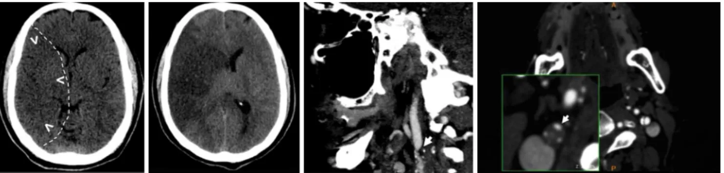
Of the 50 patients included in the stroke group, 7 (7/50, 14.0%) had normal intracranial and extracranial vessels. Imaging indings that were compatible with ILAD (Figure 2) were documented in 7 patients (7/50, 14.0%), while 12 (12/50, 24.0%) had only cervical carotid atherosclerosis and 10 (10/50, 20.0%) had concurrent ILAD and extracranial atherosclerosis. he remaining 14 patients (14/50, 28.0%) had atherothrombosis
Figure 1. A 56-year-old man presented within 60 minutes of a left hemiplegia. (A) Initial non-contrast CT with subtle
or cardioemboli with thrombi in the intracranial system. Of the 56 patients included in the non-stroke group, only 2 (2/56, 3.5%) had concurrent intra- and extra-cranial atherosclerosis, none had only intracranial disease, and 34 (34/56, 60.8%) had isolated extracranial disease (32 irrelevant stenosis and 2 rel
-evant stenosis). Twenty patients (20/56, 35.7%) had unremark
-able CTAs (X2 test: p = 0.01; Table 2).
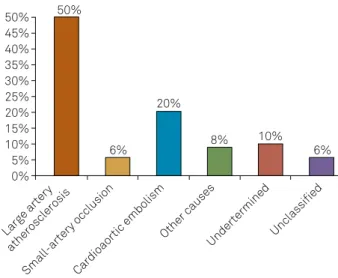
Based on the inal classiication using the SSS-TOAST al
-gorithm, our patients were divided as: 25 (25/50, 50.0%) with large artery disease, 10 (10/50, 20.0%) with cardioemboli (Figure 3), 5 (5/50, 10.0%) with undetermined causes, 3 (3/50, 6.0%) with small-artery disease, 3 (3/50, 6.0%) with more than one evident mechanism, and 4 (4/50, 8.0%) with other causes (all of them cervical artery dissection) (Figure 4).
After reviewing the follow-up imaging, we classiied the patients in the stroke group based on topographic radiolog
-ic patterns of brain infarction (OCSP classi-ication). Forty-three (43/50, 86.0%) had territorial infarcts, of which 27 had limited, 7 had large, and 9 had malignant territorial infarcts; 3 (3/50, 6.0%) had centrum oval infarcts; 3 (3/50, 6.0%) had lacunar infarcts, and 1 (1/50, 2.0%) had a watershed infarct (Figure 5). When we analyzed the centrum oval infarcts, we
found that all of them (3/3, 100.0%) were related to large ar
-tery atherosclerosis, while the lacunar infarcts (3/3, 100.0%) were due to small-artery occlusion. he only watershed in
-farct (1/1, 100.0%) was caused by cervical arterial dissection. By correlating the OCSP and SSS-TOAST classiications we observed that cervical carotid atherosclerosis caused large or malignant brain infarctions in 6 patients (6/25, 24.0%), including 5 patients (5/6, 83.3%) with concurrent ILAD and extracranial disease. Large or malignant infarc
-tions were also diagnosed in 6 patients with cardioemboli etiology (6/10, 60.0%) and in 2 patients with cervical arterial dissections (2/4, 50.0%).
DISCUSSION
Extracranial carotid atherosclerotic disease has been re
-ported as the major risk factor for stroke, and the main mech
-anism is related to artery-to-artery embolization from either an atherosclerotic plaque or an acute occlusion of the carotid artery with distal propagation of the thrombus19. Accordingly, cervical carotid atherosclerotic disease was demonstrated Figure 2. An 80-year-old woman presented within 120 minutes of onset of left hemiplegia caused by ILAD. (A) Axial CTA MIP image demonstrated at least 50% stenosis in the right MCA. Dotted line demonstrated a more extensive ischemic area according to the follow-up exam; (B) Tridimensional (3D) view of the extracranial right carotid arteries only demonstrated parietal irregularities that corresponds a smalls calciied non-stenotic plaques; (C) Noncontrast CT demonstrated subtle effacement of the right insula (arrowheads). The dotted line demonstrated a more extensive ischemic area according to the follow-up exam; and (D) Imaging follow-up after 3 days demonstrated a large ischemic stroke in the MCA territory.
Table 1. Extracranial atherosclerosis in both Groups (stroke and non-stroke).
Relevant Stenosis Total (%) (n = 106)
Stroke Group (%) (n = 50)
Non-stroke Group (%)
(n = 56) p
Present 19 15 (30) 4 (7) 0.01*
Absent 87 35 (70) 52 (93) 0.14
Type of plaque Total (%) (n = 49) Stroke Group (%) (n = 17) Non-stroke Group (%) (n = 32) p
Vunerable 20 10 (59) 10 (31) 0.01*
Stable 29 7 (41) 22 (69) 0.21
on CTA in the majority of our patients, and it was the main risk factor for stroke with half of them presenting relevant stenosis or vulnerable plaques on cervical arteries.
Luminal narrowing is the standard parameter used for reporting the severity of carotid atherosclerosis20. However, plaque morphology also plays an important role as it directly correlates with the risk of embolism and occlusion, thus re
-sulting in cerebral ischemia21.
Several studies have highlighted that MDCTA ind
-ings closely correlate with those from digital angiography for the degree of stenosis, which suggests that the sensitiv
-ity of MDCTA in evaluating the degree of stenosis may be comparable to that of angiography but with a lower level of risk22,23,24,25. he majority of the patients from our series dem
-onstrated some degree of cervical artery abnormalities that were detected on MDCTA (64.0% in both groups). Our study conirmed that MDCTA was able to detect relevant stenosis on cervical carotid arteries more commonly in patients with brain infarcts (30.0% versus 7.0%) whose had the etiology of
their ischemic stroke deined in the same moment as the di
-agnosis of the brain infarction.
Moreover, MDCTA detected vulnerable plaques, even in cases of non-relevant stenosis, in both groups. Although this inding was likely involved in the pathophysiology of the inal infarct, further studies are necessary to conirm this. he rel
-evance of the abnormal arteries on MDCTA in deining the
stroke mechanism in the stroke group was well supported by our results. he relevance of cervical carotid abnormalities in patients from the non-stroke group (relevant stenosis or vul
-nerable plaques) remains uncertain as these indings could be related to either transient ischemic attacks or minor in
-farcts with absent brain lesions or abnormalities not detect
-able on brain CT.
In patients with non-relevant abnormalities, the percent
-ages of plaques were the same in both groups, but analyzing the characteristics of the plaques on cervical CTA showed that the percentage of the vulnerable plaque type was high
-er in the stroke group (59.0% versus 21.0%), which corrobo
-rated data aggregated by Guptaet al. in a meta-analysis that suggested an association of these types of plaques with an increased risk of stroke26. Considering these characteristics, our indings corroborated that it is important to examine plaque morphology in addition to the lumen obstruction11.
Large artery atherosclerosis was the main stroke type in our group of patients, and it involved 50.0% of the cases. Limited infarction was the most common imaging inding. his inding demonstrates a slightly higher prevalence of ca
-rotid atherosclerotic disease as a cause for stroke than those previously postulated2,20,27. A more precise elucidation of the role of vulnerable plaques in non-relevant stenosis and their implications in the absence of concurrent brain infarcts de
-tectable on CT remains uncertain.
Figure 3. A 66-year-old man presented within 90 minutes of onset of right hemiplegia and aphasia. (A) Coronal CTA MIP image with unremarkable indings of the cervical carotid arteries; (B) Noncontrast CT demonstrates subtle effacement of the left insular cortex (arrowheads). Dotted line demonstrated a more extensive ischemic area according to the follow-up exam; and (C) Imaging follow-up after 2 days conirmed a large infarct in the MCA territory. A cardiac source of emboli was conirmed.
Table 2. Intra and extracranial indings in both Groups (stroke and non-stroke).
Total (%) (n = 106)
Stroke Group (%) (n = 50)
Non-stroke Group (%)
(n = 56) p
Normal vessels 24 07 (14.0) 20 (35.8) 0.01*
Atherothrombosis or cardioemboli 14 14 (28.0) 00 (00.0) 0.01*
ILAD 07 07 (14.0) 00 (00.0) 0.01*
Relevant cervical disease 46 09 (18.0) 02 (03.5) < 0.05*
Irrelevant cervical disease 03 (06.0) 32 (57.2) 0.01*
Extra and intracranial 12 10 (20.0) 02 (03.5) < 0.05*
Atherosclerosis MCA and ICA disease have been associ
-ated with racial diferences. While MCA disease is often re
-ported in Asians, ICA disease is frequently found in western populations28. Manet al.29 found that 72% of a Chinese cohort of patients had ILAD, which difered from the indings in our patient group, in which 34% had ILAD; 14% had ILAD and 20.0% had ILAD along with cervical arterial disease. hus, similar to the results from other countries of the western world, our Brazilian patients population more frequently had ICA disease as a well-deined stroke subtype. Despite that, ILAD was largely documented in our series of patients with hyperacute stroke that was isolated or in association with extracranial atherosclerosis, which suggested a relationship between the severity of atherosclerosis in Brazilian patients and the causative factor of clinical stroke. In a majority of our cases, ILAD caused a limited infarct that was similar to cervi
-cal atherosclerosis. Otherwise, patients who had these con
-comitant conditions developed a large or malignant infarct more frequently, which conlicted with the data reported by Manet al.21.
Considering the OCSP classifications in our subgroup of patients, territorial infarcts prevailed (86.0%). Of these, 32.0% were large or malignant infarcts, which exhibited a higher frequency of clinical deterioration, a minimum chance of better outcome, and a high mortality rate. In pa
-tients with large infarcts, the frequency of cardioembolic disease was clearly higher than in those with small brain lesions. Hypothetically, because cardioembolic thrombi tend to be larger, they might stop proximally in the intra
-cranial circulation.
his study’s strengths include the use of a new CT tech
-nique that is safe and minimally invasive and that is able to evaluate stroke mechanisms and vascular etiology. his protocol is reproducible because MDCTA is widely avail
-able and often applic-able in hyperacute settings in emer
-gency rooms.
This study had several limitations, including a small sample size, experience based on a single institution, re
-striction to MCA infarcts, and absence of clinical informa
-tion. The use of CT for follow-up imaging in our patients might limit the detection of brain lesions, particularly transient ischemic attacks or minor infarcts, and the ex
-tension of brain infarcts, which is useful for defining their topography and subtypes. CT has been used in similar re
-ports that have evaluated intracranial and cervical steno
-sis in hyperacute brain infarcts11,30. Although the use of dif
-fusion-weighted imaging has been advocated to be more accurate in detecting brain ischemia and exploring stroke mechanisms, this technique is not included as part of the routine workup in patients with hyperacute stroke12,29,31.
Brain CT is preferred in our institutional protocol for eval
-uating hyperacute brain ischemia in the anterior intracra
-nial circulation.
Despite these limitations, this study provided unique data on the relationship between infarct patterns on brain imaging and the likely mechanisms of stroke in a cohort of Brazilian patients.
In conclusion, MDCTA is useful for the etiologic exam
-ination of patients with acute ischemic strokes in order to plan future clinical trials for the prevention and treatment of this disorder in Latin American patients. Our data conirmed that patients with suspected hyperacute brain infarcts in the MCA territory could beneit from having MDCTA added to the imaging protocol as a fast, minimally invasive, widely available, and reliable tool for the early determination of the main etiologic factors.
This study suggested that artery-to artery emboliza
-tion was the most common stroke mechanism in patients with relevant cervical carotid stenosis. Conversely, the ab
-sence of cervical carotid abnormalities favored the car
-dioembolic mechanism with larger brain infarcts. ILAD seems to be a common etiology for stroke, and it corre
-lated with the severity of atherosclerosis in this series of Brazilian patients.
0% 10% 25% 40%
15% 30% 45%
5% 20% 35%
50% 50%
6% 6%
20%
8% 10%
Large artery atherosclerosis
Small-artery occlusio n
Cardioaortic embolism
Other causes
UnderterminedUnclassified
Figure 4. Final classiication (SSS-TOAST) of brain infarctions.
0% 10% 25% 40%
15% 30% 45%
5% 20% 35%
50% 54%
18%
2% 14%
8%
6%
Malignant
Limited Large
Centrum ovaleDeep perforato r
Watershed
60%
References
1. Rothwell PM, Eliasziw M, Gutnikov SA, Fox AJ, Taylor DW, Mayberg MR et al. Analysis of pooled data from the randomised controlled trials of endarterectomy for symptomatic carotid stenosis. Lancet. 2003;361(9352):107-16. http://dx.doi.org/10.1016/S0140-6736(03)12228-3 2. Kolominsky-Rabas PL, Weber M, Gefeller O, Neundoerfer
B, Heuschmann PU. Epidemiology of ischemic stroke subtypes according to TOAST criteria: incidence, recurrence, and long-term survival in ischemic stroke subtypes: a population-based study. Stroke. 2001;32(12):2735-40. http://dx.doi.org/10.1161/hs1201.100209
3. Amarenco P, Bogousslavsky J, Caplan LR, Donnan GA, Hennerici MG. Classification of stroke subtypes. Cerebrovasc Dis. 2009;27(5):493-501. http://dx.doi.org/10.1159/000210432 4. Bamford J, Sandercock P, Dennis M, Burn J, Warlow C.
Classification and natural history of clinically identifiable subtypes of cerebral infarction. Lancet. 1991;337(8756):1521-6. http://dx.doi.org/10.1016/0140-6736(91)93206-O
5. Tei H, Uchiyama S, Ohara K, Kobayashi M, Uchiyama Y, Fukuzawa M. Deteriorating ischemic stroke in 4 clinical categories classified by the Oxfordshire Community Stroke Project. Stroke. 2000;31(9):2049-54. http://dx.doi.org/10.1161/01.STR.31.9.2049 6. Lovett JK, Coull AJ, Rothwell PM. Early risk of recurrence
by subtype of ischemic stroke in population-based incidence studies. Neurology. 2004;62(4):569-73. http://dx.doi.org/10.1212/01.WNL.0000110311.09970.83 7. Oliveira Filho J, Martins SC, Pontes Neto OM, Longo A, Evaristo
EF, Carvalho JJ et al. Guidelines for acute ischemic stroke treatment: part I. Arq Neuropsiquiatr. 2012;70(8):621-9. http://dx.doi.org/10.1590/S0004-282X2012000800012 8. Ay H, Furie KL, Singhal A, Smith WS, Sorensen AG, Koroshetz
WJ. An evidence-based causative classification system for acute ischemic stroke. Ann Neurol. 2005;58(5):688-97. http://dx.doi.org/10.1002/ana.20617
9. Yamamoto FI. Ischemic stroke in young adults: an overview of etiological aspects. Arq Neuropsiquiatr. 2012;70(6):462-6. http://dx.doi.org/10.1590/S0004-282X2012000600014 10. White H, Boden-Albala B, Wang C, Elkind MS, Rundek
T, Wright CB et al. Ischemic stroke subtype incidence among whites, blacks, and Hispanics: the Northern Manhattan Study. Circulation. 2005;111(10):1327-31. http://dx.doi.org/10.1161/01.CIR.0000157736.19739.D0 11. Silva DA, Woon FP, Lee MP, Chen CP, Chang HM, Wong MC. South
Asian patients with ischemic stroke: intracranial large arteries are the predominant site of disease. Stroke. 2007;38(9):2592-4. http://dx.doi.org/10.1161/STROKEAHA.107.484584
12. Lee PH, Oh SH, Bang OY, Joo SY, Joo IS, Huh K. Infarct patterns in atherosclerotic middle cerebral artery versus internal carotid artery disease. Neurology. 2004;62(8):1291-6. http://dx.doi.org/10.1212/01.WNL.0000120761.57793.28 13. Scharf J, Brockmann MA, Daffertshofer M, Diepers M,
Neumaier-Probst E, Weiss C, et al. Improvement of sensitivity and interrater reliability to detect acute stroke by dynamic perfusion computed tomography and computed tomography angiography. J Comput Assist Tomogr. 2006;30(1):105-10. http://dx.doi.org/10.1097/01.rct.0000187417.15321.ca 14. Pacheco FT, Rocha AJ, Littig IA, Maia Junior AC, Gagliardi RJ.
Multiparametric multidetector computed tomography scanning on suspicion of hyperacute ischemic stroke: validating a standardized protocol. Arq Neuropsiquiatr. 2013;71(6):349-56. http://dx.doi.org/10.1590/0004-282X20130037
15. Nandalur KR, Baskurt E, Hagspiel KD, Phillips CD, Kramer CM. Calcified carotid atherosclerotic plaque is associated less with ischemic symptoms than is noncalcified
plaque on MDCT. Am J Roentgenol. 2005;184(1):295-8. http://dx.doi.org/10.2214/ajr.184.1.01840295
16. Wong KS, Huang YN, Gao S, Lam WW, Chan YL, Kay R. Intracranial stenosis in Chinese patients with acute stroke. Neurology. 1998;50(3):812-3. http://dx.doi.org/10.1212/WNL.50.3.812 17. Lee DK, Kim JS, Kwon SU, Yoo SH, Kang DW. Lesion patterns and
stroke mechanism in atherosclerotic middle cerebral artery disease: early diffusion-weighted imaging study. Stroke. 2005;36(12):2583-8. http://dx.doi.org/10.1161/01.STR.0000189999.19948.14
18. Rovira A, GrivÉ E, Rovira A, Alvarez-Sabin J. Distribution territories and causative mechanisms of ischemic stroke. Eur Radiol. 2005;15(3):416-26. http://dx.doi.org/10.1007/s00330-004-2633-5 19. Saba L, Sanfilippo R, Pirisi R, Pascalis L, Montisci R, Mallarini G.
Multidetector-row CT angiography in the study of atherosclerotic carotid arteries. Neuroradiology. 2007;49(8):623-37.
http://dx.doi.org/10.1007/s00234-007-0244-y
20. Magge R, Lau BC, Soares BP, Fischette S, Arora S, Tong E et al. Clinical risk factors and CT imaging features of carotid atherosclerotic plaques as predictors of new incident carotid ischemic stroke: a retrospective cohort study. AJNR Am J Neuroradiol. 2013;34(2):402-9. http://dx.doi.org/10.3174/ajnr.A3228 21. Dawkins AA, Evans AL, Wattam J, Romanowski CA,
Connolly DJ, Hodgson TJ et al. Complications of cerebral angiography: a prospective analysis of 2,924 consecutive procedures. Neuroradiology. 2007;49(9):753-9. http://dx.doi.org/10.1007/s00234-007-0252-y
22. Ballotta E, Da Giau G, Renon L. Carotid plaque gross morphology and clinical presentation: a prospective study of 457 carotid artery specimens. J Surg Res. 2000;89(1):78-84. http://dx.doi.org/10.1006/jsre.1999.5809 23. Boussel L, Cakmak S, Wintermark M, Nighoghossian N, Loffroy R,
Coulon P et al. Ischemic stroke: etiologic work-up with multidetector CT of heart and extra- and intracranial arteries. Radiology. 2011;258(1):206-12. http://dx.doi.org/10.1148/radiol.10100804 24. Lucas EM, Sánchez E, Gutiérrez A, Mandly AG, Ruiz E, Flórez
AF et al. CT protocol for acute stroke: tips and tricks for general radiologists. Radiographics. 2008;28(6):1673-87. http://dx.doi.org/10.1148/rg.286085502
25. Prokop M. Multislice CT angiography. Eur J Radiol. 2000;36(2):86-96. http://dx.doi.org/10.1016/S0720-048X(00)00271-0
26. Gupta A, Baradaran H, Schweitzer AD, Kamel H, Pandya A, Delgado D et al. Carotid plaque MRI and stroke risk: a systematic review and meta-analysis. Stroke. 2013;44(11):3071-7. http://dx.doi.org/10.1161/STROKEAHA.113.002551
27. Ois A, Cuadrado-Godia E, Rodríguez-Campello A, Giralt-Steinhauer E, Jiménez-Conde J, Lopez-Cuiña M et al. Relevance of stroke subtype in vascular risk prediction. Neurology. 2013;81(6):575-80. http://dx.doi.org/10.1212/WNL.0b013e31829e6f37
28. Lee SJ, Cho SJ, Moon HS, Shon YM, Lee KH, Kim DI et al. Combined extracranial and intracranial atherosclerosis in Korean patients. Arch Neurol. 2003;60(11):1561-4. http://dx.doi.org/10.1001/archneur.60.11.1561
29. Man BL, Fu YP, Chan YY, Lam W, Hui AC, Leung WH et al. Lesion patterns and stroke mechanisms in concurrent atherosclerosis of intracranial and extracranial vessels. Stroke. 2009;40(10):3211-5. http://dx.doi.org/10.1161/STROKEAHA.109.557041
30. Silva DA, Woon FP, Pin LM, Chen CP, Chang HM, Wong MC. Intracranial large artery disease among OCSP subtypes in ethnic South Asian ischemic stroke patients. J Neurol Sci. 2007;260(1-2):147-9. http://dx.doi.org/10.1016/j.jns.2007.04.020