DOI: 10.1590/0004-282X20150179
ARTICLE
Persistent Sydenham’s chorea is not associated
with sustained lymphocyte dysfunction
A coreia de Sydenham persistente não está associada à disfunção sustentada de linfócitos
Karen Cecília de Lima Torres1, Natália Pessoa Rocha2, Vítor Bortolo de Rezende1, Walderez Ornelas Dutra3,
Kenneth John Gollob4, Francisco Cardoso5, Antonio Lucio Teixeira2,5
Sydenham’s chorea (SC) is the most common cause of chorea in developing countries1. Apart from chorea, SC
pa-tients can exhibit other motor symptoms such as dysarthria and decreased muscle tone. Psychiatric syndromes can also
be present, including attention deicit hyperactivity disorder,
and obsessive-compulsive disorder2,3.
SC is the late neurological manifestation of Group A beta-hemolytic streptococcal oropharynx infection and one of the major criteria for the diagnosis of rheumatic fever4.
Despite the well-known relationship between streptococ-cal infection and rheumatic fever, the precise pathogenesis of SC remains a matter of debate. One of the most attractive
hypotheses proposes that SC is an autoimmune disorder resulting from cross-reactive antibodies against basal gan-glia neurons. Autoimmune mechanisms underlying SC pathophysiology are strongly supported by a range of evi-dence. Studies have reported increased levels of cytokines
in serum and cerebrospinal luid of SC patients in com -parison with controls5,6, as well as the presence of
circulat-ing anti-basal ganglia antibodies (ABGA) in SC patients5,7.
Chorea and other motor symptoms of acute SC improve af-ter immune-modulatory therapy8. In vitro studies showed
that SC autoantibodies may inluence neuronal cell signal -ing, impairing CNS functioning9,10. Furthermore, rats receiving
1Fundação Osvaldo Cruz, Centro de Pesquisas René Rachou, Belo Horizonte MG, Brazil;
2Universidade Federal de Minas Gerais, Faculdade de Medicina, Laboratório Interdisciplinar de Investigação Médica, Belo Horizonte MG, Brazil;
3Universidade Federal de Minas Gerais, Instituto de Ciências Biológicas, Departamento de Morfologia, Belo Horizonte MG, Brazil;
4Hospital Santa Casa de Belo Horizonte, Programa de Pós-graduação em Biomedicina e Medicina do Instituto de Ensino e Pesquisa, Belo Horizonte MG, Brazil;
5Universidade Federal de Minas Gerais, Faculdade de Medicina, Departamento de Clínica Médica, Belo Horizonte MG, Brazil.
Correspondence: Antônio Lúcio Teixeira; Faculdade de Medicina da UFMG, Laboratório Interdisciplinar de Investigação Médica; Avenida Prof. Alfredo Balena, 190 / sala 281; 30130-100 Belo Horizonte MG, Brasil; E-mail: altexr@gmail.com
Conflict of interest to declare: There is no conlict of interest to declare.
Support: Conselho Nacional de Desenvolvimento Cientíico e Tecnológico (CNPq), Fundação de Amparo à Pesquisa do Estado de Minas Gerais (Fapemig). Received 08 June 2015; Received in inal form 11 June 2015; Accepted 01 July 2015.
ABSTRACT
The mechanisms involved in the symptoms of Sydenham’s chorea (SC) remain obscure. Taking into account the autoreactive antibody-mediated hypothesis of SC pathogenesis, the persistence of chorea may be associated with increased levels of B1 lymphocytes and other lymphocyte subsets. We evaluated lymphocyte subsets, including B1 and T cells, in patients with remitted (RSC) and persistent (PSC) SC by low cytometry. Our results showed neither difference in the frequency of T and B lymphocytes subpopulations nor in their activation and functional states. These indings undermine the view of PSC as a sustained cytotoxic cellular-mediated condition. Alternative mechanisms may explain the pathogenesis of PSC.
Keywords: Sydenham chorea, autoimmunity, lymphocytes, B1 cells.
RESUMO
Os mecanismos subjacentes aos sintomas da coreia de Sydenham (CS) permanecem desconhecidos. Considerando-se a hipótese de que a patogênese da CS é mediada por anticorpos autorreativos, a persistência da coreia está provavelmente associada a níveis aumentados de linfócitos B1 e outros subtipos de linfócitos. No presente trabalho, foram avaliados subtipos de linfócitos B e T em pacientes com CS em remissão (CSR) e persistente (CSP), por citometria de luxo. Nossos resultados demonstraram que não há diferença na frequência das subpopulações de linfócitos T e B circulantes e no peril de ativação e estado funcional dessas células. Esses resultados enfraquecem a hipótese de que a CSP seja uma condição imune sustentada mediada por células citotóxicas. São necessários estudos que investiguem mecanismos alternativos que expliquem a patogênese da CSP.
intra-striatal sera from SC patients displayed higher number of contralateral rotations after apomorphine (dopamine ago-nist) injection than those receiving control sera11. Together
these indings suggest that ABGA may cause motor changes
probably by stimulating dopaminergic receptors in the basal ganglia.
SC is traditionally regarded as a self-limited disorder with spontaneous remission after a course of 6 to 9 months
(remitted SC; RSC). Nevertheless, a signiicant number of SC
patients remains with chorea on long-term follow-up (i.e.
over 2 years), what is called persistent SC (PSC). Proposed mechanisms involved in the persistence of chorea include ir-reversible basal ganglia damage during the acute phase of the disease or sustained pathological immune response12.
B1 cells are a subset of B lymphocytes that produce
an-tibodies that are frequently auto-reactive. he frequency of
circulating B1 cells is very low in healthy subjects but tends to be elevated in patients with autoimmune diseases13.
Considering the auto-reactive antibody-mediated hypothesis of SC pathogenesis, PSC may be associated with increased frequency of circulating B1 cells. Accordingly, the current study aimed at evaluating circulating lymphocyte subsets and their activation and functional states in RSC and PSC in comparison with control subjects.
METHOD
Subjects
Fourteen subjects over 16 years-old were recruited for this study: 5 PSC patients (M/F, 4/1; mean age ± SD, 24.7 ± 8.6); 9 RSC patients (M/F, 5/4; mean age ± SD, 25.4 ± 11.5) and 12 age and gender-matched healthy individuals (M/F, 8/4; mean age ± SD, 24.0 ± 5.8).
SC was diagnosed in patients fulilling the modiied Jones
Criteria for rheumatic fever14 after the careful exclusion of
alternative causes of chorea4. At diagnosis, all patients had
ecocardiographic signs of carditis represented by mild
mi-tral insuiciency. he deinition for persistent SC was chorea
lasting more than 2 years regardless of the use of antichore-ic drugs12. Control subjects had no current clinical disease
and/or previous history of rheumatic fever.
Flow cytometry analyses
Blood was aseptically collected in heparinized tubes. Peripheral blood mononuclear cells (PBMC) were obtained using a Ficoll-Hypaque (Sigma-Aldrich, St. Louis, MO, USA) gradient. Cells (2 x 10⁵) were stimulated with anti-CD3
mono-clonal antibodies (1 mg/mL) (BD Biosciences, San Jose, CA,
USA) and anti-CD28 monoclonal antibodies (0.5 mg/mL) (BD Biosciences) in RPMI 1640 (Sigma-Aldrich) supplemented with 5% heat-inactivated human serum (Sigma-Aldrich), 1 mM of L-glutamine and antibiotics 200U of penicillin (Sigma-Aldrich). Cultures were harvested following 18h of stimulation.
Cells were then stained with luorescein isothiocyanate
(FITC) and phycoerytrin (PE)-labeled antibody solutions for
20 min at 4°C. hen, PBMC were washed with 0.1% sodium azide PBS (Sigma-Aldrich), and ixed with 2% formaldehyde in PBS. he antibodies used for staining were speciic mono -clonal antibodies directed to CD4, CD8, CD5, CD19, CD25, CD69 and CD45RO surface antigens. All antibodies used were from BD Biosciences.
FITC and PE-labeled immunoglobulin isotype control
antibodies were included in all experiments. he stained cells were acquired using a FACScan low cytometer with
an air-cooled argon laser (BD Biosciences). Analyses were
performed using CellQuest (BD Biosciences) and FlowJo
(Tree-Star Inc., Ashland, OR, USA) software, in order to per-form the representative dot plots. Leukocytes were analyzed
for the frequencies of surface markers expression. he fre -quency of positive cells was analyzed inside the lymphocyte gate. Limits for the quadrant markers were always set based on negative populations and isotype controls.
Results are shown as means ± standard deviations (SD). Differences among groups were tested using Kruskal-Wallis test with Dunn’s Multiple Comparison post-test. Statistical analyses were performed using
GraphPad Prism (GraphPad Software, La Jolla, CA) with a
significance level of α set at 0.05.
RESULTS
Initially we evaluated the frequency of B and T lymphocytes.
We found no diferences between RSC patients, PSC patients
and controls regarding the percentages of B cells (CD19+)
(Figure 1A: mean ± SD, RSC = 9.10 ± 4.33%; PSC = 10.57 ± 3.41% and controls = 8.51 ± 2.74), and B1 lymphocytes (CD19+ CD5+) (Figure 1B: RSC = 0.54 ± 0.23%, PSC = 0.57 ± 0.25% and con
-trols = 0.54 ± 0.21%). he percentage of CD4+ T lymphocytes (Figure 1C: RSC = 46.59 ± 10.84%; PSC = 50.73 ± 4.71% and controls = 41.91 ± 10.39%) and CD8+ T lymphocytes (Figure 1D: RSC = 16.82 ± 6.68%, PSC = 17.45 ± 5.12% and controls = 19.81 ± 8.63%) were also similar among the three
evaluated groups.
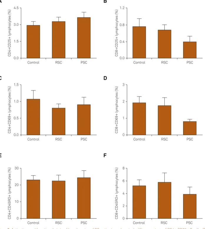
To investigate lymphocyte activation and functional
proiles, we analyzed the expression of the surface markers
of recent and chronic activation (CD69 and CD25, respec-tively) in CD4+ and CD8+ T lymphocytes (Figure 2). Here
again, we found no diferences among the three evaluated groups of subjects (PSC, RSC and controls). he frequencies of CD4+CD25+ T cells (Figure 2A) were RSC = 3.30 ± 1.18%, PSC = 3.65 ± 1.06% and controls = 2.95 ± 1.16%. he percentages of CD8+CD25+ T cells are given in Figure 2B (RSC = 0.68 ± 0.37%; PSC = 0.40 ± 0.30% and controls = 0.76 ± 0.65%). Figure 2C shows the percentages of CD4+CD69+ T cells (RSC = 0.80 ± 0.38%; PSC = 0.90 ± 0.50% and controls = 1.07 ± 0.90%). Lastly,
Figure 1D (RSC = 1.76 ± 1.42%; PSC = 0.80 ± 0.30% and con
-trols = 1.91 ± 1.32%).
We also evaluated a surface marker of memory (CD45RO)
in CD4+ and CD8+ T lymphocytes. No diferences were found among groups. he percentages of CD4+CD45RO+
and CD8+CD45RO+ memory T cells are given in
Figure 1E (RSC = 22.34 ± 10.16%; PSC = 24.32 ± 8.85% and controls = 21.02 ± 10.19%) and Figure 1F (RSC = 5.76 ± 4.53%; PSC = 3.85 ± 2.61% and controls = 4.79 ± 3.33%), respectively.
Additionally, we found no diferences when analyz -ing PBMC cultured under the presence or not of polyclonal (anti-CD3/CD28) stimuli (data not shown).
DISCUSSION
We designed this study primarily to investigate a pos-sible role of lymphocytes in the development and/or
persistence of chorea in SC. We found no diferences among
groups in all of the evaluated immunological parameters.
To the best of our knowledge, this is the irst study assessing
lymphocyte subsets in SC.
More specifically, we were interested in assessing CD4+ and CD8+ T lymphocytes as well as B1 cells in SC
patients in comparison with asymptomatic subjects. While CD4+ T cells are involved in the orchestration of immune response, CD8+ T cells can promote target cells destruction15. Changes in lymphocyte profile may be
asso-ciated with a series of autoimmune disorders. In the case of SC, this is particularly relevant for B1 cells that are in-volved in antibody-mediated autoimmune conditions13.
Our results failed to demonstrate any significant differ-ence between patients and controls in the composition of lymphocyte subsets, indicating that there is no evidence of sustained lymphocyte activation in PSC. Our work group has previously described a decrease in the percent-age of CD14+ monocytes in the peripheral blood of SC patients. In addition, we observed reduced frequency of CD14+CD86+ and CD14+HLA-DR+ cells in SC, indicating that monocytes might play a role in the immune mecha-nisms underlying SC pathogenesis16.
Non-immune mechanisms may be associated with the pathogenesis of PSC. Basal ganglia structural damage may occur during the acute phase of SC, leading to persistent in-voluntary choreic movements in a subgroup of patients. In line with this hypothesis, neuroimaging studies have dem-onstrated that patients who remitted completely their cho-reic movements did not present lesions of the basal ganglia
CD19+ lymphocytes (%)
CD19+CD5+ lymphocytes (%)
Control RSC PSC Control RSC PSC
CD4+ lymphocytes (%)
15
10
5
0
60
40
20
0
CD8+ lymphocytes (%)
0.8
0.6
0.4
0.2
0.0
25
20
15
5 10
0
Control RSC PSC Control RSC PSC
Figure 1. T and B lymphocyte frequencies of SC patients and controls. (A) percentage of B (CD19+) cells expression; (B) percentage of B1 cells expression (CD19+ CD5+); (C) frequency CD4+ T lymphocytes expression; (D) percentage of CD8+ T lymphocytes expression. RSC = remitted Sydenham’s chorea; PSC = persistent Sydenham’s chorea.
A
B
after the acute phase of the disease. Conversely, patients with persistent basal ganglia lesions were more prone to develop recurrences of chorea17.
Altogether, our data suggest that the persistence of choreic involuntary movements in SC is not associated with
Figure 2. Activation and functional state of lymphocytes of SC patients and controls. (A) percentage of CD4+ CD25+ T cells; (B) percentage of CD8+ CD25+ T cells expression; (C) frequency of CD4+ CD69+ T lymphocytes expression; (D) frequency of CD8+ CD69+ T lymphocytes expression; (E) frequency of CD4+ CD45RO+ memory T cells; (F) CD8+ CD45RO+ memory T cells expression. RSC = remitted Sydenham’s chorea; PSC = persistent Sydenham’s chorea.
CD4+CD25+ lymphocytes (%) CD8+CD25+ lymphocytes (%)
Control RSC PSC Control RSC PSC
CD4+CD69+ lymphocytes (%) CD8+CD69+ lymphocytes (%)
Control RSC PSC Control RSC PSC
CD4+CD45RO+ lymphocytes (%)
4.5
3.0
1.5
0.0
1.5
1.0
0.5
0.0
30
20
10
0 CD8+CD45RO+ lymphocytes (%)
1.2
0.8
0.4
0.0
3
2
1
0
8
6
4
2
0
Control RSC PSC Control RSC PSC
A
B
C
E
D
F
persistent lymphocyte dysfunction. Specially, the percentage of B1 cells – a subset of B lymphocytes increased in
autoim-mune conditions – are not changed in RSC or PSC. herefore,
References
1. Cardoso F. Chorea: non-genetic causes. Curr Opin Neurol. 2004;17(4):433-6. doi:10.1097/01.wco.0000137533.53620.59
2. Maia DP, Teixeira AL, Quintão Cunningham MC, Cardoso F. Obsessive compulsive behavior, hyperactivity, and attention deicit disorder in Sydenham chorea. Neurology. 2005;64(10):1799-801. doi:10.1212/01.WNL.0000161840.62090.0E
3. Teixeira AL, Maia DP, Cardoso F. UFMG Sydenham’s chorea rating scale (USCRS): reliability and consistency. Mov Disord. 2005;20(5):585-91. doi:10.1002/mds.2037
4. Cardoso F, Eduardo C, Silva AP, Mota CC. Chorea in ifty consecutive patients with rheumatic fever. Mov Disord. 1997;12(5):701-3. doi:10.1002/mds.870120512
5. Church AJ, Dale RC, Cardoso F, Candler PM, Chapman MD, Allen ML et al. CSF and serum immune parameters in Sydenham’s chorea: evidence of an autoimmune syndrome? J Neuroimmunol. 2003;136(1-2):149-53. doi:10.1016/S0165-5728(03)00012-2
6. Teixeira AL, Cardoso F, Souza AL, Teixeira MM. Increased serum concentrations of monokine induced by interferon-gamma/ CXCL9 and interferon-gamma-inducible protein 10/CXCL-10 in Sydenham’s chorea patients. J Neuroimmunol. 2004;150(1-2):157-62. doi:10.1016/j.jneuroim.2004.01.013
7. Brilot F, Merheb V, Ding A, Murphy T, Dale RC. Antibody binding to neuronal surface in Sydenham chorea, but not in PANDAS or Tourette syndrome. Neurology. 2011;76(17):1508-13. doi:10.1212/WNL.0b013e3182181090
8. Teixeira AL, Maia DP, Cardoso F. Treatment of acute Sydenham’s chorea with methyl-prednisolone pulse-therapy. Parkinsonism Relat
Disord. 2005;11(5):327-30. doi:10.1016/j.parkreldis.2005.02.007
9. Kirvan CA, Swedo SE, Heuser JS, Cunningham MW. Mimicry and autoantibody-mediated neuronal cell signaling in Sydenham chorea. Nat Med. 2003;9(7):914-20. doi:10.1038/nm892
10. Teixeira AL, Guimarães MM, Romano-Silva MA, Cardoso F. Serum from Sydenham’s chorea patients modiies intracellular calcium levels in PC12 cells by a complement-independent mechanism. Mov Disord. 2005;20(7):843-45. doi:10.1002/mds.20418
11. Doyle F, Cardoso F, Lopes L, Mendes M, Dias F, Cruz L et al. Infusion of Sydenham’s chorea antibodies in striatum with up-regulated dopaminergic receptors: a pilot study to investigate the potential of SC antibodies to increase dopaminergic activity. Neurosci Lett. 2012;523(2):186-9. doi:10.1016/j.neulet.2012.06.073
12. Cardoso F, Vargas AP, Oliveira LD, Guerra AA, Amaral SV. Persistent Sydenham’s chorea. Mov Disord. 1999;14(5):805-7. doi:10.1002/1531-8257(199909)14:5<805::AID-MDS1013>3.0.CO;2-P
13. Mix E, Goertsches R, Zett UK. Immunoglobulins: basic considerations. J Neurol. 2006;253(5):v9-17. doi:10.1007/s00415-006-5002-2
14. Dajani AS, Aypub E, Bierman FZ, Bisno AL, Denny RW, Durack DT et al.. Guidelines for the diagnosis of rheumatic fever: Jones criteria, 1992 update. JAMA. 1992;268(15):2069-73. doi:10.1001/jama.1992.03490150121036
15. Jiang H, Chess L. Regulation of immune responses by T cells. N Engl J Med. 2006;354(11):1166-76. doi:10.1056/NEJMra055446
16. Torres KC, Dutra WO, de Rezende VB, Cardoso F, Gollob KJ, Teixeira AL. Monocyte dysfunction in Sydenham’s chorea patients. Hum Immunol. 2010;71(4):351-4. doi:10.1016/j.humimm.2010.01.007