DOI: 10.1590/0004-282X20150180
ARTICLE
Migraine patients consistently show abnormal
vestibular bedside tests
Pacientes enxaquecosos consistentemente apresentam testes vestibulares de
beira-de-leito anormais
Eliana Teixeira Maranhão1,2, Péricles Maranhão-Filho3, Ronir Raggio Luiz4, Maurice Borges Vincent3
In neurological and otorrinolaryngological outpatients units migraine and vertigo are the two most common complains1. Vestibular syndromes, dizziness, vertiginous
syn-dromes as benign positional paroxysmal vertigo, Ménière disease, motion sickness, cerebellar and anxiety disorders, in whom very often dizziness is present, all occur more frequently amongst migraineurs (30-50%)2,3,4. It is well known
that the equilibrium depends on an interplay between vestib-ular mechanisms, proprioception and visual inputs. Available data suggest that this circuitry could be dysfunctional in migraine. However, the issue as how, why and to which
extent vestibular changes do occur in those subjects remains
highly controversial. he objective of the present study was to test the hypothesis that bedside clinical tests speciically
designed to detect subtle vestibular dysfunctions are abnor-mal in migraine in the absence of vestibular complains.
METHOD
In this cross sectional study, 30 (ICHD-2004, 2nd Ed. Cod:1;
1.1 and 1.2) interictal phase migraine patients, sequentially
1Instituto Nacional de Câncer (INCA), Setor de Fisioterapia, Rio de Janeiro RJ, Brazil; 2American Physical Therapy Association for Vestibular Rehabilitation, Alexandria VA, USA;
3Universidade Federal do Rio de Janeiro, Hospital Universitário Clementino Fraga Filho, Serviço de Neurologia, Rio de Janeiro RJ, Brazil; 4Universidade Federal do Rio de Janeiro, Instituto de Estudos de Saúde Pública, Rio de Janeiro RJ, Brazil.
Correspondence: Eliana Teixeira Maranhão; Avenida das Américas, 1155/1705; 22631-000 Rio de Janeiro RJ, Brasil; E-mail: [email protected]
Conflict of interest: There is no conlict of interest to declare.
Received 26 June 2015; Received in inal form 12 August 2015; Accepted 01 September 2015.
ABSTRACT
Migraine and vertigo are common disorders, with lifetime prevalences of 16% and 7% respectively, and co-morbidity around 3.2%. Vestibular syndromes and dizziness occur more frequently in migraine patients. We investigated bedside clinical signs indicative of vestibular dysfunction in migraineurs. Objective: To test the hypothesis that vestibulo-ocular relex, vestibulo-spinal relex and fall risk (FR) responses as measured by 14 bedside tests are abnormal in migraineurs without vertigo, as compared with controls. Method: Cross-sectional study including sixty individuals – thirty migraineurs, 25 women, 19-60 y-o; and 30 gender/age healthy paired controls. Results: Migraineurs showed a tendency to perform worse in almost all tests, albeit only the Romberg tandem test was statistically different from controls. A combination of four abnormal tests better discriminated the two groups (93.3% speciicity). Conclusion: Migraine patients consistently showed abnormal vestibular bedside tests when compared with controls.
Keywords: migraine, vertigo, bedside tests. RESUMO
Enxaqueca e vertigem são desordens comuns, com prevalência de 16% e 7% respectivamente, e comorbidade em torno de 3,2%. Síndromes vestibulares e tonturas ocorrem mais frequentemente em enxaquecosos. Pesquisamos alterações vestibulares utilizando testes de beira-de-leito em enxaquecosos. Objetivo: Veriicar se as respostas dos relexos vestíbulo-ocular, vestíbulo-medular e risco de quedas medidas por 14 testes de beira-de-leito são diferentes comparando-se enxaquecosos sem vertigem, e controles. Método: Estudo transversal com sessenta pessoas, 30 enxaquecosos; 25 mulheres, 19-60 anos; e trinta controles saudáveis pareados por sexo e idade. Resultados: Houve tendência de pior desempenho entre enxaquecosos em quase todos testes, porém apenas o teste de Romberg tandem foi estatisticamente diferente dos controles. Uma combinação de quatro testes anormais discrimina os grupos com especiicidade de 93,3%. Conclusão: O grupo de enxaquecosos mostrou consistentemente testes vestibulares de beira-de-leito anormais quando comparados a controles.
recruited in our unit and 30 matched healthy controls (pa-tients partners and medical students) were blindly examined by the same researcher (ETM) after diagnosis. Inclusion and exclusion criteria were checked by well-trained neurologists with long headache expertise (PM-F and MBV). All tests 5-18
(Table 1), were performed during one single session at the same hospital facility. Inclusion criteria were age between 18
and 65 years; migraine for at least 2 years with a minimum frequency of 2 attacks montly; and at least 1 brain Magnetic Ressonance (MRI) or Computerized Tomography (CT) scan within normal limits. Exclusion criteria were informed or clinically evidenced pregnancy; two or more tension-type headache episodes (ICHD-2004, 2nd Ed. Cod:2; 2.1, 2.2) in the
previous week, chronic tension type headache (coded 2.3) or
Table 1. Tests for Vestibulo-ocular Relex (VOR), Vestibulo-spinal Relex (VSR) and fall risk (FR) used in clinical evaluation.
Clinical test Method Positive result
Vestibulo-Ocular Relex
Head Impulse Test (HIT)5
Clinician, facing and holding the subject’s head at arm’s length, performs a passive and unpredictable head rotational movement in a high acceleration ~ 20° to either side having the subject’s eyes ixed on the examiner’s nose. I may also be
done vertically (Figure 1A, B, C).
Corrective saccade back to the target
Head Shaking Test (HST)6
Subject’s wearing Frenzel goggle with the head positioned downwards by 30°. The examiner rapidly oscillate the head (2Hz) 20 times. When the movement stops the
clinician observes the presence of nystagmus.
Nystagmus; more than 5 beats
Dynamic Visual Acuity (DVA)7
Subject reads the lowest possible line in a ETDRS chart positioned 2 meters ahead, than the examiner manually oscillates the subject’s head horizontally at 2
Hz in each direction and the subject tries to read the same line (Figure 1D, E).
Deviation of more than 2 lines above base line
Subjective Visual Vertical (SVV)8
Looking inside a bucket, without any peripheral vision, the subject vertically redirect a line drawn on the inner bottom of the bucket under the subject’s subjective perception. Results measured outside with a pendulum hanging over a
protractor (Figure 1F, G, H).
More than 2.5° deviation from 0° to either side
Minimal Ice Test (MIT)9 Subject is itted with a Frenzel goggle and 0.2 mL iced water ( 1-3°C) is plunged in
the ear canal with the head turned to side and bended 30° forward. After 10 sec the head is turned straigth and nystagmus is observed in the computer screen.
After 5 min the same procedure is performed on the other ear.
≠ between ears responses > 25% = canal
paresis
Vestibulo Spinal Relex
Modiied Clinical Test of Sensory Interaction and Balance
(mCTSIB)10
The subject stands on a irm surface with the feet slightly apart and crossed arms, irst with eyes open and than with eyes closed, for 30 sec each. The same
manoeuvre is repeated on a high density foam (Figure 2D).
Normal and Minimal sway: grades 1 and 2. Moderate and Severe: grades 3 and 4
Romberg Tandem Test (RTT)11
Subject stand with feet slightly apart and arms folded across the chest with eyes open for 30 seconds and then with eyes closed for 30 seconds.
Marked sway, or: move the feet, open the eyes, uncross
the arms
Past Pointing Test (PPT)12
The subject is instructed to extend the arms and place the index inger of one hand on the index inger of the examiner. The eyes are then closed, the arm raised
above the head, then quickly returned to the perceived starting position. The procedure is repeated ive times (Figure 2A, B, C).
Index drift away from the target towards the
compromised side
Fukuda Test (FT)13 Subject with arms extended and eyes closed, stepping in place with examiner
counting for 50 steps (Figure 2E, F).
Moves more than 50 cm ahead and turns toward one side of more than 45°
Fall Risk
Timed Up and Go (TUG)14
Subject rises from a standard height chair with armrests, walks 3m, turns, and returns to sit in the chair. The time to complete the path is measured with a
stopwatch.
More than 13.5 sec to complete the task
5 Times Sit to Stand Test (5TSST)15
Subject seated in a chair with arms crossed under chest and feet itted on the ground. Upon request, stands up and sit down for 5 times. Time measured with a
stopwatch.
More than 10 seconds to complete the task
Forward Reaching Test (FRT)16
Subject is asked to bend the trunk and reach as far forward as possible having both feet ixed and parallel. The extent of movement is measured with a simple
yardstick.
Score 15 cm or less indicate high risk for fall
Dynamic Gait Index (DGI)17
The subject is observed during an eight-item task within a 21 feet path walking. Score of 19 or less out of 24 points
Pull Test (PT)18 The subject stands with the feet slightly spread apart and is pulled backward by
the shoulders (one or two steps backwards allowed) (Figure 2G).
More than 2-3 steps to keep the balance or fall
other headache listed in groups 3-4 (ICHD, 2004) according to clinical evaluation; circulatory, respiratory, orthopedic ab-normalities or generalized weakness/tiredness of any nature
which could inluence/preclude the clinical tests; Ménière
disease, vestibular neuritis, benign positional paroxysmal vertigo or phobic vertigo; spontaneous nystagmus; any
sen-sitive, sensorineural, motor or coordination deicits; more than 150 mL cofee, 3 tea cups, or 2 soda cans intake per day
for the last 2 days, or any amount of alcoholic beverages, to-bacco, anti-vertiginous drugs, anti-depressive, anxiolytic antiemetic, or pain-killers more than b.i.d. use. All subjects underwent a neurootological examination including the minimal ice test (MIT)9. For the MIT a diference between
right and left responses greater than 25% was considered as a canal paresis19. he Wilcoxon Signed Rank, the McNemar
chi-square, and the Student t test were used for statistical
com-parisons; p-values < 0.05 were considered signiicant. he lo -cal Ethics Committee approved this study (Protocol:159-11).
RESULTS
Participants were mostly females (83.3%). Migraine patients were aged 19-62 (median 39.3) years and controls 22-62 (median 38.9) years. Considering all tests togeth-er thtogeth-ere was a clear tendency for migraineurs to ptogeth-erform
worse than controls (p = 0.003), except for the SVV and the FRT (Table 2). he tests involving the vestibulo-ocular relex
(VOR) assessment (Figure 1) in general did not distinguish patients from healthy controls (p < 0.05).
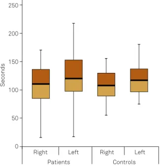
For the MIT the nystagmus duration ranged from 16 to
218 seconds in patients. he average percentage diference
between R-L ears was 20.97 ± 15.9% in the patients group and 18 ± 11.5% in controls (Figure 2).
Regarding the vestibulo-spinal relex (VSR) (Figure 3), only the RTT was statistically diferent between the two groups (p = 0.039). For the the m-CTSIB, considering only the
responses graded as 3 and 4, six migraine patients (20%) and three control subjects (10%) showed abnormal responses.
he 5TSST and the PT tests were nearly statistically dif
-ferent (p = 0,074) in contrast with other Fall Risk (FR) tests.
A combination of 4 abnormal tests distinguish patients
from controls with a sensitivity of 23.3% and a speciicity of
93.3% (Table 3).
DISCUSSION
he present study shows that vertigo-free migraine pa -tients, during the interictal phase, perform worse than controls in bedside clinical vestibular tests independently from vestibular complains.
Although various techniques may be used to address the vestibular function in migraine patients with or without
vertigo20-25, data considering vestibular bedside tests in this
context are scarce.
Marcelli et al.26 evaluated otoneurological
abnomali-ties in 22 migraineurs with vestibular symptoms as com-pared with 18 control migraineurs without vestibular
symp-toms. Seventy-three percent of the subjects in the irst
group had vestibular dysfunctions – peripheral, central or
both – contrasting with 33% in the second group. he au -thors suggest the presence of a subclinical vestibular system involvement in the vertigo-free migraine patients.
Chronic headache causes postural instability, which could be the result of a sensory information rearrangement leading to greater vestibular system contribution paralleled by a reduced visual component in the equilibrium control24.
If this is true, migraineurs would be comparatively more dependent on vestibular functions for postural control, jus-tifying the present results.
he tests we used to evaluate the VOR were not statistical
-ly diferent comparing the two groups. he response evoked
by high accelerations (HIT) is controlled by a variety of dif-ferent sensory and motor systems apart from the vestibular
system. he VOR response can change within few minutes,
pointing to a VOR plasticity7, but this is probably not
suf-icient to overcome the inadequate vestibular inputs and
generate adequate responses. It is possible that chronic
mi-graineurs develop the same phenomena identiied in subjects
with loss of vestibular function such as hidden saccades or other strategies to compensate a putative vestibular failure. Likewise, patients may increase the gain of the cervico-ocular
relex, and possibly the smooth pursuit system.
he DVA clearly tended to show worse results amongst patients although the SVV assessment was quite similar in both groups. Neck stifness could putatively interfere with
the DVA, as it has been shown that neck motility may be im-paired in migraine27.
MIT is a clean and quick monothermal bedside test, es-sentially a low-frequency stimulus caloric test that can detect vestibular impairment not apparent during higher-frequency head rotation28,29. In accordance with the other tests, data also
point to a vestibular instability in migraineurs, as relected
by a greater right-to-left asymmetry and a greater response spread amongst patients.
Apropos the VSR tests, Akdal et al.30 carried out a balance
and gait comparative study in 30 patients with vestibular migraine, 26 migraine without vertigo subjects, and 30 individuals whithout migraine. Among other tests and scales
they also used the CTSIB and the DGI, concluding that ves
-tibular migraine suferers, deined according to the Lempert and Neuhauser criteria4, as well as the migraineurs without
vertigo, presented subnormal static and dynamic balance,
as well as increased fall risk (FR). Góes31 used a
stabilomet-ric test to assess the static equilibrium of migraine patients
interictally. he stabilometric parameters did not difer
patients showed a signiicantly greater lateral oscillation
than migraineurs without aura. In our study, the RTT was the most sensitive test, pointing to a dysfunction of the static
equilibrium in migraineurs. Given the clearly unfavorable re -sult for RTT it is possible that the dysfunction in migraine
would be greater in the VSR mechanisms specially involving
proprioceptive impulses. In the m-CTSIB test, when the sub
-ject is placed on the foam, the proprioceptive inluence to the postural relex drops by approximately 60%7. Although the
patients had no anteroposterior imbalance predominance, the migraneurs had three times more abnormal tests than
controls, indicating that in migraine the vestibular inlu -ence on the posture control is comparatively dysfunctional. It remains to be explained wheather this is secondary to
im-paired neck and/or labyrith abnormal relexes in migraine, as
they could modulate the muscular tonus. Stretch relexes re
-ceive important contribution from vestibulo-spinal respons-es, proprioceptive signs from the trunk and neck, or both,
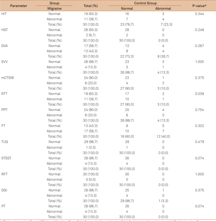
Table 2. Distribution of the tests for the Vestibulo-ocular Relex, Vestibulo-spinal Relex and Fall Risk in 30 migraine patients and 30 controls, paired for age and sex.
Parameter Group Total (%) Control Group P-value*
Migraine Normal Abnormal
HIT Normal 19 (63.3) 16 3 0.344
Abnormal 11 (36.7) 7 4
Total (%) 30 (100.0) 23 (76.7) 7 (23.3)
HST Normal 28 (93.3) 28 0 0.248
Abnormal 2 (6.7) 2 0
Total (%) 30 (100.0) 30 (100.0) 0 (0.0)
DVA Normal 17 (56.7) 13 4 0.267
Abnormal 13 (43.3) 9 4
Total (%) 30 (100.0) 22 (73.3) 8 (26.7)
SVV Normal 26 (86.7) 23 3 1.000
Abnormal 4 (13.3) 3 1
Total (%) 30 (100.0) 26 (86.7) 4 (13.3)
mCTSIB Normal 24 (80.0) 23 1 0.375
Abnormal 6 (20.0) 4 2
Total (%) 30 (100.0) 27 (90.0) 3 (10.0)
RTT Normal 19 (63.3) 17 2 0.039
Abnormal 11 (36.7) 10 1
Total (%) 30 (100.0) 27 (90.0) 3 (10.0)
PPT Normal 24 (80.0) 20 4 0.754
Abnormal 6 (20.0) 6 0
Total (%) 30 (100.0) 26 (86.7) 4 (13.3)
FT Normal 13 (43.3) 8 5 0.302
Abnormal 17 (56.7) 10 7
Total (%) 30 (100.0) 18 (60.0) 12 (40.0)
TUG Normal 29 (96.7) 29 0 0.479
Abnormal 1 (3.3) 1 0
Total (%) 30 (100.0) 30 (100.0) 0 (0.0)
5TSST Normal 26 (86.7) 26 0 0.074
Abnormal 4 (13.3) 4 0
Total (%) 30 (100.0) 30 (100.0) 0 (0.0)
RFT Normal 30 (100.0) 30 0 1.000
Abnormal 0 (0.0) 0 0
Total (%) 30 (100.0) 30 (100.0) 0 (0.0)
DGI Normal 26 (86.7) 25 1 0.375
Abnormal 4 (13.3) 4 0
Total (%) 30 (100.0) 29 (96.7) 1 (3.3)
PT Normal 26 (86.7) 26 0 0.074
Abnormal 4 (13.3) 4 0
Total (%) 30 (100.0) 30 (100.0) 0 (0.0)
visual stimulus, and vonluntary responses32. herefore, it is
possible the subnormal vestibular function in migraine could
increase the FR by stretch relexes impairment.
In patients, the FR tests pointed to a subtle fall tendency.
Only one patient showed increased FR according to the TUG
test. Four migraneurs (13.3%), showed increased FR in the
5TSST, while none of the controls exceeded this test time lim -its (10 and 14.2 seconds, according to age). According to the FRT none of the sixty examined subjects showed an increased
FR, indicating that the vestibular postural dysfunctions in migraine cannot be assessed by this test. Observing the FRT response in association with other vestibular-spinal tests we conclude that the anterior tilt does not seem to be
particu-larly afected in migraine.
Given the clearly abnormal result for RTT in migraine pa
-tients, it is possible this disease impairs mainly VSR mecha
-nisms, specially involving proprioceptive impulses. Since the
RTT preferably tests a side-to-side control, our results are in
line with the indings of Góes31, in which the laleral
oscilla-tion was greater in migraineurs examined by stabilometry.
In the DGI, 13.3% of patients had a score of less than 19
points, i.e. suggestive of increased FR, while only one control
(3.3%) scored for increased FR. Since this test requires dif -ferent skills, it may be more sensitive than other FR
assess-ment methods. he PT also shows that migraineurs present postural diiculties in the anteroposterior direction. he an
-ticipatory postural relexes are specially important in the an
-teroposterior instability provoked by pulling the patient. he
corporal shift deterioration is an important factor for gait de-sarrangement in the elderly33, but this cannot justify the
com-paratively worse PT results in migraineurs as they were not
old. he anteroposterior instability in migraine seems to be
better assessed by the PT than by the FRT.
he weaknesses of this work are the relatively small sam -ple, the relatively lower age of patients for FR assessment, the lack of a migraine with vestibular symptoms group and other primary headache groups, and a lack of subsequent testing
to conirm results consistency. Due to the high variation of
the so-called “vestibular migraine” and the pathophysiolog-ical uncertainties related to this diagnosis we believe such patients cannot be selected by reliable criteria yet. Ideally,
longitudinal and interventional studies speciically designed
to assess vestibular dysfunction in migraine should be
per-fomed. However, we are conident data are convincing to
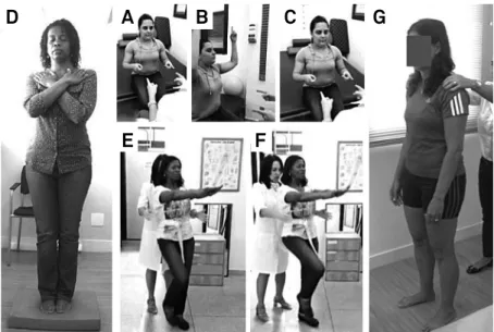
Figure 1. Vestibulo-ocular relex tests. Head impulse test (A, B, C); dynamic visual acuity (D, E); subjective visual vertical (F, G, H).
A
B
C
D
E
F
G
H
Figure 2. Vestibulo-spinal relex tests. Past pointing (A, B, C); m-CTSIB step 4 (D); Fukuda test (E, F). fall risk: pull test (G).
A
E
F
References
1. Brant T, Dieterich M, Strupp M. Vertigo and dizziness: common complains. London: Springer; 2009.
2. Shepard NT, Asher A. Non-vestibular dizziness and imbalance: suggestions for patients with migraine and mal de débarquement disequilibrium. In: Herdman SJ. Vestibular rehabilitation. 3rd ed. Philadelphia: E.A. Davis; 2007. p. 460-2. (Contemporary perspectives in rehabilitation).
3. Baier B, Winkenwerder E, Dieterich M. “Vestibular migraine”: effects of prophylactic therapy with various drugs: a restropective study. J Neurol 2009;256(3):436-42. doi:10.1007/s00415-009-0111-3
4. Lempert T, Neuhauser H. Epidemiology of vertigo, migraine and vestibular migraine. J Neurol. 2009;256(3):333-8. doi:10.1007/s00415-009-0149-2
5. Halmagyi GM, Curthoys IS. A clinical sign of canal paresis. Arch Neurol. 1988;45(7):737-9. doi:10.1001/archneur.1988.00520310043015
6. Asawavichiangianda S, Fujimoto M., Mai M, Desroches H, Rutka J. Signiicance of head-shaking nystagmus in the evaluation of the dizzy patient. Acta Otolaryngol Suppl. 1999; 540:27-33. doi:10.1080/00016489950181152
7. Herdman SJ, Clendaniel RA. Vestibular rehabilitation. 4h ed. Philadelphia: FA Davis; 2014.
8. Zwergal A, Rettinger N, Frenzel C, Dieterich M, Brandt T, Strupp M. A bucket of static vestibular function. Neurology. 2009;7(19)2:1689-92. doi:10.1212/WNL.0b013e3181a55ecf
9. Linthicum Jr FH, Churchill D. Vestibular test results in acoustic tumor cases. Arch Otolaryng. 1968;88(6):604-7. doi:10.1001/archotol.1968.00770010606007
10. Shumway-Cook A, Horak FB. Assessing the inluence of sensory interaction of balance: suggestion from the ield. Phys Ther. 1986;66(10):1548-50.
11. Baloh RW, Honrubia V. Clinical neurophysiology of the vestibular system. 3rd ed. New York: Oxford University Press; 2001.
12. Barany R, 1910 apud Devin L, McCaslin, Dundas JA, Jacobson GP. The bedside assessment of the vestibular system. In: Jacobson GP, Shepard NT. Balance function assessment and management. San Diego: Plural; 2008. p. 90.
13. Fukuda T. The stepping test: two phases of the labyrinthine relex. Acta Otolaryngol[Stockh]. 1959;50(1-2):95-108. doi:10.3109/00016485909129172
14. Podsiadlo D, Richardson S. The timed “up & go”: a test of basic functional mobility for frail elderly persons. J Am Geriatr Soc. 1991;39(2):142-8. doi:10.1111/j.1532-5415.1991.tb01616.x
15. Guralnik JM, Simonsick EM, Ferrucci L, Glynn RJ, Berkman LFet al. A short physical performance battery assessing lower extremity function: association with self-reported disability and prediction of mortality and nursing home admission. J Gerontol. 1994;49(2):M85-94. doi:10.1093/geronj/49.2.M85
16. Weiner DK, Duncan PW, Chandler J, Studenski SA. Functional reach: a marker of physical frailty. J Am Geriatr Soc. 1992;40(3):203-7. doi:10.1111/j.1532-5415.1992.tb02068.x
17. Shumway-Cook A, Baldwin M, Olissar NL, Gruber W. Predicting the probability of falls in community dwelling older adults. Phys Ther. 1997;77(8):812-9.
18. Hall CD. Assessment of gait and balance. In: Herdman SJ, Clendaniel RA. Vestibular rehabilitation: a competency-based course. Atlanta: Emory Physical Therapy Association; 2010.
19. Brandt T, Strupp M. General vestibular testing. Clin Neurophysiol. 2005;116(2):406-26. doi:10.1016/j.clinph.2004.08.009
20. Bolay H, Bayazit YA, Gunduz B, Ugur AK, Akçali D, Altunyay S et al. Subclinical dysfunction of cochlea and cochlear efferents in migraine: an otoacoustic emission study. Cephalalgia. 2008;28(4):309-17. doi:10.1111/j.1468-2982.2008.01534.x
21. Dash AK, Panda N, Khandelwal G, Lal V, Mann SS. Migraine and audiovestibular dysfunction: is there a correlation? Am J Otolaryngol. 2008;29(5):295-99. doi:10.1016/j.amjoto.2007.09.004
Seconds
250
200
150
100
50
0
Right Left Right Left
Patients Controls
Figure 3. MIT. Patients and controls average nystagmus duration of caloric induced nystagmus in seconds. Box and whiskers plot showing the results of the MIT in seconds. Notice the grater response variability amongst patients. Box right (right ear), left (left ear).
Table 3. Power rating of discrimination scores between migraineurs and controls for different cutoff points.
Cutoff points* Sensitivity Speciicity
1 96.7% 200%
2 70.0% 60.0%
3 50.0% 86.7%
4 23.3% 93.3%
5 20.0% 100.0%
6 10.0% 100.0%
* Positive for values higher or equal to score that counts the number of abnormal results in 13 tests.
demonstrate that migraineurs present subclinical vestibular
dysfunctions that may be detected at the bedside. his is in
line with the high prevalence of vertigo and unbalance com-plains amongst migraineurs.
In conclusion, migraine patients consistently showed worse vestibulo-ocular, vestibulo-spinal and FR bedside tests
as compared with controls. he results indicate that the ves -tibular function is impaired subclinically in migraine without vertigo complains. Bedside tests are suitable to detect such
22. Baier B, Stieber N, Dieterich M. Vestibular-evoked myogenic potentials in vestibular migraine. J Neurol. 2009;256(9):1447-54. doi:10.1007/s00415-009-5132-4
23. Arriaga MA, Chen DA, Hillman TA, Kunschner L, Arriaga RY. Visually enhanced vestibulo-ocular relex: a diagnostic tool for migraine vestibulopathy. Laryngoscope. 2006;116(9):1577-9. doi:10.1097/01.mlg.0000231308.48145.f6
24. Çelebisoy N, Gokcay F, Sirin H, Biçak N. Migrainous vertigo: clinical, oculographic and posturographic indings. Cephalalgia. 2008;28(1):72-7. doi:10.1111/j.1468-2982.2007.01474.x
25. Teggi R, Colombo B, Bernasconi L, Bellini C, Comi G, Bussi M. Migrainous vertigo: results of caloric testing and stabilometric indings. Headache. 2009;49(3):435-44. doi:10.1111/j.1526-4610.2009.01338.x
26. Marcelli V., Furia T., Marciano E. Vestibular pathways involvement in children with migraine: a neuro-otological study. Headache. 2010; 50(1):71-6. doi:10.1111/j.1526-4610.2009.01454.x
27. Tali D, Menahem I, Vered E, Kalichman L. Upper cervical mobility, posture and myofascial trigger points in subjects with episodic
migraine: case-control study. J Bodyw Mov Ther. 2014;18(4):569-75. doi:10.1016/j.jbmt.2014.01.006
28. Schmäl F, Lübben B, Weiberg K, Stoll W. The minimal ice water caloric test compared with established vestibular caloric test procedures. J Vestib Res. 2005;15(4):215-24.
29. Brevern M, Zeise D, Neuhauser H, Clarke AH, Lempert T. Acute migrainous vertigo: clinical and oculographic indings. Brain. 2005;128(2):365-74. doi:10.1093/brain/awh351
30. AkdalG., BalciB. Evaluation of balance in vestibular migraine and migraine patients without history of vertigo. Neurology. 2013;80(meeting abstracts 1):261.
31. Góes CPQF. Avaliação do cerebelo na enxaqueca. Um estudo com estabilometria e ressonância magnética [thesis]. Rio de Janeiro: Universidade Federal do Rio de Janeiro; 2010.
32. Davidoff RA. Skeletal muscle tone and the misunderstood stretch relex. Neurology. 1992;42(5):951-63. doi:10.1212/WNL.42.5.951