DOI: 10.1590/0004-282X20150188
ARTICLE
Anterior temporal lobectomy versus selective
amygdalohippocampectomy in patients with
mesial temporal lobe epilepsy
Lobectomia temporal anterior versus amigdalohipocampectomia seletiva para epilepsia
de lobo temporal mesial
Fábio A. Nascimento1, Luana Antunes Maranha Gatto2, Carlos Silvado3, Maria Joana Mäder-Joaquim4, Marlus Sidney Moro2, Joao Candido Araujo2
It is estimated that one third of patients with seizures
have medically intractable epilepsy (MIE) – deined as fail -ure of two antiepileptic medications given at appropri-ate doses1,2. Temporal lobe epilepsy (TLE) is the most
com-mon form of MIE. he most frequent pathologic substrate
related to this condition is sclerosis and atrophy of the hippo-campus – disease named mesial temporal sclerosis (MTS) 3.
Presently, there are multiple approaches to resection of TLE;, the most common being the standard anterior temporal lo-bectomy (ATL) and selective amygdalohippocampectomy
1University of Toronto, Toronto Western Hospital, Division of Neurology, Toronto, Ontario, Canada;
2Universidade Federal do Paraná, Hospital de Clinicas, Departamento de Neurocirurgia, Curitiba PR, Brazil;
3Universidade Federal do Paraná, Hospital de Clinicas, Departamento de Neurologia, Serviço de Epilepsia e Eletroencefalograma, Curitiba PR, Brazil;
4Universidade Federal do Paraná, Hospital de Clinicas, Departamento de Neurologia, Curitiba PR, Brazil.
Correpondence: Fábio Augusto Nascimento e Silva; 5W-445; 399, Bathurst St. M5T 2S8; Toronto, Ontario, Canada; E-mail: nascimento.fabio.a@gmail.com
Conflict of interest: There is no conlict of interest to declare.
Received 25 May 2015; Received in inal form 31 August 2015; Accepted 22 September 2015.
ABSTRACT
Objective: To contribute our experience with surgical treatment of patients with mesial temporal lobe epilepsy (mTLE) undergoing anterior temporal lobectomy (ATL) or selective amygdalohippocampectomy (SelAH). Method: This is a retrospective observational study. The sample included patients with medically refractory mTLE due to unilateral mesial temporal sclerosis who underwent either ATL or SelAH, at Hospital de Clinicas – UFPR, from 2005 to 2012. We report seizure outcomes, using Engel classiication, cognitive outcomes, using measurements of verbal and visuospatial memories, as well as operative complications. Result: Sixty-seven patients (33 ATL, 34 SelAH) were studied; median follow-up was 64 months. There was no statistically signiicant difference in seizure or neuropsychological outcomes, although verbal memory was more negatively affected in ATL operations on patients’ dominant hemispheres. Higher number of major complications was observed in the ATL group (p = 0.004). Conclusion: Seizure and neuropsychological outcomes did not differ. ATL appeared to be associated with higher risk of complications.
Keywords: temporal lobe epilepsy, amygdalo-hippocampal epilepsy, anterior temporal lobectomy, neuropsychological tests, seizures, postoperative complications.
RESUMO
Objetivo: Contribuir com nossa experiência para o tratamento cirúrgico de pacientes com epilepsia do lobo temporal mesial submetidos a lobectomia temporal anterior (LTA) ou amigdalohipocampectomia seletiva (AHS). Método: Estudo retrospectivo observacional. Foram incluídos pacientes com epilepsia refratária devido a esclerose mesial temporal unilateral, submetidos a LTA ou AHS no Hospital de Clínicas – UFPR, entre 2005-2012. Foram comparados os resultados cognitivos (análises de memórias verbal e visuoespacial), controle de crises (Engel) e complicações cirúrgicas. Resultados: Sessenta e sete pacientes (33 LTA, 34 AHS) foram estudados; o período de acompanhamento médio foi de 64 meses. Não houve diferença no controle das crises ou resultado neuropsicológico, mas a memória verbal foi mais negativamente afetada nos pacientes submetidos à LTA no hemisfério dominante. Maior número de complicações graves ocorreu no grupo de LTA (p = 0.004). Conclusão: Controle de crises e resultados neuropsicológicos não diferiram. LTA pareceu estar associada a um maior risco cirúrgico.
(SelAH). In general, and regardless of the subtype of surgical approach, patients with MTS achieve postoperative seizure freedom in 59-89% of the times4. Studies have been focusing
on the comparison of the two procedures, in terms of seizure and/or neurocognitive outcome, although a consensus is far from being reached.
In this context, this study contributes the Hospital de Clínicas – UFPR experience, as a reference hospital in Brazil,
for the treatment of epilepsy. his is a retrospective observa -tional study assessing seizure and neurocognitive outcomes, as well as postoperative complications, in patients diagnosed with MTS who were submitted to either ATL or SelAH at the Hospital de Clínicas from 2005 and 2012.
METHOD Subjects
his is a retrospective observational study. Patients with
medically refractory TLE due to unilateral MTS who un-derwent resective surgical therapy (either ATL or SelAH) at Hospital de Clínicas – UFPR from 2005 to 2012 were
includ-ed. Data was collected by chart review. he study was ap -proved by the local regulatory board. Informed consent was obtained from every individual involved in this research.
A total of 212 patients diagnosed with medically refrac-tory MTS were submitted to resective surgery at our centre. Of these, sixty-seven met the inclusion criteria and were, in-cluded in this study (33 ATL; 34 SelAH). Median follow-up was 64 months. For the interest of statistical convenience,
we analysed only the data up to the ifth year of follow-up.
Demographic and clinical features of the patients can be seen at Table 1. Both groups were well matched as depicted
in Table 1; therefore, these groups can be deined as being
epidemiologically homogenous.
All patients had pre-surgical workup including video-EEG long-term monitoring, magnetic resonance imaging (MRI),
and neuropsychological evaluation – if required, we performed
additional tests such as invasive EEG, Wada test, SPECT and/or PET-CT –, as well as postoperative neuropsychological testing,
and at least two years of follow-up after surgery. he exclusion
criteria were as follows: (1) age less than 14 years, and (2) se-vere cognitive delay or mental retardation (these would not be able to be assessed by the same neuropsychological tests, what would preclude a reliable singular comparison).
Selection of patients to this study was not inluenced by
genre, ethnicity, social/economical/cultural status, or comor-bidities – including psychiatric disorders (such as depression,
psychosis, and anxiety), since these conditions are frequently
concomitant in patients with TLE5.
Surgical approach
As part of the teaching hospital Hospital de Clínicas – UFPR, our Epilepsy Surgical Program has two
attending neurosurgeons that alternate weekly. One surgeon systematically uses the ATL approach, whereas the other sys-tematically uses the SelAH approach. Patients were assigned
to a surgeon, and consequently a surgical approach based on which week the operation was booked for. herefore, a reli -able comparison between the outcomes of the two surgical approaches is permitted because the patient distribution can be considered as random and the two groups can be consid-ered as homogenous.
Operative techniques (ATL and SelAH)
Surgical treatment for TLE secondary to MTS aims to re-move mesial temporal structures. Partial resection (approxi-mately 3 cm in length) of the hippocampus, amygdala, and parahippocampal gyrus is performed, followed by total re-section of the uncus.
In the ATL approach, the resection involves, in addition
to the mesial structures, the supericial neocortical tempo
-ral gyri. hese gyri should be removed even if they are not
known to be epileptogenic, based on the observation that these structures (despite being healthy) would propagate seizure activity from temporal mesial sites6. Since more
tis-sue is resected, some experts argue that the ATL technique
confers higher chances of medium/long-term seizure con-trol (Figures 1 and 2).
he SelAH technique, on the other hand, spares the
neocortical gyri. Concerning the possible accesses, through which the resection of the hippocampus and adjacent structures is performed, they can be transsylvian, tran-sinsular, subtemporal, or transcortical – via white matter
on the middle temporal gyrus. he latter is the most used
worldwide – and also at our service –, due to lower
opera-tive risks. he rationale behind choosing the SelAH remains
on the fact that the main origin of epileptogenic activity lies on the mesial portion of the temporal lobe; therefore,
addi-tional resection of supericial cortex would not inluence on
seizure control7. Moreover, this technique, for sparing the
temporal neocortex, in theory results in less postoperative
cognition deicits (including memory, language, and behav -iour) (Figures 3, 4, and 5).
Complications
Recent systematic review on complications of epilepsy
surgery proposed a slightly diferent classiication of opera -tive complications8. According to this study, minor
complica-tions would be the ones that resolved within three months after surgical intervention, whereas major complications are those that persisted beyond this period of time. Although this
classiication has been frequently used by other researchers9,
we believe it would not be suitable for this study, given that it rates events exclusively by their duration.
procedure, as well as increase the risk of death (lethal
poten-tial). With this deinition, all major vascular accidents, as well
as severe infections, are considered major postoperative
com-plications. Complications that do not it the above-mentioned
criteria are classiied as minor. In terms of neurological deicits
after procedure, we divided them into two groups, transitory
and permanent, based on a 3-month cut of (transitory if the deicit resolves completely before 3 months postoperatively;
permanent if it does not resolve by the 3rd month).
Neuropsychological testing
All patients were evaluated prior to and at least 6 months following surgery. Individuals with severe cognitive delay or mental retardation – due to the im-possibility of them being assessed by standard neuropsy-chological tests –, as well as those who were not tested post-operatively, were excluded.
he neuropsychological protocol used by our center’s
Epilepsy Service investigates visuospatial and verbal
memo-ries. he former was evaluated by the Rey-Osterrieth Complex Figure Test (ROCF). he parameter of most importance for this study, provided by the ROCF test, was our patients’ delayed recall ability. he verbal memory was evaluated by the Rey Auditory Verbal Learning Test (RAVL). From this test’s results, we could measure our patients’ long-term verbal memory. Table 1. Patient demographics and relevant clinical information.
SelAH(n = 34) n (%) ATL(n = 33) n (%) p-value Total
Gender
Female 14 (41.18) 16 (48.48) 0.627 30
Male 20 (58.82) 17 (51.52) 37
Age at surgery (Ø) 33.4 37.6 0.136 35.46
Age at irst seizure (Ø) 8.3 10.2 0.286 9.23
Duration of epilepsy (Ø) 25.4 27.4 0.552 26.38
Side of resection
Nondominant 15 (44.12) 18 (54.55) 0.467 33
Dominant 19 (55.88) 15 (45.45) 34
Handedness
Right 30 (88.24) 30 (90.91) 1 60
Left 3 (8.82) 2 (6.06) 5
Ambidextrous 1 (2.94) 1 (3.03) 2
Education level (Ø) 8.2 8 0.778 8.1
Systemic comorbidities 6 11 - 17
History of CNS infection 2 2 - 4
History of severe head trauma 2 2 - 4
Family history of epilepsy 2 (5.88) 1 (3.13) 1 3
Additional presurgical workup needed
Wada test 3 (8.82) 1 (3.03) 0.614 4
PET-CT 1 (2.94) 3 (9.09) 0.356 4
SPECT 2 (5.88) 1 (3.03) 1 3
Sphenoidal EEG 1 (2.94) 3 (9.09) 0.288 4
Number of AEDs at time of surgery
1 3 (8.82) 3 (9.09) 0.585 6
2 11 (32.35) 9 (27.27) 20
3 15 (44.12) 19 (57.58) 34
4 5 (14.71) 2 (6.06) 7
Histopathology
Mesial sclerosis 15 29 44
Normal 1 2 3
Not performed. Aspiration only 19 1 20
Hemorrhage only 0 1 1
Admission period (days) 8.7 11.5 0.537 10
Ø: average, in years; CNS, central nervous system; AED: antiepileptic drug; SelAH: selective amygdalohippocampectomy; ATL: anterior temporal lobectomy.
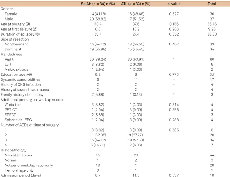
T1
T2
T3
SF
PH
H
T1-weighted coronal MRI; resection is demarcated with gray-colored line. ATL: anterior temporal lobectomy; H: hippocampus; PH: parahippocampal gyrus; T1, T2 e T3: superior, middle, and inferior temporal gyri; SF: Sylvian issure.
Longitudinal neuropsychological evolution (in terms of verbal and visuospatial memories), prior and after surgery, was analysed using both (a) raw psychometric data and (b)
Z-scores. he irst method (a) considered cognitive improve -ment, worsening, or stability comparing neuropsychologi-cal tests raw measurements to the standard deviation of the
control group (the population sample). he second method (b) classiied improvement or worsening according to any change on the classiication of memory based on the Z-score. his score was calculated based on raw neuropsychological
clinical measurements, the mean of the control (population´s sample of Curitiba) for the cognitive test, and the standard
deviation (SD) of the control. We then classiied each patient,
both at the pre and postoperative periods, as having nor-mal memory (Z-score greater than -1.26) or deicits (mild, Z-score ranging from -1.6 to -1.26; moderate, from -2.2 to -1.6; or severe, less than -2.2)10.
RESULTS
Neuropsychological outcome
In pursuance of a broad overview, we compared mem-ory performances of all patients before and after surgery (using the Z-score classification), regardless of the surgical
technique. We found that in terms of verbal memory,
17.9% of patients improved, 23.9% worsened, and 58.2% had no change. In respect of visual memory, 16.7% im-proved, 10.6% worsened, and 72.7% had no change. In the following paragraphs, the results on more detailed analy-ses are shown.
Conforming to the irst analytic method (a), there were no diferences in the evolution of neuropsychological perfor
-mance, in regards to both visuospatial memory (p = 0.182) and verbal memory (p = 0.386), for the two surgical approaches. Similarly, no diferences were found in regards to the side
operated on (Table 2).
We essentially found the same results when using the analytic method (b). Considering the time points prior and
PH
T3
T2
T1
H
SF
WM
T1-weighted coronal MRI. The black line corresponds to the corridor of dissection (with a 3 cm incision) through T2 to the mesial structures. SelAH: selective amygdalohippocampectomy; WM: white matter; H: hippocampus; PH: parahippocampal gyrus. T1, T2 e T3: superior, middle, and inferior temporal gyri; SF: Sylvian issure.
Figure 3. Transcortical access in the SelAH approach.
A
B
T3
T3
T1
T1
SF
SF
LV
LV
T2
T2
(A) Final surgical cavity, after resection and hemostasis. (B) View after suture of the cortical incision. SelAH: selective amygdalohippocampectomy; SF: Sylvian issure; LV: Labbé vein.
Figure 4. SelAH, operative site.
T1
T1-weighted coronal MRI; grey arrow targets the site of status-post right ATL. T2, T3, hippocampus, and parahippocampal gyrus were resected; amygdala was partially removed. ATL: anterior temporal lobectomy.
after surgery, there were no diferences in the distribution of categories both in terms of visual memory (p = 0.117), and of verbal memory (p = 0.817). Of note, in the ATL group,
when looking exclusively at the tests done prior to
opera-tion, less patients had visual memory classiied as normal in relation to the SelAH group (p = 0.027). However, as
shown above, considering the evolution before and after
surgery, this observation did not result in diferences for
either group (Tables 3 and 4).
Further, analyses were done separately for the domi-nant and the nondomidomi-nant subsets. Focusing on the ATL
group, speciically the individuals operated on the dominant
hemisphere, 60 percent of them had normal verbal
memo-ry preoperatively; this igure dropped to 20 percent after
surgery. Still concerning the ATL on the dominant side, 20
percent of patients had severe verbal memory deicit prior to
the ATL; after the procedure, 53.3% of patients were assessed
as having severe verbal memory deicit. In regards to individ -uals who underwent ATL on the nondominant hemisphere, the verbal memory persisted the same without relevant change. Finally, in the whole ATL group, including dominant and nondominant, the visuospatial memory remained with no alteration throughout (Table 3).
When focusing on the SelAH group, on the other hand,
there was no important diference in regards to evolution
of cognitive status (including both verbal and visuospatial memories) – regardless of the side of operation (dominant versus nondominant) (Table 4).
After interpreting all these data and results, it was con-cluded that the only subgroup of patients that experienced
a relevant cognitive deicit after surgery was the set of sub -jects who underwent ATL on the dominant hemisphere. Postoperatively, these patients showed an important decline in cognition, exclusively in verbal memory.
Seizure outcome
he evaluation of postoperative mid/long-term seizure
control was done based on the Engel rating scale11, which
is the most commonly used outcome classiication for epi -lepsy surgery patients12. his evaluation occurred, for every
Table 2. Comparison of longitudinal change (pre and postoperative) in neuropsychological performance, between ATL and SelAH groups, using standard deviation variation.
Verbal memory Visuospatial memory Verbal memory Verbal memory
SelAH n (%) ATL n (%) SelAH n (%) ATL n (%) ND side n (%) Dom side n (%) ND side n (%) Dom side n (%)
Improvement 7 (20.6) 5 (15.2) 4 (12.1) 8 (24.2) 7 (21.2) 5 (14.7) 5 (15.2) 7 (20.6)
Worsening 9 (26.5) 14 (42.4) 2 (6.1) 0 11 (33.3) 12 (35.3) 0 2 (5.9)
Stability 18 (52.9) 14 (42.4) 27 (81.8) 25 (75.8) 15 (45.5) 17 (50.0) 28 (84.8) 25 (73.5)
Total 34 33 33 33 33 34 33 34
p-value 0.386 0.182 0.784 Not applicable
SelAH: selective amygdalohippocampectomy; ATL: anterior temporal lobectomy; Dom: Dominant; ND: Nondominant; n: number of patients. Note: Chi-square tests were applied.
Table 3. Comparison of side of operation (dominant vs. nondominant) in the ATL group in relation to longitudinal change (pre and postoperative) in neuropsychological performance using Z-score for classiication.
ATL
Verbal memory Visuospatial memory
Pre Post Pre Post
ND n (%) Dom n (%) ND n (%) Dom n (%) ND n (%) Dom n (%) ND n (%) Dom n (%)
Normal 11 (61.11) 9 (60.00) 14 (77.78) 3 (20.00) 13 (72.22) 9 (60.00) 15 (83.33) 11 (73.33) Mild 2 (11.11) 2 (13.33) 1 (5.56) 1 (6.67) 3 (16.67) 2 (13.33) 2 (11.11) 2 (13.33) Moderate 2 (11.11) 1 (6.67) 1 (5.56) 3 (20.00) 2 (11.11) 4 (26.67) 1 (5.56) 2 (13.33) Severe 3 (16.67) 3 (20.00) 2 (11.11) 8 (53.33)
Total 18 15 18 15 18 15 18 15
ATL: anterior temporal lobectomy; Dom: Dominant; ND: Nondominant; n: number of patients. Note: Chi-square tests were applied. Mild, moderate and severe refer to degrees of memories deicits.
H
PH
T3
T1
T1-weighted coronal MRI. MRI coronal T1 sequence. Black arrow targets the site of status-post right SelAH, mesial structures were appropriately resected; SelAH: selective amygdalohippocampectomy; H: hippocampus; PH: parahippocampal gyrus.
patient, at the 6th month after procedure, then annually up
until the last clinical visit.
Table 5 shows, by percentage, the Engel rate at each eval-uation for every patient in the study. Further, we categorized
Engel rates I and II as ‘satisfactory’, and plotted the percent
-age of patients ( from both groups, side to side) itting the ‘satisfactory’ category, at the diferent clinical evaluations
(Table 6). Regardless of the surgical approach, the number of patients who were rated Engel I or II was persistently higher than 70% throughout the 5-year period.
After a mean follow-up of 64 months, 82% of all pa-tients had a satisfactory (Engel I or II) outcome, and 51.18%
remained seizure-free (Engel Ia) throughout the irst 5 years
after procedure. In spite of the fact that seizure-freedom (Engel Ia) seemed to be reached more rapidly after SelAH
than ATL during the irst years after surgery, when we look at the entire follow-up of 5 years there are no statistically signii
-cant diferences between the two groups.
Complications
Table 7 summarizes our descriptive classiication (major
or minor) of all surgical complications, and also distinguishes
which of them resulted in neurological deicits (classiied as
being transitory or permanent).
Overall, 19 patients (15 ATL; 4 SelAH), or 28.35%,
devel-oped general complication(s) after surgery. here was not
any death. In the SelAH group, there were two major and two minor complications; also in this set of patients, six
de-veloped postoperative neurological deicits, only one being
permanent. In contrast, in the ATL group, there were 13
ma-jor and two minor complications; neurological deicits were
nine, two being permanent. Comparing the major
compli-cations between the two techniques, rates are higher in the ATL group (p = 0.004). In regards to complications resulting in neurological deicits, there was no signiicant diference between the both approaches (p = 0.370). Besides complica -tions, we believe that patient satisfaction is also important
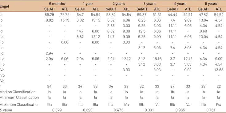
Table 5. Postoperative seizure outcome, according to Engel’s classiication, at predetermined time points.
Engel 6 months 1 year 2 years 3 years 4 years 5 years
SelAH ATL SelAH ATL SelAH ATL SelAH ATL SelAH ATL SelAH ATL
Ia 85.29 72.72 64.7 54.54 58.82 54.54 59.37 51.51 44.44 51.51 47.82 54.54
Ib 8.82 15.15 8.82 15.15 8.82 6.06 6.25 6.06 7.4 9.09 13.04 4.54
Ic - - - - 5.88 3.03 6.25 3.03 11.11 6.06 4.34 4.54
Id - - 14.7 6.06 8.82 9.09 12.5 6.06 11.11 - 8.69
-IIa - - 8.82 12.12 14.7 9.09 6.25 9.09 11.11 6.06 13.04 4.54
IIb - 6.06 - 6.06 - 3.03 - - -
-IIc - - - 3.12 3.03 7.4 3.03 4.34 4.54
IId 2.94 - - -
-IIIa 2.94 6.06 2.94 6.06 2.94 12.12 3.12 15.15 3.7 12.12 4.34 9.09
IIIb - - - 3.12 3.03 3.7 3.03 4.34 4.54
IVa - - - 3.03 - 3.03 - 9.09 - 13.63
IVb - - -
-IVc - - -
-n 34 33 34 33 34 33 32 33 27 33 23 22
Median Classiication Ia Ia Ia Ia Ia Ia Ia Ia Ib Ia Ib Ia
Minimum Classiication Ia Ia Ia Ia Ia Ia Ia Ia Ia Ia Ia Ia
Maximum Classiication IIIa IIIa IIIa IIIa IIIa IVa IIIb IVa IIIb IVa IIIb IVa
p-value 0.379 0.393 0.473 0.331 0.965 0.761
SelAH: selective amygdalohippocampectomy; ATL: anterior temporal lobectomy. Statistical analysis performed with the Mann-Whitney test; p < 0.05. Engel classes were considered as ordinal scales variables (points) ranging from 1 point (for Class Ia) to 13 points (for Class IVc). The numbers refer to the percentage of patients scored in each subgroup in Engel’s Classiication (ENGEL et al, 1993).
Table 4. Comparison of side of operation (dominant vs. nondominant) in the SelAH group in relation to longitudinal change (pre and postoperative) in neuropsychological performance using Z-score for classiication.
SelAH
Verbal memory Visuospatial memory
Pre Post Pre Post
ND n (%) Dom n (%) ND n (%) Dom n (%) ND n (%) Dom n (%) ND n (%) Dom n (%)
Normal 13 (86.67) 8 (42.11) 9 (60.00) 12 (63.16) 12 (85.71) 18 (94.74) 12 (85.71) 18 (94.74)
Mild 1 (6.67) 1 (5.26) 0 (0.00) 1 (5.26)
Moderate 0 (0.00) 2 (10.53) 2 (13.33) 1 (5.26) 2 (14.29) 1 (5.26) 2 (14.29) 1 (5.26) Severe 1 (6.67) 8 (42.11) 4 (26.67) 5 (26.32)
Total 15 19 15 19 14 19 14 19
to consider. For that, we collected patients’ perception af
-ter procedure: 61 of the 67 patients (91%) were satisied, and
would undergo surgery again, if necessary.
DISCUSSION
he aim of this study was to compare the results of two techniques, ATL or SelAH, for patients diagnosed with medi -cally refractory mesial TLE secondary to unilateral MTS, with-out any additional structural lesion. Quite a few studies have
tried to identify the best surgical technique to treat patients
with TLE, few of them, however, included patients with uni-lateral MTS as the sole underlying pathology13,14,15. With this
goal, we carefully selected a series of patients diagnosed with unilateral MTS who were operated by one of our two attend-ing neurosurgeons. Although there was no randomization per se, both groups were epidemiologically homogenous on most variables. Final data analysis was divided in three spheres: sei-zure outcome, cognitive outcome, and complications.
Concerning seizure outcomes, no statistically signiicant diferences in seizure outcomes after ATL and SelAH were
observed in our study. It has been described by most
ma-jor series that postoperative seizure control does not difer signiicantly between the two approaches7,13,14,15,16,17,18,19,20,21.
Table 7. Operative complications according to surgical approach.
Complications SelAH (n/N) ATL (n/N) Total
Operative site infection / Meningitis - 1 Operative site infection - 3 Meningitis 8/67
- 1 Abscesses - 2 Abscesses
- 1 Empyema + osteomyelitis
2/34 6/33
Hemorrhage and/or Hematoma - 1 Cerebellar hemorrhage 2/67
- 1 Operative site hematoma
0 2/33
Cerebrospinal luid (CSF) related - 1 CSF leak - 2 CSF leaks 4/67
- 1 Subdural hygroma
1/34 3/33
Ischemic cerebrovascular accident (iCVA)
- 1 iCVA affecting left internal capsule = permanent hemiparesis
- 1 Left, transitory MCA ischemia (vasospasm) 4/67
- 1 Ischemia in the left MCA territory = permanent deicits - 1 Ischemia in the right MCA territory due to
edema = hemiparesis
1/34 3/33
Neurological deicit - 3 III CN palsies - 4 III CN palsies 15/67
- 2 Transitory dysnomias - 2 Transitory dysphagias - 1 Permanent hemiplegia - 1 Transitory hemiparesis
- 1 Permanent hemiparesis - 1 Permanent hemiplegia
6/34 9/33
Transitory 5 7 12/67
permanent 1 2 3/67
0 - 1 Ventilator-associated pneumonia 1/67
Systemic infection 1/33
SelAH: selective amygdalohippocampectomy; ATL: anterior temporal lobectomy; n: number of complications; N: number of patients submitted to surgical approach; MCA: middle cerebral artery; CN: cranial nerve.
Table 6. Comparison of postoperative seizure control between ATL and SelAH groups according to satisfactory (Engel I and II) or unsatisfactory (Engel III and IV) categories at predetermined time points.
Assessment Engel SelAH ATL p-value
n % n %
6 months Satisfactory 33 97.1 31 93.9 0.613 Unsatisfactory 1 2.9 2 6.1
Total 34 100.0 33 100.0 1 year Satisfactory 33 97.1 31 93.9 0.613
Unsatisfactory 1 2.9 2 6.1 Total 34 100.0 33 100.0 2 years Satisfactory 33 97.1 28 84.8 0.105
Unsatisfactory 1 2.9 5 15.2 Total 34 100.0 33 100.0 3 years Satisfactory 32 100.0 33 100.0 1
Unsatisfactory 0 0.0 0 0.0 Total 32 100.0 33 100.0 4 years Satisfactory 25 92.6 25 75.8 0.162
Unsatisfactory 2 7.4 8 24.2 Total 27 100.0 33 100.0 5 years Satisfactory 21 91.3 16 72.7 0.135
Unsatisfactory 2 8.7 6 27.3 Total 23 100.0 22 100.0
Nonetheless, three studies have found better seizure-control in patients submitted to ATL4,22,23. Similarly, two recent
sys-tematic reviews concluded that ATL is associated with a re-duced rate of seizure recurrence compared to SelAH24,25.
In other words, a consensus on this matter has not yet
been reached. here are a number of reasons that contrib -ute to this lack of agreement. Few studies performed to date have aimed to compare seizure control after ATL and SelAH at one single center, due to the fact surgical programs gen-erally choose one of these approaches to be used. Hence, comparisons can only be done with the collaboration of at least two centers. Further, ATL and SelAH groups usually
have unequal number of patients. Also an issue, follow-up pe
-riods are frequently of just a couple of years4,18. Finally, most
studies include individuals with TLE due to several diferent
pathologies, such as tumors, malformations of cortical de-velopment, MTS, etc.; rarely do studies limit the recruitment to patients with MTS as the only epileptogenic source18. All
these observations help create many potential biases, which ultimately weaken methodology strength14.
Along these lines, our study tried to minimize all these potential methodological pitfalls. All operations were performed at the same center, where ATL and SelAh were alternated every week. We carefully recruited similar
num-ber of patients for both groups. hese individuals had similar
clinical presentation, unilateral MTS on MRI without addi-tional lesions, unilateral epileptiform discharges on EEG, and were homogenous on epidemiological variables. Further, our mean follow-up was more than 5 years, which is long
com-pared to the majority of studies in this ield.
Regarding neuropsychological outcomes, our study did
not observe statistically signiicant diferences between the
two surgical approaches. However, we did note slight superi-ority on postoperative verbal memory in patients submitted to SelAH. Based on our analysis, the most important predic-tor of worse postoperative cognitive status was surgery on
the dominant hemisphere, regardless of the technique.
As with seizure control, there is not yet a consensus in terms of cognitive outcomes after ATL and SelAH. Numerous
studies concluded that there are no diferences between the
two approaches, including the systematic review conducted
by Hu et al.16,25,26. Nevertheless, many other studies claim that
SelAH confers lower cognitive morbidity, mostly involving language and verbal memory. In fact, many of these studies agree that ATL is particularly cognitively harmful when per-formed on the dominant hemisphere6,7,23,27,28,29 – an
observa-tion that was also noted in our series.
In terms of operative complications, we found a high rate
of general complications. his high rate resulted essentially
from major complications, especially in the ATL group. In
fact, there was a statistically signiicant diference in ma -jor complications between the ATL and SelAH groups. We
could not ind published studies that speciically compared
operative complications between ATL and SelAH, therefore
our subgroup data could not be compared to other centers’
results. However, in comparison to complications in
epi-lepsy surgery in general (including diferent approaches to
resective surgery, and invasive EEG implantation), our rate of total complications is categorically higher8. Notably,
de-spite the relatively high overall complication rate, 91% of all
operated patients were satisied with having had epilepsy surgery and would undergo it again if necessary. his high
satisfaction rate in our patients after epilepsy surgery is in agreement with other recent studies30. his inding reinforc
-es the importance of surgical therapy in epilepsy – when well indicated.
We believe at least three reasons could address our inci-dence of operative complications. First, given that our center
is a teaching-hospital, residents’ learning curve could have potentially afected the complication rate. Also, all patients
needed to be followed for at least 2 years after surgery and have their charts thoroughly completed. As a result, these criteria could have selected a biased sample of patients who
needed closer follow-up and care. hirdly, there is a possibil -ity that operative complications are under reported both in charts and in the literature.
We acknowledge that this study has limitations, basi-cally because it is retrospective, and the number of enrolled patients is small. Another limitation lies in the fact that we incorporated data from the experience of two surgeons (one of them systematically performing ATL and the other SelAH); thus, the experience and skills of each surgeon should be taken into consideration when interpreting our study re-sults. Nonetheless, although individual studies may not have
enough power to detect diferences that are statistically and clinically signiicant, these investigations are necessary and essential to produce reliable and important signiicant medi -cal evidence when analysed on a broad perspective.
Acknowledgements
he authors gratefully acknowledge Ms. Yvonne DeWit
for proofreading the manuscript.
References
1. Junna MR, Buechler R, Cohen-Gadol AA, Mandrekar J, Christianson T, Marsh WR et al. Prognostic importance of risk factors for temporal lobe epilepsy in patients undergoing surgical treatment. Mayo Clinic Proc. 2013;88(4):332-36. doi:10.1016/j.mayocp.2013.01.011
3. Ramey WL, Martirosyan NL, Lieu CM, Hasham HA, Lemole GM Jr, Weinand ME. Current management and surgical outcomes of medically intractable epilepsy. Clin Neurol Neurosurg. 2013;115(12):2411-8. doi:10.1016/j.clineuro.2013.09.035
4. Bate H, Eldridge P, Varma T, Wieshmann UC. The seizure outcome after amygdalohippocampectomy and temporal lobectomy. Eur J Neurol. 2007;14(1):90-4. doi:10.1111/j.1468-1331.2006.01565.x
5. Gilliam F, Hecimovic H, Sheline Y. Psychiatric comorbidity, health, and function in epilepsy. Epilepsy Behav. 2003;4 Suppl 4:26-30. doi:10.1016/j.yebeh.2003.10.003
6. Helmstaedter C, Reuber M, Elger CC. Interaction of cognitive aging and memory deicits related to epilepsy surgery. Ann Neurol. 2002;52(1):89-94. doi:10.1002/ana.10260
7. Paglioli E, Palmini A, Portuguez M, et al. Seizure and memory outcome following temporal lobe surgery: selective compared with nonselective approaches for hippocampal sclerosis. J Neurosurg. 2006;104(1):70-8. doi:10.3171/jns.2006.104.1.70
8. Hader WJ, Tellez-Zenteno J, Metcalfe A, et al. Complications of epilepsy surgery: A systematic review of focal surgical resections and invasive EEG monitoring. Epilepsia. 2013;54(5):840-7. doi:10.1111/epi.12161
9. Salanova V, Markand O, Worth R. Temporal lobe epilepsy surgery: outcome, complications, and late mortality rate in 215 patients. Epilepsia. 2002;43(2):170-4. doi:10.1046/j.1528-1157.2002.33800.x
10. Strauss E, Sherman EMS, Spreen O. Compendium of
neuropsychological tests: administration, norms and commentary. 3rd ed. New York: Oxford University Press; 2006.
11. Engel J Jr, Van Ness PC, Rasmussen T. Outcome with respect to epileptic seizures. In: Engel J Jr, editor. Surgical treatment of the epilepsies. New York: Raven; 1993. p. 609-21.
12. Schomer DL, Black PM. A 24-year-old woman with intractable seizures: review of surgery for epilepsy. JAMA. 2008;3;300(21):2527-38. doi:10.1001/jama.2008.709
13. Tanriverdi T, Olivier A. Cognitive changes after unilateral
cortico-amygdalohippocampectomy unilateral selective-amygdalohippoca mpectomy mesial temporal lobe epilepsy. Turk Neurosurg. 2007;17(2):91-9.
14. Tanriverdi T, Olivier A, Poulin N, Andermann F, Dubeau F. Long-term seizure outcome after mesial temporal lobe epilepsy surgery: corticalamygdalohippocampectomy versus selective amygdalohippocampectomy. J Neurosurg. 2008;108(3):517-24. doi:10.3171/JNS/2008/108/3/0517
15. Wendling AS, Hirsch E, Wisniewski I, Davanture C, Ofer I, Zentner J et al. Selective amygdalohippocampectomy versus standard temporal lobectomy in patients with mesial temporal lobe epilepsy and unilateral hippocampal sclerosis. Epilepsy Res. 2013;104(1-2):94-104. doi:10.1016/j.eplepsyres.2012.09.007
16. Tanriverdi T, Dudley RW, Hasan A, Al Jishi A, Al Hinai Q, Poulin N et al. Memory outcome after temporal lobe epilepsy surgery: corticoamygdalohippocampectomy versus selective amygdalohippocampectomy. J Neurosurg. 2010;113(6):1164-75. doi:10.3171/2009.10.JNS09677
17. Yang XL, Lu QC, Xu JW, Wang GS, Liu Q. Predictors of outcome in the surgical treatment for epilepsy. Chin Med J (Eng). 2011;124(24):4166-71.
18. Arruda F, Cendes F, Andermann F, Dubeau F, Villemure JG, Jones-Gotman M et al. Mesial atrophy and outcome after
amygdalohippocampectomy or temporal lobe removal. Ann Neurol. 1996;40(3):446-50. doi:10.1002/ana.410400314
19. Lee T, Mackenzie RA, Walker AJ, Matheson JM, Sachdev P. Effects of left temporal lobectomy and amygdalohippocampectomy on memory. J Clin Neurosci. 1997;4(3):314-9.
doi:10.1016/S0967-5868(97)90098-9
20. Morino M, Uda T, Naito K, Yoshimura M, Ishibashi K, Goto T et al. Comparison of neuropsychological outcomes after selective amygdalohippocampectomy versus anterior temporal lobectomy. Epilepsy Behav. 2006;9(1):95-100. doi:10.1016/j.yebeh.2006.04.017
21. Grivas A, Schramm J, Kral T, Lehe M, Helmstaedter C, Elger CE et al. Surgical treatment for refractory temporal lobe epilepsy in the elderly: seizure outcome and neuropsychological sequels compared with a younger cohort. Epilepsia. 2006;47(8):1364-72. doi:10.1111/j.1528-1167.2006.00608.x
22. Mackenzie RA, Matheson J, Ellis M, Klamus J. Selective versus non-selective temporal lobe surgery for epilepsy. J Clin Neurosci. 1997;4(2):152-4. doi:10.1016/S0967-5868(97)90064-3
23. Clusmann H, Kral T, Fackeldey E, Blümcke I, Heimstaedter C, Oertzen J et al. Lesional mesial temporal lobe epilepsy and limited resections: prognostic factors and outcome. J Neurol Neurosurg Psychiatry. 2004;75(11):1589-96. doi:10.1136/jnnp.2003.024208
24. Josephson CB, Dykeman J, Fiest KM, Liu X, Sadler RM, Jette N et al. Systematic review and meta-analysis of standard vs selective temporal lobe epilepsy surgery. Neurology. 2013;80(18):1669-76. doi:10.1212/WNL.0b013e3182904f82
25. Hu WH, Zhang C, Zhang K, Meng FG, Chen N, Zhang JG. Selective amygdalohippocampectomy versus anterior temporal lobectomy in the management of mesial temporal lobe epilepsy: a meta-analysis of comparative studies: a systematic review. J Neurosurg. 2013;119(5):1089-97. doi:10.3171/2013.8.JNS121854
26. Mansouri A, Fallah A, McAndrews MP, Cohn M, Mayor D, Andrade D et al. Neurocognitive and seizure outcomes of selective amygdalohippocampectomy versus anterior temporal lobectomy for mesial temporal lobe epilepsy. Epilepsy Res Treat. 2014;2014:ID 306382. doi:10.1155/2014/306382
27. Helmstaedter C, Grunwald T, Lehnertz K, Gleiβner U, Elger CE. Differential involvement of left temporolateral and temporomesial structures in verbal declarative learning and memory: evidence from temporal lobe epilepsy. Brain Cogn. 1997;35(1):110-31. doi:10.1006/brcg.1997.0930
28. Helmstaedter C, Richter S, Roske S, Oltmanns F, Schramm J, Lehmann TN. Differential effects of temporal pole resection with amygdalohippocampectomy versus selective amygdalohippocampectomy on material-speciic memory in patients with mesial temporal lobe epilepsy. Epilepsia. 2008;49(1):88-97. doi:10.1111/j.1528-1167.2007.01386.x
29. Kneebone AC, Lee GP, Wade LT, Loring DW. Rey Complex Figure: igural and spatial memory before and after temporal lobectomy for intractable epilepsy. J Int Neuropsychol Soc. 2007;13(4):664-71. doi:10.1017/S1355617707070828