4 9 0 4 9 0 4 9 0 4 9 0 4 9 0
Botura et al
Aortopulmonary window
Arq Bras Cardiol 2001; 77: 490-2.
ULTRAMED-MEDTAC Imagem em Medicina
Mailing address: Marcelo Piazzalunga – Rua Santos, 646/102 – 86020-020 – Londrina, PR – Brazil - E-mail: marcelop@cardiol.br
English version by Stela Maris C. e Gandour
Evander Moraes Botura, Marcelo Piazzalunga, Flavio Barutta Jr, Douglas S. Grion, Milton F. Neves Fo,
Ricardo Ueda
Londrina, PR - Brazil
Aortopulmonary Window and Double Aortic Arch. A Rare Association
Case Report
We report the case of a 27-year-old male patient with dyspnea on physical exertion. Clinical assessment and va-rious tests led to the diagnosis of aortopulmonary window and double aortic arch. According to a literature search, this may be the first report on such association.
Aortopulmonary window, also know as aortopulmonary fenestration and aortopulmonary septal defect, is a rare anomaly 1,2, accounting for 0.13% to 0.15% of the cases of
con-genital heart defect referred to a specialized center. Aortopul-monary window is an opening between the aorta and the pulmonary trunk. Two distinct separated semilunar valves must exist to establish the diagnosis of aortopulmonary win-dow, and this defect should be differentiated from truncus arte-riosus. The opening varies in size and is located adjacent to the semilunar valves or closer to the origin of the right pulmonary artery. The diagnosis is usually made early when major defects are present because of the significant left-to-right shunt 1,2.
Double aortic arch 3-5 is usually an isolated anomaly; it may,
ho-wever, occur in association with other defects, tetralogy of Fal-lot and transposition of the great arteries being the most com-mon6. Its origin is persistency of structures present in
em-bryonic life, namely the 4th branchial arch on both sides. When
tracheoesophageal compression exists, the diagnosis is usual-ly established during the first weeks of life 4,5. The anomalies
most commonly associated with aortopulmonary window are the following: aortic origin of the right pulmonary artery, inter-ruption of the aortic arch 1,2,6-8, tetralogy of Fallot, anomalous
origin of the right coronary artery 8, and the right aortic arch.
No report exists in the literature about the association of aorto-pulmonary window and a double aortic arch.
Case Report
The patient is a 27-year-old male who sought the
car-diology outpatient care clinics in August 1999 complaining of dyspnea on strenuous and moderate exertion. He repor-ted no significant clinical or surgical antecedent in his previ-ous history.
On physical examination, the patient was in good con-dition, anicteric, cyanotic, with healthy coloring, and club-bing of his fingers.
His blood pressure was 120/90mmHg, his heart rate was 75bpm, the cardiac rhythm was regular, and the 2nd
car-diac sound was loud on the 2nd left intercostal space. The
ab-domen was normal, and the rest of the physical examination did not reveal any abnormalities.
The electrocardiogram revealed sinus rhythm and sig-nificant hypertrophy of the right chambers (fig. 1). The chest X-ray in the pulmonary artery revealed an increase in the caliber of the hilar arterial vessels more evident to the left, and bulging of the middle arch. The echocardiogram showed hypertrophy of the right ventricle with systolic dila-tion and dysfuncdila-tion, and tricuspid insufficiency with right ventricular systolic pressure estimated as 100 mm Hg (sig-nificant pulmonary hypertension).
Pulmonary hypertension of unknown origin was the clinical diagnosis established.
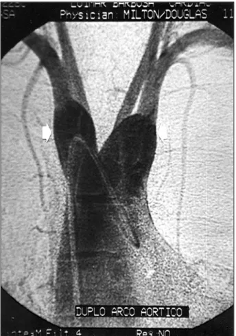
The hemodynamic study revealed severe pulmonary hypertension (110mmHg), aortopulmonary window, double aortic arch (fig. 2), and normal coronary arteries.
Nuclear magnetic resonance showed a wide aortopul-monary window (40mm in its major diameter), significant di-lation of the ascending aorta and of the central pulmonary arteries, and complete double aortic arch (fig. 3). Significant tracheoesophageal compression was observed at the level of the double aortic arch.
Contrast radiography of the esophagus confirmed the extrinsic compression.
Discussion
Some characteristic findings in this patient are particu-larly noteworthy. In addition to the rarity of the incidence of these 2 diseases occurring alone, this may be the first report
Arq Bras Cardiol 2001; 77: 490-2.
Botura et al Aortopulmonary window
4 9 1 4 9 1 4 9 1 4 9 1 4 9 1
ever made of their association. Furthermore, the late ap-pearance of significant symptoms in this patient is also important.
An aortopulmonary window 1,2 is usually wide,
cau-sing important symptoms in the first weeks or months of life, which result from the significant left-to-right shunt, and these symptoms are similar to those observed in the pre-sence of large ventricular septal defect or a wide arterial ca-nal. Our patient had cyanosis, dyspnea on moderate exerti-on, due to existing pulmonary hypertensiexerti-on, in addition to clubbing of his fingers, which is a characteristic finding of
central cyanosis (usually secondary to cyanotic congenital heart defect or pulmonary disease with hypoxia). If no surgi-cal correction occurs, pulmonary vascular disease deve-lops early in the first year of life.
Diagnosis of this anomaly and distinguishing it from arterial canal and persistent truncus arteriosus are made with two-dimensional echocardiography 8, but the definitive
identification of aortopulmonary window and associated malformations may require hemodynamic study and selec-tive angiocardiography. Anomalous origin of the coronary arteries may occur in approximately 5% to 10% of the pa-tients with aortopulmonary window, is difficult to diagnosis preoperatively (because the high perfusion pressure resul-ting from the aortopulmonary communication favors good coronary flow), and is an example of the importance of the hemodynamic study in this type of patient 8. Even though
some patients may survive until adulthood with aortopulmo-nary window, most of them die early, until the 2nd decade of
life, unless surgical correction is performed. Elective surge-ry is indicated in all symptomatic infants between the 3rd and
6th months of life.
The term “vascular ring” 3-5 is used for those
malfor-mations of the aortic arch or of the pulmonary artery that show an abnormal relation with the trachea and the eso-phagus, and they represent less than 1% of congenital car-diovascular defects.
Fig. 1 – Electrocardiogram showing sinus rhythm and significant hypertrophy of the right chambers.
Fig. 2 – Hemodynamic study showing double aortic arch (arrows).
4 9 2 4 9 2 4 9 2 4 9 2 4 9 2
Botura et al
Aortopulmonary window
Arq Bras Cardiol 2001; 77: 490-2.
1. Brooks MM, Heymann MA. Aortopulmonary window. In: Emmanouilides GC, Riemenschneider TA, Allen HD, et al, (eds): Moss and Adams’ Heart Disease in Infants, Children, and Adolescents, Including the Fetus and Young Adult (5th
ed.). Baltimore: Willians & Wilkins, 1994: 764-8.
2. Fyler DC. Aortopulmonary window. In: Fyler DC, (ed): Nadas’ Pedriatic Car-diology. Philadelphia: Hanley & Belfus, 1992: 693-5.
3. Weinberg PM. Aortic arch anomalies. In: Emmanouilides GC, Riemenschneider TA, Allen HD, et al (eds): Moss and Adams’ Heart Disease in Infants, Children, and Adolescents, Including the Fetus and Young Adult (5th ed). Baltimore:
Wil-lians & Wilkins, 1994: 810-37.
4. Kocis KC, Midgley FM, Ruckman RN. Aortic arch complex anomalies: 20-year experience with symptoms, diagnosis, associated cardiac defects, and surgical re-pair. Pediatr Cardiol 1997; 18: 127-32.
5. Valletta EA, Pregarz M, Bergamo-Andreis IA, Boner AL. Tracheoesophageal
References
compression due to congenital vascular anomalies (vascular rings). Pediatr Pul-monol 1997; 24: 93-105.
6. Gloss G, Delgado Leal F, Vazquez G, Calderon-Colmenero J, Buendia A. The aorto-pulmonary window. A reporter of 4 cases. Arch Inst Cardiol Mex 1994; 63: 149-52. 7. Terrapon M, Schneider P, Friedli B, Cox JN. Aortic arch interruption type a with aortopulmonary fenestration in an offspring of a chronic alcoholic mother (“fetal alcohol syndrome”). Helvet Paediatr Acta 1997; 32: 141-8.
8. Soares AM, Atik E, Cortez TM, et al. Janela aortopulmonar. Análise clínico-ci-rúrgica de 18 casos. Arq Bras Cardiol 1999; 73: 59-66.
9. Weinberg PM, Hubbard AM, Fogel MA. Aortic arch and pulmonary artery ano-malies in children. Semin Roentgenol 1998; 3: 262-80.
10. Lee ML, Wang JK, Wu MH, Lue HC, Chiu IS, Chang CI. Clinical implications of isolated double aortic arch and its complex with intracardiac anomalies. Int J Cardiol 1998; 63: 205-210.
The most common and severe “vascular ring” is the one produced by a double aortic arch in which the 4th embryonic left
and right aortic arches persist. In the most common type of double aortic arch, a left arterial ligament or duct exists, and both arches are patent, the right being larger than the left.
The symptoms 4 produced by vascular rings result
from the anatomical constriction of the trachea and esopha-gus. They usually appear early in the complete double aortic arch, and consist mainly of respiratory difficulty, cyanosis (especially associated with feeding), stridor, and dysphagia. The electrocardiogram is normal unless associated car-diovascular anomalies exist. Contrast radiography of the esophagus is a useful screening procedure and usually shows a prominent posterior indentation at the level of the “vascular rings”. Selective angiography delineates the anato-my of the aorta and its branches, and the course of the major pulmonary arteries. Computerized axial tomography and nu-clear magnetic resonance 9,10, however, show the spatial
re-lation between the “vascular ring” and the trachea and eso-phagus more clearly, allowing better surgical programming.
The severity of the symptoms and anatomy of the malformation are the major factors for establishing the ap-propriate treatment.
In the present case, in addition to the lack of a report in the medical literature about the association of these di-seases, this being probably the first case ever reported, the long symptom-free survival of our patient is notable in the presence of a wide aortopulmonary window and complete double aortic arch with significant tracheoesophageal com-pression, which are anomalies that usually cause important and early symptoms.
The difficulty in making a diagnosis with the initial echocardiography was mainly due to the presence of severe pulmonary hypertension with equalization of systemic and pulmonary pressures, and consequent low flow through the defect.
