1 7 4 Arq Bras Oftalmol. 2012;75(3):174-7
Artigo Original |
Original articleINTRODUCTION
Adherence to manufacturers’ recommendations for the single use of phacoemulsiication accessories has been the standard practice. Therefore, in developing countries, with the increasing quantity of pha coemulsiication procedures performed, there has been a corres ponding increase in the number of disposable phaco accessories that have been purchased and used. This increase has occurred si ABSTRACT
Purpose: To determine the incidence of Piry virus contamination among surgical instruments used with disposable accessories for phacoemulsiication during se quential surgeries.
Methods: An experimental model was created with 4 pigs’ eyes that were conta minated with Piry virus and 4 pigs’ eyes that were not contaminated. Phacoemul siication was performed on the eyes, alternating between the contaminated and noncontaminated eyes. From one surgery to another, the operating ields, gloves, scalpel, tweezers, needles, syringes, tips and bag collector from the phacoemulsi i cation machine were exchanged; only the hand piece and the irrigation and aspira tion systems were maintained.
Results: In the collector bag, three samples from the contaminated eyes (3/4) were positive, and two samples from the noncontaminated (2/4) eyes were also positive; at the tip, one sample from the contaminated eyes (1/4) and two samples of the non contaminated eyes (2/4) yielded positive results. In the irrigation system, one sample from a noncontaminated eye (1/4) was positive, and in the aspiration system, two samples from contaminated eyes (2/4) and two samples from noncontaminated eyes (2/4) were positive. In the gloves, the samples were positive in two samples from the noncontaminated eyes (2/4) and in two samples from the contaminated eyes (2/4). In the scalpel samples, three contaminated eyes (3/4) and none of the noncontaminated eyes (0/4) were positive; inally, two samples from the anterior chambers of the noncontaminated eyes gathered after surgery were positive.
Conclusions: In two noncontaminated eyes, the presence of genetic material was detected after phacoemulsiication surgery, demonstrating that the transmission of the genetic material of the Piry virus occurred at some point during the surgery on these noncontaminated eyes when the hand piece and irrigation and aspiration systems were reused between surgeries.
Keywords: Phacoemulsiication; Equipment reuse; Arboviruses; Equipment conta mination
RESUMO
Objetivo: Determinar a incidência de contaminação com o vírus Piry en tre os
instru-mentos cirúrgicos e acessórios usados durante cirurgias sequenciais de f acoemulsificação.
Métodos: Um modelo experimental foi realizado com quatro olhos de porcos que foram contaminados com o vírus Piry e quatro olhos de porcos não contaminados. A fa coemulsificação foi realizada alternando um olho contaminado para outro olho não contaminado. Entre as cirurgias, os campos de operação, luvas, bisturi, pinças, agulhas, seringas, pontas e bolsa coletora foram trocados, mantendo somente a caneta e os sistemas de irrigação e aspiração do facoemulsificador.
Resultados: No saco coletor, três amostras de olhos contaminados (3/4) foram positivos, e duas amostras de olhos não contaminados (2/4) também foram positivos; na ponta do facoemulsiicador, uma amostra dos olhos contaminados (1/4) e duas amostras de olhos não contaminados (2/4) apresentaram resultados positivos. No sistema de irrigação, uma amostra de um olho não contaminado (1/4) foi positivo, e no sistema de aspiração, duas amostras de olhos contaminados (2/4) e duas amostras de olhos não contaminados (2/4) foram positivos. Nas luvas, as amostras foram positivos em dois olhos não contaminados (2/4) e duas amostras de olhos contaminados (2/4). Nas amostras de bisturi, três olhos contaminados (3/4) e nenhum dos olhos não contami-nados (0/4) foram positivos e, finalmente, duas amostras da câmara anterior dos olhos não contaminados (2/4) reunidos após a cirurgia foram positivos.
Conclusões: Em dois olhos não contaminados, a presença de material genético foi de-tectado após a cirurgia de facoemulsificação, demonstrando que a transmissão do material genético do vírus Piry ocorreu em algum ponto durante a cirurgia para estes olhos não contaminados, quando a caneta de facoemulsificação e o sistema de irrigação e aspiração foram reutilizados entre as cirurgias.
Descritores: Facoemulsificação; Reutilização de equipamento; Arbovírus; Contaminação de equipamentos
multaneously with the implementation of costreduction initiatives precipitated by increased demand, which have caused increases in supply costs to undergo close scrutiny.
Although routine procedures enable maximum sterility, a signi icant source of contamination may be the internal tubing of auto mated surgical equipment contiguous to the operating ield and in aqueous communication with the patient’s eye, which efectively
Viral contamination during sequential phacoemulsification surgeries
in an experimental model
Contaminação viral durante cirurgias sequenciais de facoemulsiicação em um modelo experimental
RobeRto Pinto Coelho1, tatiana VannuCCi GaRCia2, JayteR SilVa Paula2, antonio auGuSto VelaSCoe CRuz3, eduaRdo Melani RoCha2, luiz tadeu MoRaeS FiGueiRedo4, MaRia louRdeS VeRoneSe RodRiGueS3
Submitted for publication: October 25, 2011 Accepted for publication: March 11, 2012
Study carried out at Department of Ophthalmology and Otorhinolaryngology, Faculdade de Me dicina de Ribeirão Preto USP Ribeirão Preto.
1 Physician, Cataract Service, Department of Ophthalmology and Otorhinolaryngology, Faculdade de Medicina, Universidade de São Paulo USP Ribeirão Preto (SP), Brazil.
2 Physician, Department of Ophthalmology and Otorhinolaryngology, Faculdade de Medicina, Uni versidade de São Paulo USP Ribeirão Preto (SP), Brazil.
3 Professor, Department of Ophthalmology and Otorhinolaryngology Faculdade de Medicina, Uni versidade de São Paulo USP Ribeirão Preto (SP), Brazil.
4 Professor, Virology Department, Faculdade de Medicina, Universidade de São Paulo USP Ribeirão Preto (SP), Brazil.
Funding: No specific financial support was available for this study.
Disclosure of potential conflicts of interest: R.P.Coelho, None; T.V.Garcia, None; J.S.Paula, None; A.A.V. e Cruz, None; E.M.Rocha, None; L.T.M.Figueiredo, None; M.L.V.Rodrigues, None.
Correspondence address: Roberto Pinto Coelho. Departamento de Oftalmologia, Otorrinolaringologia e Cirurgia de Cabeça e Pescoço, Hospital das Clínicas de Ribeirão Preto Universidade de São Paulo. Av. Bandeirantes, 3900 Ribeirão Preto (SP) 14049900 Brazil
Coelho RP, et al.
1 7 5
Arq Bras Oftalmol. 2012;75(3):174-7 contaminates the aspiration luid(13). Contamination of the aspiration
luid may seem normal to a certain extent because it has been shown that during and at the conclusion of intraocular operations, bacteria are present on the conjunctiva and in the anterior chamber(4,5).
It has been suspected that relux from the internal tubing repre sented a potential source of endophthalmitis, but outlow lines have not been investigated. Therefore, the signiicance of this inding has remained unclear. Recently, contamination of the internal tubing has also been conirmed in vitrectomy machines(6).
In this study, we hypothesized that disposable phaco accessories used in cataract surgery can be a vector of transmission of infectious sys temic and ocular diseases caused by various microorganisms, such as viruses, bacteria and prions, when a part of the accessory is not ex changed between phaco surgeries.
METHODS
This study was previously approved by the local University Hos pital Ethics Committee/Institutional Review Board. To assess possible instrument contamination and transmission to subsequent eyes, we performed ocular inoculation with thePiryvirus, which is easily cultu red in the laboratory and demonstrates extremely high multiplication rates and low infectivity and pathogenicity for humans.
The seed stock of the Piry virus, strain BeAn 41191 (LD50=108,5),
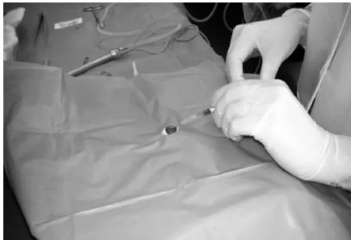
was obtained from the brain macerates of an infected mice. Each pig eye was inoculated with 10 7.36 LD 50 of Piry virus (23,000,000 virus particles dose lethal to 50% of the mice used to quantitate the virus), contained in 300 μL. This volume of viral solu tion, diluted 1:10 was injected into one enucleated pig eye by corneal paracentesis with an insulin syringe (Figure 1).
The experimental model was established in 4 pigs’ eyes that were contaminated and in 4 pigs’ eyes that were not contaminated. The contaminated eyes were identiied as C2, C3, C4 and C5. Another pig’s eye (C1) was inoculated with 300 μL of diluted MEM; that eye was used as a negative control for the experiments (Figure 1). The noncontaminated eyes were identiied as N1, N2, N3, N4 and N5. The phacoemulsiication was performed using a Universal Model II® phaco system (Alcon Laboratories) and was performed by alternating between contaminated and noncontaminated eyes.
After each surgery, surgical drapes and the operated eye were removed; the tip, the bag used to collect the phaco luids and all of the instruments used during surgery (e.g., forceps, needles, syringes, gloves and 2.75 mm scalpels) were exchanged. The new eye to be operated upon was placed in the new operative ield, and only the handpiece used for phacoemulsiication, without the tip, and the irrigation and aspiration tubing remained.
Seven sites were sampled for virus assessment: the anterior cham ber, phacoemulsiication tip, irrigation tubing, aspiration tu bing, scal pel (2.75 mm), bag collector and gloves (Figure 2). The sam ples were washed and/or aspirated with approximately 0.5 mL of saline solution (NaCl 0.9%) connected to a 5 mL syringe and stored in an Eppendorf tube. These samples were taken from the collection, preserved in ice and, inally, stored in a freezer at 70 °C until the reac tions were processed.
For the detection of viral RNA, RNA was initially extracted from the samples using the RTP® DNA/RNA Virus Mini Kit (INVITEK, Ger many), the product of which was iltered from the centrifuged sam ples. Reverse transcription, which generates complementary DNA from the viral genomic RNA that is then ampliied by primers for PCR (nested RT PCR), was used to amplify part of the G glycoprotein gene of the Piry virus and to produce ampliication products (amplicons). Amplicons obtained from the nestedPCR were visualized on 2.0% agarose gel, loaded with the sample and 25 μL of dye. The gels were electrophoresed at 100 volts and were then treated with a solution containing 0.5 mg/ml ethidium bromide, washed with water and observed under ultraviolet (UV) light.
RESULTS
S
AMPLESFROMTHEANTERIORCHAMBERBEFOREPHACOEMULSIFICATIONIt was possible to detect the presence of amplicons in all of the eyes (4/4) infected by the Piry virus, but none of the noncontami nated eyes.
S
AMPLESFROMTHECOLLECTORBAGAmong the samples collected from the collector bag after the surgeries, it was possible to detect the presence of amplicons in three (3/4) of the contaminated eyes and two (2/4) of the noncon taminated eyes.
S
AMPLESFROMTHETIPIt was possible to detect the presence of amplicons in the tip of one (1/4) sample from the contaminated eyes subjected to surgery. Among the noncontaminated eyes, it was possible to detect the presence of amplicons in two (2/4) samples.
Samples from the Irrigation Tubing It was possible to detect the presence of amplicons in none (0/4) of the contaminated eyes and in one (1/4) sample from the noncontaminated eyes.
S
AMPLESFROMTHEASPIRATIONTUBINGIt was possible to detect the presence of amplicons in samples from two (2/4) contaminated eyes and two (2/4) noncontaminated eyes.
Viral contamination during sequential phacoemulsification surgeries in an experimental model
1 7 6 Arq Bras Oftalmol. 2012;75(3):174-7
S
AMPLESFROMTHEGLOVEIt was possible to detect the presence of amplicons in two (2/4) samples that were from contaminated eyes and in two (2/4) samples from noninfected eyes.
S
AMPLESFROMTHE2.75
MMSCALPELThree (3/4) samples from the scalpels used on infected eyes and no samples (0/4) from the scalpels used on uncontaminated eyes revealed the presence of amplicons.
S
AMPLESFROMTHEANTERIORCHAMBERAFTERPHACOEMULSIFICATIONAfter the surgeries, no (0/4) samples obtained from the anterior chambers of contaminated eyes subjected to phacoemulsiication were positive. Amplicons were detected by RTnested PCR in two (2/4) of the four noncontaminated eyes.
The results of presence amplicons at sites sampled for virus as sessment in the contaminated eyes and the noncontaminated eyes were summarized in table 1.
DISCUSSION
Bacterial and fungal contamination of the automated surgical equi p ment used during routine cataract surgery has been repor ted(7,8). Investigations have shown that residual debris in reused pha
coemulsiication probes can be a potential source of postphacoe mulsiication endophthalmitis(911).
Viral contamination may be associated with the reuse of auto mated surgical equipment during cataract surgery. This association is particularly true for the risk associated with ophthalmic surgery and even more so in the case of cataract surgery, a procedure mostly per formed in elderly patients who have very high rates of HCV in fection(1214). An association between HCV infection and ophthalmic
surgery, mostly performed for cataracts, has been reported(15). Addi
tionally, diferent types of viral contamination after corneal trans plantation have been reported, including the rabies virus(16), prion
Creu t zfeldtJakob disease (CJD)(17), hepatitis B(18) and the acqui red
immunodeiciency virus(19). Notably, HIVRNA was detected in the
aqueous humor and subretinal luid of an HIV carrier with a rhegma togenous retinal detachment(20).
Factors are possibly involved as well, including skilled surgeons performing cataract surgery in only a few minutes, the high demand for surgeries(21) (especially in the public health system), the low price
paid for surgery in the government health system, the high cost of cassettes (many of which sufer structural damage when uncoupled from the phaco, preventing their resterilization), and only a small number of hospitals having fast sterilization machines, thus creating
a delay in the sterilization process. Additionally, some surgeons may even use the same tape irrigation and aspiration for many surgeries in one day, changing only the handpiece or exchanging only the tip, and using the same tape and pen for multiple surgeries.
In this context, we created an experimental model using pig eyes artiicially contaminated with a virus for comparison with uncontami nated eyes. We chose the Piry vesiculovirus because it is easily grown in a laboratory, its replication is fast and it easily produces a large number of copies.
The irst two eyes (C1 and N1) were set aside as negative controls, and no positive results were found in any of their samples, as expec ted. Of the samples from the anterior chambers of contaminated eyes before surgery (C2, C3, C4, C5), all of them showed positive results; of the samples from noncontaminated eyes (N2, N3, N4, N5), none of them showed positive results, demonstrating that contamination was eicient and that there were no falsepositive results.
The detection of amplicons in the bag collector after surgery on two uncontaminated eyes (eye samples N3 and N5) and in two sam ples from tips used on uncontaminated eyes (eye samples N3 and N5) corroborates the possibility that viral particles were retained somewhere in the phacoemulsiier and that the routes could have been the internal irrigation or aspiration pen or even the methods of irrigation and aspiration.
Among the samples from the irrigation route, positive results we re observed in one eye (N2), as the means of irrigation brought the low of luid into the eye; this result could only be positive if there was a relux of luid during the procedure or if there was contamination during the handling of the equipment. In the samples from gloves, we obtained two positive contaminated eyes (C3 and C4) and two positive noncontaminated eyes (N2 and N3), and we believe that while manipulating the pen, it was contaminated with another sterile glove, so there was transfer of viral genetic material to this glove, which was expected.
Thus, we observed that all of the components of phacoemulsiier (tip, roads, irrigation and aspiration, collector bag) and the instru ments connected to it, such as the knife and glove, became infected after one or more surgery.
CONCLUSION
Considering the large proportion of the general population un der going surgery or other invasive procedures, the present results stress the importance of complying with universal precautions and implementing eicient maintenance and sterilization methods for medical instruments. Ideally, disposable materials should be used with care, particularly during phacoemulsiication, because of the increased risk of infection due to the high turnover of patients.
Table 1. Summarizes the results in the contaminated eyes and non-contaminated eyes. Detection of amplicons of ~130 bp at the site collected
C1 N1 C2 N2 C3 N3 C4 N4 C5 N5
I Anterior chamber before phaco + + + +
II Collector bag + + + + +
III Tip + + +
IV Irrigation tubing +
V Aspiration tubing + + + +
VI Glove + + + +
VII 2.75mm scalpel + + +
VIII Anterior chamber after phaco n N n + n + n
Coelho RP, et al.
1 7 7
Arq Bras Oftalmol. 2012;75(3):174-7 REFERENCES
1. Kattan HM, Flynn HW Jr, Plugfelder SC, Forster RK. Nosocomial endophthalmitis sur vey. Current incidence of infection after intraocular surgery. Ophthalmology. 1991; 98(2):22738.
2. Plugfelder SC, Flynn HW Jr. Infectious endophthalmitis. Infect Dis Clin North Am. 1992;6(4):85973.
3. O’Brien TP, Green WR. Endophthalmitis. In: Mandell GL, Bennett JE, Dolin R, editors. Mandell, Douglas and Bennett’s Principles and practice of infectious disease. 4th ed.
New York: Churchill Livingstone; 1995. p.11209.
4. Ariyasu RG, Nakamura T, Trousdale MD, Smith RE. Intraoperative bacterial contamina tion of the aqueous humor. Ophthalmic Surg. 1993;24(6):36773; discussion 3734. 5. Mistlberger A, Ruckhofer J, Raithel E, Muller M, Alzner E, Egger SF, et al. Anterior
cham ber contamination during cataract surgery with intraocular lens implantation. J Cataract Refract Surg. 1997;23(7):10649. Comment in: J Cataract Refract Surg. 2003; 29(8):14656; author reply 1446.
6. Janknecht P, Kappstein I. Bacterial contamination of the pressure receiver of a vitrec tomy machine. Ophthalmic Surg Lasers. 1997;29(4):3457.
7. Mino de Kaspar H, Grasbon T, Kampik A. Automated surgical requires routine di sin fection of vacuum control manifold to prevent postoperative endophthalmitis. Ophthal mo lo gy. 2000;107(4):68590. Comment in: Ophthalmology. 2001;108(4):6367. 8. Kappstein I, Schneider CM, Grundmann H, Scholz R, Janknecht P. Longlasting conta mination of a vitrectomy apparatus with Serratia marcescens. Infect Control Hosp Epidemiol. 1999;20(3):1925.
9. Leslie T, Aitken DA, Barrie T, Kirkness CM. Residual debris as a potential cause of post phacoemulsiication endophthalmitis.Eye (Lond). 2003;17(4):50612.
10. Baskaran M, Rao SK, Ramana Kumar PJ, Vijaya L, Madhavan HN. Postphacoemulsii cation endophthalmitisrole of residual debris in the handsets used for surgery. Eye (Lond.) 2004;19(1):1156. Comment on: Eye (Lond). 2003;17(4):50612.
11. Rao SK, Baskaran M, Kumar PJ, Vijaya L, Madhavan HN. Debris in phacoemulsiication han dsets. A potential cause of endophthalmitis after cataract surgery? Ind J Ophthalmol.
2004;52(1):801. Comment in: Indian J Ophthalmol. 2004;52(3):2601; author reply 2612.
12. Bellentani S, Tiribelli C, Saccoccio G, Sodde M, Fratti N, De Martin C, et al. Prevalence of chronic liver disease in the general population of northern Italy: the Dionysos study. Hepatology. 1994;20(6):14429.
13. Strofolini T, Menichelli M, Taliani G, Dambruoso V, Poliandri G, Bozza A, et al. High pre valence of hepatitis C virus infection in a small central Italian town: lack of evidence of parenteral exposure. Ital J Gastroenterol. 1995;27(5):2358.
14. Guadagnino V, Strofolini T, Rapicetta M, Costantino A, Kondili LA, Menniti Ippolito F, et al. Prevalence, risk factors and genotype distribution of hepatitis C virus infection in the general population: a communitybased survey in southern Italy. Hepatology. 1997;26(4):100611.
15. Mele A, Spada E, Sagliocca L, Ragni P, Tosti M, Gallo G, et al. Risk of parenterally trans mitted hepatitis following exposure to surgery or other invasive procedures: results from the hepatitis surveillance system in Italy. Hepatology. 2001;35(2):2849. 16. Houf S, Burton R, Wilson R, Henson T, London WT, Baer GM, et al. Humantohuman
transmission of rabies virus by corneal transplant. N Engl J Med. 1979;300(11):6034. 17. Lang CJ, Heckmann JG, Neundorfer B. CreutzfeldtJakob disease via dural and corneal
transplants. J Neurol Sci. 1998;160(2):12839.
18. Hoft R, Plugfelder S, Foster R, Ullman S, Polack FM, Schif ER. Clinical evidence for he patitis B transmission resulting from corneal transplantation. Cornea. 1997;16(2): 1327. 19. Schwarz A, Hofmann F, LageStehr J, Teqzess Am, Ofermann G. Human immunode iciency virus transmission by organ donation. Outcome in cornea and kidney re cipients. Transplantation. 1987;44(1):214.
20. Kashiwagi K, Gohdo T, Sato S, Iijima H, Tsukahara S. Detection of HIVRNA in aqueous hu mor and subretinal luid in an HIV carrier with rhegmatogenous retinal detach ment. Jpn J Ophthalmol. 2000;44(6):6879.
21. Centers for Disease Control. Update: universal precautions for prevention of trans mission of human immunodeiciency virus, hepatitis B virus, and other bloodborne pa thogens in healthcare settings. MMWR Morb Mortal Wkly Rep. 1988; 37(24):37782, 3878.