Severity of Angiographic Coronary Obstruction and the
Apolipoprotein E Polymorphism in Acute Coronary Syndromes
Arlisa Monteiro de Castro Dias, Amália Faria dos Reis, Claudia Guerra Murad Saud, Maria das Graças Leitão
Chilinque, Rafaela Ferreira Leite, Rosemery Nunes Cardoso Abdalah, Mariana Ferreira Fiqueiredo, Georgina
Severo Ribeiro, Carlos Augusto Cardozo de Faria
Hospital Universitário Antonio Pedro (HUAP) da Faculdade de Medicina da Universidade Federal Fluminense (UFF), Niterói, RJ - Brazil
Summary
Background: There is evidence of the association between the apolipoprotein E (APOE) and coronary disease; however, there are controversies.
Objective: To evaluate the association between the number of coronary vessels with significant obstruction defined by angiography, the APOE polymorphism and clinical variables.
Methods: This was a cross-sectional, multicenter study with 207 patients (138 men), with acute coronary syndrome (ACS), in the city of Niteroi, state of Rio de Janeiro, Brazil, who underwent coronary angiography and genotype determination for the APOE *2*3*4 polymorphism by the Restriction Fragment Length Polymorphism (RFLP) method.
Results: The frequency of the alleles was APOE *2 - 6.8%, *3 - 82.5%, *4 - 10.7%. Regarding the number of affected vessels, 27% of patients presented monoarterial obstruction, 33.8% biarterial and 39.1% triarterial and/or left coronary trunk. The degree of multivascular lesion did not correlate with the presence of the *4 allele (p= 0.78), but with age ≥ 55 years (p=0.025), being an ex-smoker (p=0.004) and dyslipidemia (p=0.05) at the multivariate analysis and also with previous coronary artery disease (CAD) (p=0.05), diabetes (p=0.038) and metabolic syndrome (p=0.021) at the univariate analysis. The prevalence of dyslipidemia, diabetes and systemic arterial hypertension (SAH) was elevated regarding similar studies, with progressive increases in the prevalence of SAH (p=0.59) and diabetes (p=0.06), according to the number of affected vessels.
Conclusion: The APOE polymorphism was not associated with the number of coronary vessels with significant obstruction at any age range. On the other hand, age ≥ 55 years, being an ex-smoker and dyslipidemia associated with the multivascular lesion. (Arq Bras Cardiol 2009; 93(2):XXX-XXX)
Key words: Apolipoprotein E2; acute coronary syndrome; polymorphism, genetic; coronary angiography.
Mailing address: Arlisa Monteiro de Castro Dias •
Praça da República 111, Centro, 22.220-350, Rio de Janeiro, RJ - Brazil E-mail: [email protected]
Manuscript received April 27, 2008; revised manuscript received August 10, 2008; accepted September 09, 2008.
Introduction
The apolipoprotein E (APOE), a plasma protein that comprises the structure of several lipoproteins (kilomicron, VLDL-cholesterol, IDL-cholesterol and HDL-cholesterol), acts as a receptor-binding factor. It has an acknowledged role in the transportation of cholesterol and other lipids from peripheral tissues to the liver, so they can be metabolized1.
The most studied isoforms are the three first described ones: apolipoprotein E2, E3 and E4, codified by the alleles (epsilon 2, 3 an 4). The frequency of the *2, *3, *4 alleles is estimated as being around 11%, 72% and 17% in the general population, respectively1. The *4 allele has been found with
higher prevalence in the African populations2, and has been
associated to higher plasma levels of total cholesterol and LDL-cholesterol and lower serum levels of APOE1; therefore,
due to these influences, it has been studied as a possible risk factor for coronary disease3.
In spite of the ethnical differences in the studied populations, Wilson et al3 and Song et al4 found, in their meta-analyses,
increased risk for significant coronary artery disease (CAD) in the presence of *4 allele. However, when certain populations are studied separately, as demonstrated in the studies by Kolovou et al5,6 in Greece and Muros & Ferrer7 in Spain, the
association between the presence of the *4 allele and CAD can be negative. Recently, an important study published by Morgan et al8 evaluated 84 genetic polymorphisms in acute
myocardial infarction (AMI) and did not report any significant association with APOE polymorphism.
Two Brazilian studies9,10 did not find any association
between the presence of *4 allele and CAD; however, Salazar et al11 observed an association between the *4 allele
We chose to study this subject in the city of Niteroi, as it was the only city in the state of Rio de Janeiro where the rate of mortality due to ischemic coronary disease was higher than the one by cerebrovascular disease, from 1980 to 200012 and
due to the fact that circulatory diseases were the main cause of death and hospital admission in the last ten years13.
With the objective of evaluating the existence of an association between this APOE polymorphism, clinical variables and the number of affected coronary vessels with significant obstruction defined by coronary angiography, we selected all patients that had undergone coronary angiography during hospital admission due to ACS in the city of Niteroi (RJ) from a prospective cohort study carried out in partnership between the Universidade Federal Fluminense (UFF) and the
Universidade Federal do Rio de Janeiro (Federal University of Rio de Janeiro)13. One of the objectives of this cohort study,
of which methodology and epidemiological aspects were published by Reis et al13, was to study the association of APOE
polymorphisms, angiotensin- and angiotensinogen-converting-enzyme with clinical and laboratory outcomes that had been pre-defined in patients with ACS, given the small number of Brazilian publications that have reported on these aspects. This research group has dedicated since 2003 to the study of epidemiological and genetic aspects of the ACS in cities of the state of Rio de Janeiro, within the lines of research of the Post-Graduation course in Cardiovascular Sciences at UFF. The results of the other analyses carried out with this cohort study population will be published subsequently, as they are not part of the objective of the present study.
Methods
Three public hospitals and two private ones in the city of Niteroi, state of Rio de Janeiro, Brazil, participated in the prospective cohort study, with patients’ enrollment occurring between June 2004 and June 2005. This study was approved in 2003 by Ethics Committee in Research of the School of Medicine of UFF and the National Commission on Ethics in Research. All patients signed the Free and Informed Consent Form. Men and women aged ≥ 20 years, consecutively admitted with a diagnosis of ACS: AMI with or without ST-segment elevation or unstable angina were included in the study13. This was a cross-sectional study that selected
the 234 patients from the original prospective study, who had undergone coronary angiography during hospital stay. Of these, 207 presented at least one coronary artery with significant obstruction (≥ 70%), who were selected for the statistical analysis of the present study.
The collected blood samples were sent to the Laboratory of PCR and Molecular Biology of the Service of Clinical Pathology of
Hospital Universitário Antonio Pedro (UFF) for laboratory analyses and identification of the genetic polymorphism of APOE.
The samples were collected in tubes with EDTA for genomic DNA isolation, which was carried out through the saline precipitation method14. After the extraction,
the integrity of the samples was analyzed by submarine electrophoresis (BIO RAD electrophoresis system) in 0.8% agarose gel with TBE 1X buffer (90 Mm Tris-HCl, 90 mM boric acid and 2 mM EDTA), stained with ethidium bromide
and stored at 4°C for posterior polymorphism analysis. The DNA samples were submitted to the Polymerase-Chain Reaction (PCR) and evaluated by electrophoresis in a 2% agarose gel. The APOE genotype determination was carried out through the Restriction Fragment Length Polymorphism (RFLP) method15.
The 220 bp amplicon obtained from the PCR was treated with a unit of HhaI restriction enzyme, according to the manufacturer’s instructions, producing DNA fragments of different sizes and allowing the identification of the genotypes.
After the digestion, the samples were evaluated in a 10% polyacrylamide gel in a Hoefer SE 400 vertical electrophoresis system by Amersham Biosciences. As size indicator, a 25 base-pair (bp) marker was used, applied together with the samples, to help the visualization of the fragments of 91, 83, 72, 48, 38 and 34 bp. The identification of the APOE genotypes was based on the following profile: E2E2 – fragments of 91, 83, 38 bp; E3E3 – 91,48, 38, 34 bp; E4E4 – 72, 48, 38, 34 bp; E2E3 – 91, 83, 48, 38, 34 bp ; E2E4 – 91, 83, 72, 48, 38 bp; E3E4 – 91, 72, 48, 38, 34 bp.
The coronary angiography results were discussed with the hemodynamicists and cardiologists from the teams responsible for the hospital admission units, who were blinded to the results of the genetic analysis. As the number of patients that had undergone coronary angiography and presented coronary obstruction < 70% or that presented normal results was small (11.5%), and as they could be joined in a single group, we chose to exclude them from the statistical analysis.
Arterial obstruction ≥ 70%, was considered a significant lesion, quantified or estimated by angiography16 and
multivascular disease stenosis ≥ 70% in two or more coronary systems17 or left coronary trunk lesion ≥ 50%.
We considered a vessel each one of the systems18: 1)
Descending Artery System: (DA and branches); 2) Circumflex System: (CX and branches); 3) Right Coronary System: (RC and branches). It was considered monoarterial when only one of the systems above was affected with lesions ≥ 70%; biarterial if two systems were affected and triarterial if there were lesions ≥ 70% in the three systems.
As the trunk lesion and the trivascular lesion are independent risk factors for intra-hospital adverse events16, these two groups
were evaluated together in some statistical analyses.
The diagnosis of metabolic syndrome was defined by the criteria of the World Health Organization, which allows the use of the body mass index (BMI) to evaluate obesity19.
The criteria for the diagnosis of dyslipidemia were: a) Dyslipidemia (1) – cholesterol ≥ 200 or LDL ≥ 130 or TG
≥ 150 mg/dl or history of previous dyslipidemia (lipid profile values above the normal range) associated to the previous use of hypolipemiant drugs; b) Dyslipidemia (2) – HDL < 40 or TG ≥ 150 or LDL ≥130 or Cholesterol ≥ 200 mg/dl or previous use of hypolipemiant drugs.
textbooks and scientific articles, having been detailed in a previously published article13.
Statistical analysis
Statistical significance was set at 5% and the statistical analysis was performed with the SAS® software, version 6. The
analysis of the association between the qualitative variables and the degree of affected vessels was carried out by the 2 test
and the adjusted 2 test. Tukey’s multiple comparisons test was
used to identify which degrees of coronary lesions differed among them, at a 5% level.
The analysis of association between the degree of the affected vessels and the presence of the *4 allele in the general sample and at the age range < 65 years was carried out by the 2 test and by Fisher’s exact test. The 2
test was also used to perform the statistical analysis of the correlation between coronary obstruction and the presence of metabolic syndrome.
The Logistic Regression analysis was carried out to identify the independent variables, including the APOE polymorphism, which simultaneously influenced the degree of the obstructed vessels according to the coronary angiography. The selected variables were: presence of *4 allele, age 55 years, age 60 years, male sex, BMI (kg/m2), history of previous CAD,
history of CAD in first-degree relatives, smoking, diagnosis of diabetes mellitus, ACS type, diagnosis of dyslipidemia (1) and left ventricular failure (LVF) during hospital stay. The process of factor selection was the stepwise one, at the level of 5% and 10%. Regarding age, the model was tested with age 55 years and later with age 60 years, to evaluate which one of them had a better explicative power in association with the other variables.
Results
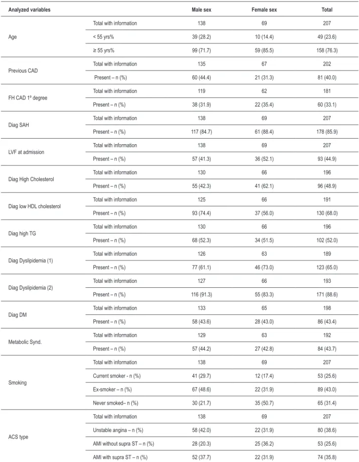
The prevalence of the studied variables in the 207 patients according to gender is shown in Table 1. The distribution of the APOE polymorphism genotypic frequencies in the sample (Table 2) was in Hardy-Weinberg equilibrium. For the statistical analysis, the genotypes were grouped in three groups: E2E2, E2E3 (*2 allele present), E3E3 (*3 allele present), E2E4, E3E4, E4E4 (*4 allele present). The patients that did not harbor the *4 allele represent the addition of the two first groups.
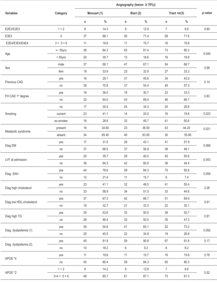
The APOE polymorphism (Table 3) was not statistically associated with the degree of the affected vessels (p=0.80), not even when the *4 allele and the *2 allele were evaluated separately (p=0.78 and p=0.52, respectively).
A significant association as observed between age 55 years with the degree of affected vessels (p=0.045). The percentage of patients aged 55 years at the monoarterial degree (64.3%) was significantly lower than in the biarterial (81%) and triarterial + trunk (80%).
There was a significant association between smoking and the degree of affected vessels (p=0.023). The percentage of ex-smokers at the triarterial degree + trunk (50.6%) and biarterial (45.7%) was significantly higher than in the monoarterial degree (28,6%).
The group of patients with triarterial degree + trunk (55.6%) presented a LVF proportion at hospital admission that was significantly higher than the monoarterial degree (35.7%), with p=0.043.
The diagnosis of diabetes mellitus increased progressively when correlated with the degree of affected vessels: 31% at the monoarterial, 43% at the biarterial and 51.9% at the triarterial + trunk (p=0.06). The diagnosis of systemic arterial hypertension (SAH) followed a similar pattern: 78% at the monoarterial, 84.3% at the biarterial and 92.6% at the triarterial + trunk (p=0.059).
The diagnosis of dyslipidemia (1) and (2), total cholesterol increase, low HDL-cholesterol and increase in triglycerides did not present a significant association with the number of affected vessels.
Metabolic syndrome presented a significant association with the degree of affected vessels, being more prevalent in the triarterial + trunk group, than in the monoarterial group (p=0.012).
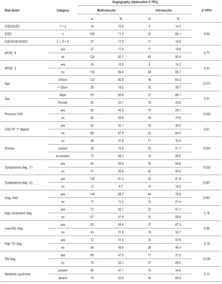
A new analysis was carried out with the objective of comparing the monoarterial with the multivascular groups (biarterial + triarterial + trunk) and verifying whether they were statistically different under the point of view of the analyzed variables (Table 4). When the classification was performed with two degrees of coronary obstruction severity (Table 4), the multivascular degree presented a significant association with the variables age ≥ 55 years (p=0.025), presence of previous CAD (p=0.05), ex-smokers (p=0.004), diabetes mellitus (p=0.038) and dyslipidemia (1) (p=0.05). However, the degree of affected vessels still did not present a significant association with APOE polymorphism.
Differently from what was observed at the analysis with three degrees of affected vessels (Table 3), in this analysis there was no significant association between the mono and multivascular degrees and the metabolic syndrome variable (p=0.12). The diagnosis of SAH increased proportionally to the degree of the angiographic lesion severity (univascular = 78.6% and multivascular = 88.7%), although it did not reach statistical significance (p=0.061).
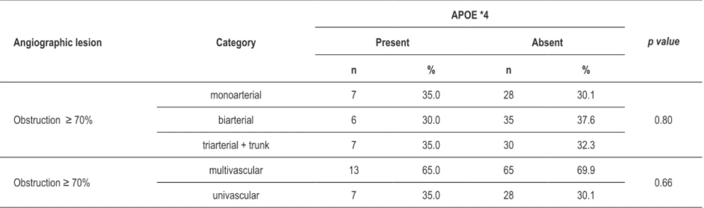
As shown in Table 5, when the subgroup of patients aged < 65 years was analyzed, no association was observed between the degree of affected vessels and the presence of *4 allele, in cases with three degrees of severity (p=0.80) , as well as with two degrees (p=0.66).
Tables 6 and 7 present the parameters for the significant variables selected by the Logistic Regression method at the level of 5% and 10%, respectively. The parameters are: coefficient, standard error, p value and relative risk (RR), with its respective 95% confidence interval (CI) for the occurrence of multivascular lesion.
Table 1 – Description of the qualitative variables
Analyzed variables Male sex Female sex Total
Age
Total with information 138 69 207
< 55 yrs% 39 (28.2) 10 (14.4) 49 (23.6)
≥ 55 yrs% 99 (71.7) 59 (85.5) 158 (76.3)
Previous CAD
Total with information 135 67 202
Present – n (%) 60 (44.4) 21 (31.3) 81 (40.0)
FH CAD 1º degree
Total with information 119 62 181
Present – n (%) 38 (31.9) 22 (35.4) 60 (33.1)
Diag SAH
Total with information 138 69 207
Present – n (%) 117 (84.7) 61 (88.4) 178 (85.9)
LVF at admission
Total with information 138 69 207
Present – n (%) 57 (41.3) 36 (52.1) 93 (44.9)
Diag High Cholesterol
Total with information 130 66 196
Present – n (%) 55 (42.3) 41 (62.1) 96 (48.9)
Diag low HDL cholesterol
Total with information 125 66 191
Present – n (%) 93 (74.4) 37 (56.0) 130 (68.0)
Diag high TG
Total with information 130 66 196
Present – n (%) 68 (52.3) 34 (51.5) 102 (52.0)
Diag Dyslipidemia (1)
Total with information 126 63 189
Present – n (%) 77 (61.1) 46 (73.0) 123 (65.0)
Diag Dyslipidemia (2)
Total with information 127 66 193
Present – n (%) 116 (91.3) 55 (83.3) 171 (88.6)
Diag DM
Total with information 133 65 198
Present – n (%) 58 (43.6) 28 (43.0) 86 (43.4)
Metabolic Synd.
Total with information 129 63 192
Present – n (%) 57 (44.2) 27 (42.8) 84 (43.7)
Smoking
Total with information 138 69 207
Current smoker - n (%) 41 (29.7) 12 (17.4) 53 (25.6)
Ex-smoker – n (%) 67 (48.6) 22 (31.9) 89 (43.0)
Never smoked– n (%) 30 (21.7) 35 (50.7) 65 (31.4)
ACS type
Total with information 138 69 207
Unstable angina – n (%) 58 (42.0) 22 (31.9) 80 (38.6)
AMI without supra ST – n (%) 28 (20.3) 25 (36.2) 53 (25.6)
AMI with supra ST – n (%) 52 (37.7) 22 (31.9) 74 (35.8)
When analyzing at the level of 10% (table 7), we observed that the ex-smoker (RR 3.37), diagnosis of dyslipidemia (1) (RR 2.11) and age 55 years (RR 1.96) were significant to explain the degree of multivascular lesion, whereas the non-smoker presented a borderline p value.
Discussion
In this studied population, there was a predominance of the male sex (66.7%), mean age was 63 years, the most frequent type of ACS was acute myocardial infarction (62%), the most prevalent degree of lesion was the multivascular one (72.9%) and the percentage of patients with previous CAD was 40.1%. In a Brazilian study carried out in São Paulo20, which
evaluated 1,413 patients with unstable angina submitted to coronary angioplasty, the predominance of the male sex was also observed (62%), with a mean age of 61 years; however, the frequency of multivascular lesions was lower (42%). Other authors in a study carried out in patients with Ami in the city of Niteroi21 found almost the same percentage of male sex
patients (66.8%), also with a lower percentage of multivascular disease (54.9% in men and 63.3% in women). In the present study, the percentages of the coronary obstruction type did not significantly differ regarding sex (Table 4).
The high prevalence of multivascular lesion (72.9%), found in patients that underwent coronary angiography in the present study demonstrates the correct indication of the examination. On the other hand, the low percentage of patients with univascular lesion might have influenced the results, considering that the influence of the APOE polymorphism might not have been demonstrated due to the lack of of a representative group of patients with a lower extension of the disease.
In the present study, 43.4% of the patients met the diagnostic criteria of diabetes mellitus and 86% of SAH. These findings characterize a population with a high prevalence of the mentioned diseases, considering the findings of other studies with CAD patients Duarte et al22 observed a prevalence of 56%
of SAH in patients with ACS that presented adverse events during hospital admission and of 62% in patients without events. At the AFIRMAR23 study, which evaluated risk factors
in ACS in 1,279 Brazilian patients, the prevalence of SAH was even lower (54.34%), as well as of diabetes (21.6%).
The diagnosis of dyslipidemia (2) showed a prevalence of 88.6%, much higher than the one observed by Duarte et al22,
also in a Brazilian study with similar criterion (60.5%). Regarding the current smoking, the prevalence found (31,5%) was similar to the prevalence observed in the study by Peixoto et al21 (34.1%) and lower than the prevalence of
41.5% described by Piegas et al23.
The metabolic syndrome was diagnosed in 44.9% of the total sample and in 47.6% of the patients aged < 65 years. These results are a little higher than the ones found in another Brazilian study related to ACS, in which 40% f the patients were classified as having metabolic syndrome24.
We observed that the most significant variables to explain the multivascular degree at the logistic regression analysis were: 1) ex-smoker and dyslipidemia (1) at the level of significance of 5% and 2) ex-smoker, dyslipidemia (1) and age ≥ 55 years at the level of 10%. The ex-smoker was more significant than the current smoker, which might be explained by the fact that ex-smokers are older and probably stopped smoking due to a previous diagnosis of CAD or other pathologies that contraindicate smoking, whereas the current smoker, due to the fact of being younger, has not been exposed long enough to risk factors to develop multivascular lesions.
In this studied population, the prevalence of the *4 allele (10.7%) was similar to the prevalence of the allele in healthy populations such as the Portuguese (9%)25, the Italian
(10%), the Mexican (10.2%)26, the Spanish (9%)2, the elderly
population of Rio Grande do Sul that descends from Italians (11%)27, as well as the patients with CAD in Sao Paulo (9%)10.
On the other hand, the frequency of the allele was lower than the one observed in northern European countries, such as Ireland (22%)28 and lower than the one found in the town
of Ouro Preto, state of Minas Gerais, Brazil (20%)29, where
the African contribution for the formation of the population was higher.
No association was found in the present study between the presence of the *4 allele and a higher angiographic severity. These findings differ from the ones found by Lehtinen et al30,
Wang et al31 and Ye et al32, who also evaluated the APOE
genotype regarding the number of vessels with significant lesions and observed an increase in the frequency of the *4 allele in cases with multivascular lesions. It is worth mentioning that Wang (op. cit.) evaluated only three individuals aged < 65 years.
Even when the subgroup of younger individuals ( 65 years) was analyzed separately, the severity of the coronary lesions did not present a significant association with the presence of the *4 allele. The study by Salazar et al11 in São Paulo found
Table 2 – Description of apolipoprotein E genotypes observed in the sample
Variable Category n %
APOE
1(E2E2) 2 1.0
2(E2E3) 22 10.6
3(E2E4) 4 1.9
4(E3E3) 145 70.0
5(E3E4) 31 15.0
6(E4E4) 3 1.4
E2E2/E2E3 1+2 24 11.6
E3E3 4 145 70.0
E2E4/E3E4/E4E4 3+5+6 38 18.4
*4
yes 38 18.4
no 169 81.6
Table 3 - Analysis of the variables according to three degrees of affected vessels
Variables Category
Angiography (lesion ≥ 70%))
p value Monoart (1) Biart (2) Triart +tr(3)
n % n % n %
E2E2/E2E3 1 + 2 8 14.3 9 12.9 7 8.6 0.80
E3E3 4 37 66.1 50 71.4 58 71.6
E2E4/E3E4/E4E4 3 + 5 + 6 11 19.6 11 15.7 16 19.8
Age >– 55yrs 36 64.3 63 81.4 73 80.3 0.045
< 55yrs 20 35.7 13 18.6 16 19.8
Sex male 37 66.1 47 67.1 54 66.7 0.99
fem 19 33.9 23 32.9 27 33.3
Previous CAD yes 16 29.1 31 45.6 34 43.0 0.14
no 39 70.9 37 54.4 45 57.0
FH CAD 1st degree yes 18 36.0 19 30.7 23 33.3 0.83
no 32 64.0 43 69.4 46 66.7
Smoking
no 17 30.4 24 34.3 24 29.6
current 23 41.1 14 20.0 16 19.8 0.023
ex-smoker 16 28.6 32 45.7 41 50.6
Metabolic syndrome present 18 34.60 23 36.50 43 44.20 0.021
absent 34 65.40 40 63.50 30 55.80
Diag DM yes 17 31.5 28 43.1 41 51.9 0.066
no 37 68.5 37 56.9 38 48.1
LVF at admission yes 20 35.7 28 40.0 45 55.6 0.043
no 36 64.3 42 60.0 36 44.4
Diag. SAH yes 44 78.6 59 84.3 75 92.6 0.059
no 12 21.4 11 15.7 6 7.4
Diag high cholesterol yes 23 41.1 32 48.5 41 55.4 0.26
no 33 58.9 34 51.5 33 44.6
Diag low HDL-cholesterol yes 37 67.3 42 66.7 51 69.9 0.91
no 18 32.7 21 33.3 22 30.1
Diag high TG yes 30 53.6 33 50.0 39 52.7 0.91
no 26 46.4 33 50.0 35 47.3
Diag. dyslipidemia (1) yes 30 54.6 41 65.1 52 73.2 0.092
no 25 45.5 22 34.9 19 26.8
Diag. dyslipidemia (2) yes 45 81.8 59 90.8 67 91.8 0.17
no 10 18.2 6 9.2 6 8.2
APOE *4 yes 11 19.6 11 15.7 16 19.8 0.78
no 45 80.4 59 84.3 65 80.3
APOE *2 1 + 2 8 14.2 9 12.8 7 8.6 0.52
3+4 + 5 + 6 48 85.7 61 87.1 73 91.3
Table 4 - Analysis of variables according to two degrees of affected vessels
Risk factor Category
Angiography (obstruction ≥ 70%)
p value Multivascular Univascular
n % n %
E2E2/E2E3 1 + 2 16 10.6 8 14.3
0.69
E3E3 4 108 71.5 37 66.1
E2E4/E3E4/E4E4 3 + 5 + 6 27 17.9 11 19.6
APOE *4
yes 27 17.9 11 19.6
0.77
no 124 82.1 45 80.4
APOE *2
yes 16 10.5 8 14.2
0.42
no 135 89.4 48 85.7
Age
≥55yrs 122 80.8 36 64.3
0.013
< 55yrs 29 19.2 20 35.7
Sex
Male 101 66.9 37 66.1
0.91
Female 50 33.1 19 33.9
Previous CAD
yes 65 44.2 16 29.1
0.050
no 82 55.8 39 70.9
CAD HF 1st degree
yes 42 32.1 18 36.0
0.61
no 89 67.9 32 64.0
Smoker
no 48 31.8 17 30.4
0.004
current 30 19.9 23 41.1
ex-smoker 73 48.3 16 28.6
Dyslipidemia diag. (1)
yes 93 69.4 30 54.6
0.050
no 41 30.6 25 45.5
Dyslipidemia diag. (2)
yes 126 91.3 45 81.8
0.061
no 12 8.7 10 18.2
Diag. HAS
yes 134 88.7 44 78.6
0.061
no 17 11.3 12 21.4
High cholesterol diag.
yes 73 52.1 23 41.1
0.16
no 67 47.9 33 58.9
Low HDL diag.
yes 93 68.4 37 67.3
0.88
no 43 31.6 18 32.7
High TG diag.
yes 72 51.4 30 53.6
0.78
no 68 48.6 26 46.4
DM diag.
yes 69 47.9 17 31.5
0.038
no 75 52.1 37 68.5
Metabolic syndrome
present 66 47.1 18 34.6
0.12
absent 74 52,9 34 65,4
different results; however, the population was younger, with a mean age of around 48 years and consisted of women, only. In our sample, only 6 (2.8%) of the women aged < 65 years harbored the *4 allele. Thus, it was not possible to establish comparisons.
The outcome of our study, in relation to the fact that the APOE polymorphism was not considered an independent risk factor for the severity of the angiographic lesions in both sexes, was similar to the case-control studies carried out in the state of Sao Paulo by Mansur et al9 and Souza et al10. The study by
Mansur et al.9 also compared within the group with coronary
disease (with or without AMI), uni- and multivascular lesion, similar to present study. The lack of association observed in the present study could be explained, in part, by the fact that the frequency of this allele was low in our sample (10.7%), as also occurred in the study by Souza et al (9%)10.The analysis
of the outcomes of these Brazilian studies together allows us to conclude that even associations that have been
well-established in the literature might not be confirmed due to population differences.
The exposition to classic risk factors starts to have a higher influence on the individual tendency to present significant coronary obstruction with aging33. The high prevalence of
risk factors observed in this study probably contributed to the high percentage of multivascular lesion found in it and it also might have exercised a higher influence on the degree of angiographic lesion than the APOE polymorphism, due to the percentage of elderly individuals in the present study (45% aged ≥ 65 years). As multivascular lesions are independent risk factors for intra-hospital adverse events16, the results
of the present study point out to the need for establishing more effective public health policies that will allow a better control of coronary risk factors, as well as the decrease in the high morbimortality due to ACS observed in this population studied in the city of Niteroi, RJ, Brazil, as previously reported by Reis et al13.
Table 5 - Analysis of the correlation between *4 allele and angiographic lesion in patients aged < 65 years
Angiographic lesion Category
APOE *4
p value
Present Absent
n % n %
Obstruction ≥ 70%
monoarterial 7 35.0 28 30.1
0.80
biarterial 6 30.0 35 37.6
triarterial + trunk 7 35.0 30 32.3
Obstruction ≥ 70%
multivascular 13 65.0 65 69.9
0.66
univascular 7 35.0 28 30.1
Table 6 – Signiicant variable results at the 5% level for the multivascular degree by Logistic Regression
Signiicant variable Coeficient Standard error p value RR 95% CI
1) Current smoke reference 0.004
Ex-smoker 1.350 0.411 0.001 3.86 1.72 8.63
No smoker 0.897 0.433 0.038 2.45 1.05 5.73
2) Diagnosis of Dyslipidemia (1) 0.763 0.351 0.03 2.14 1.08 4.26
Table 7 – Signiicant variable results at the 10% level for the multivascular degree by Logistic Regression
Signiicant variable Coeficient Standard error p value RR 95% CI
1) Current smoker reference 0.015
Ex-smoker 1.214 0.421 0.004 3.37 1.48 7.68
Non-smoker 0.735 0.446 0.100 2.09 0.87 5.00
2) Diagnosis of Dyslipidemia (1) 0.744 0.354 0.036 2.11 1.05 4.22
Considering the aforementioned facts, this APOE polymorphism had little influence on the population of the present study. The predisposition for CAD is determined by multiple genetic and environmental factors, and it is unlikely that a single genetic polymorphism is capable of causing a significant increase in the risk of developing it33. The negative result of
the present study corroborates the existing idea in the current scientific literature, of the importance of the simultaneous study of several polymorphisms, mainly in polygenic and multifactorial diseases such as the coronary disease.
We believe that the publication of these negative results is important, as they call attention to the variations that exist in different geographic regions of Brazil and the world, regarding the genetic profile and the prevalence of environmental risk factors, which, when associated, will lead to a higher or lower prevalence of CAD. These different results also indicate the need for adequacy to the local reality of risk scores, as genetic and environmental factors will influence or not the risk of CAD or its prognosis, depending on the complex inter-relation among these factors in that population.
The lack of a control group was considered one of the main limitations of the present study. However, the study data were obtained from a larger, prospective study that included patients admitted at the hospital with a diagnosis of ACS and it would not have been ethical to program a hemodynamic study in patients in whom the diagnosis of ACS had not been
confirmed, or in those with ACS considered as low clinical risk. Additionally, the study was an observational one and the research team had no influence regarding the performance or not of the hemodynamic study.
Conclusion
The apolipoprotein E polymorphism did not show an association with a higher degree of coronary lesion severity in the total sample or in the subgroup of patients aged < 65 years.
Potential Conflict of Interest
No potential conflict of interest relevant to this article was reported.
Sources of Funding
This study was partially funded by Instituto Biossocial de Volta Redonda.
Study Association
This article is part of the thesis of Master submitted by Arlisa Monteiro de Castro, from Universidade Federal Fluminense.
References
1. Curtiss LK, Boisvert WA. Apolipoprotein E and atherosclerosis. Curr Opin Lipid. 2000; 11: 243-51.
2. Corbo RM, Scacchi R. Apolipoprotein E (APOE) allele distribution in the world. Is APOE*4 a ‘thrifty’ allele? Ann Hum Genet. 1999; 63: 301-10. 3. Wilson PW, Schaefer EJ, Larson MG, Ordovas JM. Apolipoprotein E alleles
and risk of coronary disease: a meta-analysis. Arterioscler Thromb Vasc Biol. 1996; 16: 1250-5.
4. Song Y, Stampfer M, Liu S. Metanalysis: apolipoprotein E genotypes and risk for coronary heart disease. Ann Interm Med. 2004; 141: 137-47. 5. Kolovou G, Yiannakouris N, Hatzivassiliou M, Malakos J, Daskalova D,
Hatzigeorgiou G, et al. Association of apolipoprotein E polymorphism with myocardial infarction in Greek patients with coronary artery disease. Curr Med Res Opin. 2002; 18 (3): 118-24.
6. Kolovou GD, Anagnostopoulou KK, Mikhailidis DP, Panagiotakos DB, Pilatis ND, Cariolou MA, et al. Association of apolipoprotein E genotype with early onset of coronary heart disease in Greek men. Angiology. 2005; 56 (6): 663-70.
7. Muros M, Ferrer CR. Apolipoprotein E polymorphism influence on lipids, apolipoproteins and Lp(a) in a Spanish population underexpressing apo E4. Atherosclerosis. 1996; 121: 13-21.
8. Morgan TM, Krumholz HM, Lifton RP, Spertus JA. Nonvalidation of reported genetic risk factors for acute coronary syndrome in a large-scale replication study. JAMA. 2007; 297 (14): 155-6.
9. Mansur AP, Annicchino-Bizzacchi J, Favarato D, Avakian SD, Cesar LA, Ramires JA. Angiotensin-converting enzyme and apolipoproteins genes polymorphism in coronary artery disease. Clin Cardiol. 2000; 23 (5): 335-40.
10. Souza DRS, NaKachima L, Biagioni RB, NaKazone MA, Pinhel MAS, Trindade DM, et al. Relevance of apolipoprotein E4 for lipid profile of Brazilian patients
with coronary artery disease. Braz J Med Biol Res. 2007; 40 (2): 189-97. 11. Salazar LA, Hirata M, Giannini SD, Forti N, Diament J, Lima TM, et al. Seven
DNA polymorphisms at the candidates genes of atherosclerosis in Brazilian women with angiographically documented coronary heart disease. Clin Chim Acta. 2000; 300: 139-49.
12. Oliveira GMN, Klein CH, Souza e Silva NA. Mortalidade por doenças isquêmicas do coração, doenças cérebros vasculares e causas mal definidas nas regiões de saúde do estado do Rio de Janeiro, no período de 1980 a 2000. Rev SOCERJ. 2005; 18 (1): 13-22.
13. Reis AF, Salis LHA, Macrini JLR, Dias AMC, Chilinque MGL, Saud CGM, et al. Síndrome coronariana aguda: morbimortalidade e prática clínica em pacientes do município de Niterói (RJ). Rev SOCERJ. 2007; 20 (5): 360-71. 14. Salazar LA, Hirata MH, Cavalli SA, Machado MO, Hirata RDC. Optimized
procedure for DNA isolation from fresh and cryopreserved clotted human blood useful in clinical molecular testing . Clin Chem. 1998; 44 (8): 1748-50.
15. Hixon JE, Vernier DT. Restriction isotyping of human apolipoprotein E by gene amplification and cleavage with HhaI. J Lipid Res. 1990; 31: 545-7. 16. Smith SC Jr, Dove JT, Jacobs AK, Kennedy JW, Kereiakes D, Kern MJ, et al.
ACC/AHA guidelines of percutaneous coronary interventions (revision of the 1993 PTCA guidelines) - executive summary: a report of the American College of Cardiology / American Heart Association Task Force on Practice Guidelines (Committee to revise the 1993 guidelines for percutaneous transluminal coronary angioplasty). J Am Coll Cardiol. 2001; 37: 2215-39.
17. Holmes DR, Berger PB. Complex and multivessel treatment. In: Topol EJ. Textbook of interventional cardiology. 3rd ed. Philadelphia: Saunders; 1999. p. 188-208.
19. Picon PX, Zanatta CM, Gerchman T, Zelmanovitz T,Gross JL, Canani LH. Análise dos critérios de definição da síndrome metabólica em pacientes com Diabetes Melito Tipo 2. Arq Bras Endocrinol Metab. 2006; 50(2): 264-70. 20. Peixoto DS, Tanajura LFL, Sousa AGRM, Centemero MP, Chaves AJ, Maia
JP, et al. Pacientes com angina instável tratados por meio de intervenções coronarianas percutâneas no Novo Milênio: o que os caracteriza? Arq Bras Cardiol. 2007; 88 (1): 26-30.
21. Peixoto RST, Peixoto ECS, Sena MA, Tedeschi AL, Borges IP, Rachid MBF. Influência do sexo na evolução imediata e médio prazo após a intervenção coronariana percutânea primária e análise dos fatores independentes de risco para óbito e eventos. Arq Bras Cardiol. 2006; 86 (3): 211-8.
22. Duarte RD, Pellanda LC, Portal VL. Perfil inflamatório, metabólico e lipídico na síndrome isquêmica aguda: relação com eventos intra e pós-hospitalares. Arq Bras Cardiol. 2005; 84 (2): 122-9.
23. Piegas LS, Avezum A, Pereira JRC, Rossi Neto JM, Hoepfner C, Farran JA, et al. Risk factors for myocardial infarction in Brazil. Am Heart J. 2003; 146: 331-8.
24. Avezum A, Piegas LS, Pereira JCR. Fatores de risco associados com infarto agudo do miocárdio na região metropolitana de São Paulo: uma região desenvolvida em um país em desenvolvimento. Arq Bras Cardiol. 2005; 84 (3): 206-13.
25. Haddy N, Bacquer DD, Chemaly MM, Maurice ML, Ehnholm C, Evans A, et al. The importance of plasma apolipoprotein E concentration in addition to its common polymorphism on inter-individual variation in lipid levels: results from Apo Europe. Eur J Hum Genet. 2002; 10: 841-50.
26. Eichner JE, Dunn ST, Perveen G, Thompson DM, Stewart KE, Strehla BCL.
Apolipoprotein E polymorphism and cardiovascular disease: a HuGE review. Am J Epidemiol. 2002; 155 (6): 487-95.
27.Schwanke CK, Cruz IB, Leal NF, Scheibe R, Moriguchi Y, Moriguchi EH. Analysis of the association between Apolipoprotein E polymorphism and cardiovascular risk factors in an elderly population with longevity. Arq Bras Cardiol. 2002; 78 (6): 571-9.
28. Sheehan D, Bennett T, Cashman K. Apolipoprotein E gene polymorphisms and serum cholesterol in healthy Irish adults: a proposed genetic marker for coronary artery disease risk. Ir J Med Sci. 2000; 169 (1): 50-4.
29. Mendes-Lana AM, Pena GG, Freitas SN, Lima AA, Nicolato RLC, Nascimento-Neto RM, et al. Apolipoprotein E polymorphism in Brazilian dyslipidemic individuals: Ouro Preto study. Braz J Med Biol Res. 2007; 40: 49-56.
30. Lehtinen S, Lehtimäki T, Sisto T, Salenius JP, Nikkila M, Jokela H, et al. Apolipoprotein E polymorphism, serum lipids, myocardial infarction and severity of angiografphically verified coronary artery disease in men and women. Atherosclerosis. 1995; 114: 83-91.
31. Wang, XL, McCredie RM, Wilcken DEL. Polymorphisms of the apolipoprotein E gene and severity of coronary artery disease defined by angiography. Arterioscler Thromb Vasc Biol. 1995; 15 (8): 1030-4.
32. Ye S, Dunleavey L, Bannister W, Day LB, Tapper W, Collins AR, et al. Independent effects of the 219G4T and e2/e3/e4 polymorphisms in the apolipoprotein E gene on coronary artery disease: the Southampton. Atherosclerosis Study. Eur J Hum Genet. 2003; 11: 437-43.