I
Sara Sonim Garcia Benabon
Evaluation of Different Retreatment Techniques in Single-Root Teeth by using Cone Beam Computed Tomography
Universidade Fernando Pessoa Faculdade de Ciências da Saúde
III
Sara Sonim Garcia Benabon
Evaluation of Different Retreatment Techniques in Single-Root Teeth by using Cone Beam Computed Tomography
Universidade Fernando Pessoa Faculdade de Ciências da Saúde
IV
Sara Sonim Garcia Benabon
Evaluation of Different Retreatment Techniques in Single-Root Teeth by using Cone Beam Computed Tomography
“Trabalho apresentado à Universidade Fernando Pessoa como parte dos requisitos para obtenção do grau de Mestre em Medicina Dentária.” Atesto a originalidade do trabalho,
________________________________________ (Sara Sonim Garcia Benabon)
V RESUMO
Objectivo: O presente estudo teve como objetivo a parametrização do material
obturador removido durante o retratamento endodôntico, bem como a quantificação dos detritos extruídos pelos diferentes sistemas de instrumentação em análise.
Materiais e Métodos: 40 dentes monocanalares, com Tratamento Endondôntico prévio,
foram selecionados e divididos em 2 grupos (n=20) de acordo com os sistemas em teste: Reciproc 25® (VDW, Munich, Germany) e Reciproc 25® adjuvado pela lima Xp-Endo
Finisher® (FKG, La Chaux de Fonds, Switzerland). Todos os dentes foram digitalizados
pela técnica “Cone-Beam Computed Tomography”, antes e depois da remoção do material obturador. Foi calculada a área do material obturador remanescente recorrendo ao software de análise Adobe Photoshop CC 2015®, de forma a poder inferir, dados
sobre a eficácia da remoção.
Os detritos extruídos pelo Foramen Apical foram coletados para Tubos de Eppendorf, sendo que, para cada dente, a sua quantidade foi pesada.
Os dados obtidos foram colocados em tabelas do Microsoft Excel e analisados estatisticamente recorrendo ao GraphPad Prism® versão 5.00 para Windows, GraphPad Software, San Diego Califórnia. O nível de significância foi fixado em 5% para todos os testes (p < 0,05).
Resultados: Não houve diferenças significativas na remoção de material obturador
entre os sistemas em teste. A XP-Endo Finisher® originou maior quantidade de detritos extruídos.
Conclusões: A XP-Endo Finisher® não contribuíu para uma melhor remoção de material obturador e promoveu uma maior extrusão apical de detritos.
Palavras-Chave: “Retratamento Endodôntico”; “remoção de gutta-percha”; “Reciproc 25®”; “Xp Endo Finisher®“; “Tomografia computorizada de feixe cónico”
VI ABSTRACT
Objective: The present study aim to parameterize the filling material removed during
Endodontic retreatment, as well as to quantify the debris extracted by the apical foramen during the use of different systems.
Material and Methods: 40 single-canal teeth with Endodontic treatment were selected
and divided into 2 groups (n = 20) according to the systems in test: Reciproc 25® (VDW, Munich, Germany) and Reciproc 25® supplemented by XP-Endo Finisher®
(FKG, La Chaux de Fonds, Switzerland). All teeth were scanned by a Cone-Beam
Computed Tomography technique before and after the filling material’ removal. The
total area of the remaining filling material was measured using the analysis software Adobe Photoshop CC 2015®, in order to infer data about the efficiency of the technique
in test.
The debris extracted by the Apical Foramen were collected into Eppendorf Tubes and, for each tooth, its amount was weighed.
All data collected were organized into Microsoft Excel tables and, then, statistical analysed using GraphPad Prism® version 5.00 for Windows, GraphPad Software, San Diego California. The level of significance was set at 5% for all the tests (p < 0.05).
Results: There were no significant differences in the removal of filling material
between the systems in test. XP-Endo Finisher® produced more apical debris extrusion.
Conclusions: XP-Endo Finisher® did not contribute to a better removal of filling material and promoted more apical debris extrusion.
Keywords: “Endodontic retreatment”; “gutta-percha removal”; “Reciproc 25®”; “XpEndo Finisher®”; “Cone-Beam Computed Tomography”.
VII AGRADECIMENTOS
À Professora Doutora Ana Moura Teles, por todo o apoio, pelo encorajamento e pelo incentivo, pela paciência, por despertar a minha curiosidade na investigação. Por confiar em mim.
Ao Professor Doutor Duarte Guimarães, pelo incentivo, pela ajuda, por também fazer parte deste projeto tão importante.
Aos assistentes de laboratório, à Dra. Marina Remoaldo e à Dra. Rosário da Costa, pela ajuda, sem os quais este estudo não teria existido.
À Professora Augusta Silveira, por me ter encorajado, agradeço pela boa disposição, pela simpatia e ajuda quando precisei.
À minha família, por todo o apoio e pela preocupação, pela paciência, especialmente nos momentos em que o stress acumula. Agradeço por tudo o que me fez ser quem sou e continuar a crescer.
Aos meus amigos, que de maneira direta ou indireta estiveram sempre presentes, tanto para me distrair como para me ajudar e apoiar.
Aos meus colegas, Rita, Mariana e Shayan que tornaram esta fase do percurso académico mais fácil. Obrigada por todos os momentos que passamos juntos, pelo apoio e, claro, pelas noitadas de estudo em conjunto.
VIII GENERAL INDEX
INDEX OF FIGURES……….IX INDEX OF ANNEX ... X INDEX OF CHARTS ... XI INDEX OF ABBREVIATIONS... XII
INTRODUCTION... 1
MATERIALS AND METHODS ... 3
1. TYPE OF STUDY ... 3
2. IN VITRO ANALYSIS ... 3
3. PREPARATION OF THE SAMPLE ... 4
4. RETREATMENT PROCEDURES ... 7
4.1 Reciproc 25® Group ... 7
4.2 XP-Endo Finisher® Group ... 8
5. STATISTICAL ANALYSIS... 8 RESULTS ... 9 DISCUSSION ... 11 CONCLUSIONS ... 15 BIBLIOGRAPHY ... 16 ANNEX ... 18
IX Index of figures
Figure 1- Photographic illustration of the experimental setup……..………....5 Figure 2 - Teeth immersed in a warm bath at a temperature of 36°C……….……..5 Figure 3 - Incubator at 40°C with the Eppendorf Tube lids open..………..…….6 Figure 4 – Illustration of the cotton roll absorbing the eventual over flow of irrigant: (a) tooth outside the warm bath; (b) tooth inside the warm bath. ………...…7
X Index of annex
Annex 1- Ethics Committee Approval………18 Annex 2 – Crystals resulted from the evaporation of the irrigant collected in the Eppendorf Tube………...19 Annex 3 – Weighing of the Eppendorf Tube………..19
XI Index of charts.
Chart 1- Percentage of reduction of the root canal filling in two views - Bucco-Lingual (BL) and Mesio-Distal (MD)..………...9 Chart 2- Weight of apical debris extrusion in grams (g)……….10
XII Index of abbreviations
ADE- Apical Debris Extrusion CEJ- Cementoenamel Junction ET- Eppendorf Tubes
g- Grams
ISO- International Organization for Standardization MD- Mesio-Distal
mL- Milliliters mm- Millimeter
Ncm- Newtons per centimetres Ni-Ti- Nickel Titanium
NSER- Non-Surgical Endodontic Retreatment NSET- Non-Surgical Endodontic Treatment R25- Reciproc 25®
RC- Root Canal
RCS- Root Canal System Rpm – Rotation per minute WL- Working Length XP-F- XP-Endo Finisher®
1 I. INTRODUCTION
Nowadays, it is widely accepted that a natural tooth with a good prognosis does not have to be lost or replaced (Kasam & Mariswamy, 2016). Root canal (RC) treatments have saved millions of teeth that, once, have been considered lost.
Although the Non-Surgical Endodontic Treatment (NSET) is, currently, a common, safe and predictable treatment with a success rate exceeding 93% (Silva et al., 2018), 25-40% of patients require retreatment due to some failure on NSET (Kasam et al., 2016; Tarallo et al., 2018).
Non-Surgical Endodontic Retreatment (NSER) is the first choice to re-establish the health of periapical tissues when NSET doesn’t succeed. The process requires the complete removal of the filling material from the Root Canal System (RCS) for a proper microorganism clearance (Khedmat et al., 2017).
In a NSER, a good removal of material filling is crucial. It must be duly done, at a three-dimensional level: there are several steps such as cleaning, shaping and filling of the RCS that should be perfectly performed (Ozyurek & Ozsezer-Demiryurek, 2017) in order to better assure success.
A variety of techniques have been proposed to remove filling materials from the RCS, including the use of Endodontic hand files, Nickel Titanium (Ni-Ti) rotary instruments, Gates Glidden burs, heated instrument, ultrasonic instruments, laser, and the use of adjunctive solvents, among others (Kasam & Mariswamy, 2016).
An ideal instrument should be able to remove all gutta-percha and sealer in a short working time, without deforming the RC space, with a minimum level of apical debris extrusion (ADE) and no instrument separation or occurrence of other untoward events. Currently, no Endodontic Retreatment technique has been able to demonstrate all of these features (Azim et al., 2018).
2
The Reciproc (R25) is a simple technique that use only a taper instrument made off super elastic Ni-Ti M-Wire that presents greater flexibility and resistance of cyclic fatigue than conventional Ni-Ti wire (Gavini et al., 2012).
The XP-Endo Finisher® (XP-F) is a Ni-Ti MaxWire, a special alloy that is characterized
by his expansion at body temperature and presents helical movements inside the RC. The remaining filling volume is significantly reduced after its use. (Vaz-Garcia, et al., 2018). According to the manufacturer, this instrument does not remove tooth structure when activated in the RC. (De-Deus, et al., 2019).
During de NSER, the ADE (root filling materials, necrotic pulp tissue, bacteria and irrigants) can cause pain and an inflammatory reaction in the apical region (Yilmaz et
al., 2018). The amount of that debris can influence the magnitude of that reaction (Uslu, et al., 2018). The incidence of these complications is reported to range between 1.4 and
16% (Burklein et al., 2014; Jain, 2018) and, so, avoiding or decreasing the ADE from the apical foramen might be an important factor for successful Endodontic Treatment (Yilmaz, et al., 2018).
This project aimed to evaluate the effectiveness of different Endodontic Retreatment systems in its capacity of RC filling removal and to compare the amount of debris extruded from the apical foramen during the retreatment procedures.
The null hypothesis formulated were:
- higher values would be found in the amount of filling material removal in the group where the XP-F instrument was used;
3 II. MATERIALS AND METHODS
Favourable opinion for this study was obtained by the Ethics Committee of the Health Sciences Faculty of Fernando Pessoa University (Annex 1).
1. Type of Study
Cross-sectional descriptive observational study of Endodontic Retreatment Systems.
2. In vitro Analysis
This project aimed to know the effectiveness of 2 distinct Endodontic systems in their capacity to remove the filling material: R25 (VDW, Munich, Germany) alone or supplemented by the XP-F (FKG, La Chaux de Fonds, Switzerland).
Only one operator did the whole experimental process.
From a total of 224 teeth, previously extracted and endodontically treated by Students of the pre-clinical classes of Endodontics in the Health Sciences Faculty of Fernando Pessoa University, 40 were selected that fulfilled the following inclusion criteria:
• oval-shaped canals •single apical foramen • absence of dental anomaly; • absence of prosthetic crowns;
• absence of horizontal and/or vertical fractures; • teeth with closed apex;
• permanent teeth;
• teeth without signs of cracks; • presence of a single RC. • teeth displaying a good filling.
4 3. Preparation of the Sample
The selected teeth were using the same protocol – a manual technique constituted by: instrumentation of the first 2/3 of RC (“crown-down”)
instrumentation of the apical 1/3 of the RC (“step-back”) filling with lateral condensation technique.
Firstly, the sample was radiographed in a Mesio-Distal (MD) direction using a system of digital radiographies, Vista Scan® (Dürr Dental SE, Höpfigheimer, Germany) for
posterior selection of teeth that fulfil the previously mentioned inclusion criteria.
The 40 best teeth were scanned, in the same way, using a fixed support where they were helded, by 3 Shape X1® “Cone-Beam Computed Tomography” (CBCT) scanner
(3Shape Medical A/S, Copenhagen K., Denmark).
The teeth were randomly divided into 2 groups (n=20), taking into account that the 2 groups had equal number of incisors, canines and pre-molars.
After the retreatment procedure, all teeth were similarly anew exactly scanned as describe before.
The images obtained were then transferred to an image analysis system (Adobe Photoshop CC 2015®) and the remaining filling material was, then, quantified.
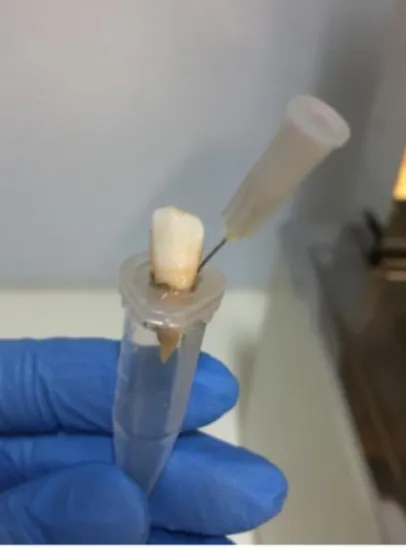
To quantify the eventual amount of ADE during the instrumentation process all teeth were placed into new Eppendorf Tubes (ET) covers. Using a warmed ball burnisher to make holes in the covers and insert the tooth until the cementoenamel junction (CEJ), so each tooth remained suspended in the interior of the ET, to collect the debris inside.
Two layers of varnish (Risqué®, São Paulo, Brazil) were placed to seal the cervical part
of the tooth around each tooth near the CEJ. Then a needle was introduced in the cover of each ET, laterally to the tooth to balance the pressure inside the tube (Figure 1).
5
Figure 1- Photographic illustration of the experimental setup.
The number of the teeth correctly identified each ET, so that they could be individually weighted and the amount of ADE could also be respectively registered.
First of all, each ET used was weighted 3 consecutive times by an analytic balance LPC-513L (VWR, Leuven, Belgic) before the instrumentation process beginning. This measurement was produced in order to obtain the mean for the initial weight.
After the preparation of the test set, each tooth was immersed in a warm bath at a temperature of 36 °C, to mimic physiologic conditions (Figure 2) in order to proceed to the filling material removal techniques in test.
6
Once finished all this procedures, each ET was again weighed as described above, so that, it could be calculate the average of ADE.
To quantify the amount of debris in the end of the procedures each ET was filled with 2,5% NaOCl until the irrigant volume performed 1.25ml.
Three controls ET (C1, C2, C3) were full with the same amount of NaOCl and previously weighed 3 times, as described before.
Then, all ET were placed in an incubator for 8 consecutive days at a constant temperature of 40°C, with the lids open, allowing the liquid to evaporate (Figure 3 and Annex 2).
Figure 3 - Incubator at 40°C with the Eppendorf Tube lids open
The ET were again weighed 3 times (Annex 3) as described above. The total amount of ADE was calculated as the difference between the pre and post retreatment weight.
7 4. Retreatment Procedures
All used retreatment systems were applied by following the manufacturer's instructions. In both experimental groups, the instruments were placed in a contra-angle hand piece in a motor Wave One® (Dentsply Maillefer, Ballaigues, Switzerland).
For each tooth, the determination of the working length (WL) was obtained by radiography in a BL (Bucco-Lingual) direction.
4.1 Reciproc 25® Group
The R25 is a single file system. The instrument R25 (25.08) was moved in the apical direction in a reciprocating motion, using a slow in-and-out pecking motion of about 3 mm in amplitude with a light apical pressure combined with brushing action against the lateral canal walls. After 3 or 4 pecking motions, the instrument was removed and cleaned. This file was used at 300rpm and torque (2N/cm). The canals were irrigated by applying a total of 2mL of 2,5% NaOCl during the instrumentation. Any irrigant that eventually overflowed through the crown was absorbed with cotton rolls. (Figure 4)
Figure 4 – Illustration of the cotton roll absorbing the eventual overflow of irrigant: (a) tooth outside the
8 4.2 XP-Endo Finisher® Group
As recommended by the manufacturer, the instrument was removed from the plastic tube immediately before the XP-F file was inserted into the RC, being the depth of this first insertion a value that would vary from 7 to 8mm. Until this point, rotation has not been activated. From this moment on, the XP-F file started to rotate slowly and gently at 800 rpm and 1 Ncm, until the WL was reached.
The instrumentation process was divided in 6 periods of 10 seconds each. In each and every period the file was removed from the RC and the tooth was irrigated. For the all 6 periods, a total of 1 mL of 2.5% NaOCl was used.
Finally, each RC was irrigated with 2 mL of 2.5% NaOCl using a syringe needle 1 mm short of the WL. Any irrigant that eventually overflowed through the crown was absorbed with cotton rolls, as described before.
5. Statistical Analysis
The data, before and after filling material removal and the amount of the ADE, were collected into Microsoft Excel tables and then statistical analysis to compare the experimental groups was performed using GraphPad Prism® version 5.00 for Windows,
GraphPad Software, San Diego California. The level of significance was set at 5% for all the tests (p < 0.05). A D'Agostino & Pearson normality test was applied to evaluate the normality of data distribution. A ANOVA (one-way) test was applied to compare whether there were significant differences between the VP and BL projections. Moreover, a Student t-test was applied to check for differences in the weight of the debris.
All tests were carried out in order to compare between the groups tested, which system was more effective.
9 III. RESULTS
Analysis of the total area revealed no statistical differences between the systems in test (p > 0.05) (Chart 1). There is no significant difference in the percentage of the filling material’ reduction, between both groups.
In addition, group R25 had no difference regarding the percentage of RC filling removal in both directions. In the MD the percentage of removal was up to 84% and in BL was 87%. On the other hand, in the group R25 supplemented by XP-F there was a higher percentage of RC filling removal in the MD direction 95% comparing to the BL 90%; however this difference had no statistical significance.
Chart 1- Percentage of reduction of the root canal filling in two views – Bucco-Lingual (BL) and
10
There is a significant difference (chart 2) between the ADE during the use of XP-F compared when it was not used (p < 0.0001).
11 IV. DISCUSSION
The success of Endodontic Retreatment can be measured by the efficiency of removing the filling material. The more filling material is expunged, the more likely it is granted that the cause of the previous treatment failure may be eliminated (Oliveira et al., 2018).
It should be emphasized that the results of this study should not be applied directly to clinical situations. In the in vitro studies, the apex of the tooth is suspended in air, while,
in vivo, the apex would be surrounded by granulomatous or periapical tissues, that could
serve as a natural barrier, restricting the amount of ADE (Kfir et al., 2017). Results may also differ because of the positive and negative pressures at the apex, (Tanalp & Güngör, 2014) as the vacuum effect, in the ET may prevent ADE (Mittal et al., 2015).
In opposition to the present study, some studies, Versiani et al., (2011) e De-Deus et al., (2019) have shown that using only rotary files for canal cleaning during endodontic retreatment is not sufficient.
In fact, the XP-F was introduced to the market as a supplementary file. As reported by it’s manufacturer, this file may be used as an universal complement file in the NSER, providing that the final file size is at least an ISO# 25 (Kfir et al., 2017).
The R25 system was originally designed for the complete preparation of the RC by using a single file and later was adapted for retreatment. The R25 has a S-shaped cross section that promote a higher cutting capacity comparing with others rotary Ni-Ti systems, as Mtwo® or WaveOne® systems, used in reciprocating motion in the purpose of filling material’ removal (Capar, et al., 2015; Alves, et al., 2016; Nevares, et al., 2016).
The studies Alves et al., (2016) and Silva et al., (2018) show that the use of the XP-F, when introduced as a supplementary technique, significantly reduces, the quantity of filling material. One of the reasons for the disparate results of the present study may be the fact that in some of the above mentioned studies had applied different protocols.
12
In this study, students of the pre-clinical classes of Endodontics performed the Endodontic Treatment. They all used the same protocol during the instrumentation and the filling material was introduced by lateral condensation technique. The previous
mentioned studies (Alves et al., 2016; Silva et al., 2018) always used the same operator
during the all procedure. That maybe a factor that potentially could interfere with results.
With lateral condensation technique is harder to fill lateral and accessory canals (DuLac
et al., 1999) and takes more time to fill the RC in comparison to the thermoplastic
gutta-percha technique (Endal, et al., 2018). To overcome this limitation, it was introduced the continuous-wave technique which uses thermoplasticized percha. Warm gutta-percha adapts, naturally, more effectively to the RCS’ irregularities (Guess, et al., 2003).
So, it can be presumed, that when using the lateral condensation technique, it would be easier to remove the filling material, if necessary. Logically, it can be further concluded that techniques users of thermoplasticized gutta-percha, in obtaining an enhanced three-dimensional adaptation to the RC, lead to a more challenging removal of the filling material.
For filling, Alves, et al. (2016) used a single cone technique without warming the gutta-percha and Silva et al. (2018) used the continuous wave technique. In both of this studies the XP-F reduced equally the volume of filling material. The present study, that used cold lateral condensation technique, did not notice any differences in the quantity of removal filling material, comparing with the studies above describe.
NaOCl was the irrigant used in this study, such as in the ones of Alves et al., (2016) and Silva et al., (2018). One of the major goals of a NSER is to eliminate the filling material and as well the, frequently present, bacteria and their by-products, aiming to achieve the most higher level of RCS’ disinfection. NaOCl is the most used irrigant to reduce the bacterial load in clinical Endodontics practice (Iandolo et al., 2017). However, the present research did not take into account the temperature of the irrigant: in fact, some articles reported that the effect of NaOCl heated can be 210 times faster compared to its use at room temperature (Woodmansey, 2005; Iandolo et al., 2017).
13
In this study, in order to mimic physiologic conditions, it was used a warm bath at a temperature of 36 °C in which the teeth were immerse for a period of 1 hour before the NSER begin. All the procedures of both groups were done at that temperature, opposite to other studies in which the teeth were only immerse in a warm bath when XP-F was used (Alves et al., 2016; Silva et al., 2018). Actually, we didn´t found any study done with the same procedure as this one. All the related articles that used a warm bath, only used it with the XP-F. This study is a more realistic simulation of the clinical practice because during all the NSER in vivo, all instruments are at a body temperature.
Although that, the NaOCl was introduce at a room temperature; nevertheless, when inserted into the RC its temperature tended to increase. In a future research the NaOCl should be heated before, in order to better avail all the potential benefits of its heating. It seemed relevant to this study to remove the filing material at a body temperature to mimic a real clinical situation. Furthermore, the studies of Alves et al., (2016) and Silva et al., (2018) kept all specimen at 37°C and 100% humidity for 2 weeks to allow the sealer that was used in the RC during filling to settle completely and to simulate oral conditions.
In the present study this was not necessary because the teeth used were sealed, at least 3 months before the NSER and kept in a proper ambient storage. So, the sealer was completely settled. Possibly, due to the fact that the teeth used for this study had been sealed for months, the NSER was more difficult to perform than in the studies of Alves
et al. (2016) and Silva et al. (2018) where filling took place only two weeks before the
NSER. The set sealer is completely done and has the greatest dimensional changes at 4 weeks (Allan et al., 2001; Ørstavik et al., 2001). Taking this in consideration, it can be concluded that the previous mentioned studies, did not wait enough time for the sealer to set correctly. That could be the fact that justifies the results not concordants between those studies and the present one, since different protocols were applied.
Another major goal of the present research was to study the amount of ADE. The study of Kfir et al. (2017), with the same purpose, makes reference to a number of elements (n=30) higher than this study. However, Alves et al. (2016) used the same number of
14
elements of the present study and, indeed, Silva et al. (2018) used less (n=10). This is not a factor that could explain the differences of results because this study used an acceptable number of elements compared to the previous mentioned studies and the sample used allowed checking the eventual significant differences between techniques.
During the NSER, besides the anatomical complexities as isthmuses, bifurcations, dumbbell-shaped canals, probably the most commonly encountered anatomical challenge may be a curved canal. (Reddy, et al., 2011).
Alves et al. (2016) used curved canals in their study. Silva et al. (2018) chosen to use oval-shaped canals as this study. So, this is not a variable of the protocol that could explain the difference between the result of the present study and the studies mentioned, because Alves et al. (2016) and Silva et al. (2018) show that the use of the XP-F, when introduced as a supplementary technique, reduces the quantity of filling material, on the contrary to the present study that used straight canals, a priori easier to deal during NSER, compared to curved ones.
According to the XP-F manufacturer, this instrument does not remove the tooth structure (De-Deus, et al., 2019). Nevertheless, it makes more ADE when used as a supplementary stage in cleaning oval canals (Kfir et al., 2017). The present study also showed that XP-F contributed to more amount of ADE, but did not contribute as a supplementary efficient file, when removal of filling material is the issue.
Among problems presented, all instrumentation systems induce ADE. Nowadays, according to the various publications available on the subject, there is no system that does not produce ADE (Bürklein et al.., 2014; Kfir et al., 2017; Hizarci et al., 2019).
The results obtained in this study alert Professionals to be aware of the potential complications associated to ADE, and to adapt the best system to each clinical case, when performing NSER, always thinking in minimising the debris extrusion.
15 V. CONCLUSIONS
After the bibliographic research in the main Scientific Research Engines, no articles published yet, states that there is a system that completely removes the filling material or that does not cause ADE. In this study, two systems were taken in consideration related to its performance in NSER.
Considering the limitations of the study, after statistical analysis of the data obtained from the clinical protocols in test, it can be concluded that:
- both techniques remove equally the filling material;
- XP-F system does not bring any improvement as a supplementary technique; - all instrumentation techniques used produce apical extrusion of debris;
- the average debris produced by the R25 technique is significantly smaller than that produced by the XP-Fsystem (p <0.001);
As the professional, in cases of NSER, should privileged instrument that removes the higher quantity of the filling material and cause less ADE, in light of the results shown by the present study, it can be concluded that it is advisory to use only the R25, as it proved to be an efficient technique.
16 VI. BIBLIOGRAPHY
Allan, N. A., Walton, R. E., & Schaffer, M. (2001). Setting times for endodontic sealers under clinical usage and in vitro conditions. Journal of Endodontics, 27(6), 421-423.
Alves, F. R., et al., (2016). Removal of root canal fillings in curved canals using either reciprocating single-or rotary multi-instrument systems and a supplementary step with the XP-Endo Finisher. Journal
of Endodontics, 42(7), 1114-1119.
Azim, A. A. et al. (2018). Comparison between Single-file Rotary Systems: Part 1—Efficiency, Effectiveness, and Adverse Effects in Endodontic Retreatment. Journal of Endodontics, 44(11), 1720-1724.
Bürklein, S. et al., (2014). Quantitative evaluation of apically extruded debris with different single‐file systems: Reciproc, F 360 and One Shape versus M two. International Endodontic Journal, 47(5), 405-409.
Cardoso Tarallo, A., et al. (2018). Influence of Working Length on Post-operative Pain after Single or Two-Visit Endodontic Treatment: A Randomised Clinical Trial. Journal of Clinical & Diagnostic Research, 12(10).
Capar, I. D., et al., (2015). Effectiveness of ProTaper Universal retreatment instruments used with rotary or reciprocating adaptive motion in the removal of root canal filling material. International endodontic
journal, 48(1), 79-83.
Dave, P., et. al., (2019). Analysis of for Root Canal Obturation Techniques with Gutta Percha: An Observational Study. Journal of Advanced Medical and Dental Sciences Research, 7(2), 1-4.
De-Deus, G. et al. (2019). Effectiveness of Reciproc Blue in removing canal filling material and regaining apical patency. International Endodontic Journal, 52(2), 250-257.
De-Deus, G., et al. (2019). Micro-CT comparison of XP-endo Finisher and passive ultrasonic irrigation as final irrigation protocols on the removal of accumulated hard-tissue debris from oval shaped-canals. Clinical Oral Investigations, 23(7), 3087-3093.
De-Deus, G., et al. (2011). The self-adjusting file optimizes debridement quality in oval-shaped root canals. Journal of Endodontics, 37(5), 701-705.
DuLac, K. A., et al., (1999). Comparison of the obturation of lateral canals by six techniques. Journal of endodontics, 25(5), 376-380.
Endal, U., et al., (2018). Evaluation of Quality and Preparation Time of Retrograde Cavities in Root Canals Filled with GuttaCore and Cold Lateral Condensation Technique. Journal of endodontics, 44(4), 639-642.
Gavini, G. et al. (2012). Resistance to flexural fatigue of Reciproc R25 files under continuous rotation and reciprocating movement. Journal of Endodontics, 38(5), 684-687.
Guess, G. M., et al., (2003). Analysis of continuous-wave obturation using a single-cone and hybrid technique. Journal of endodontics, 29(8), 509-512.
Hizarci, U., Koçak, S., Sağlam, B. C., & Koçak, M. M. (2019). Effect of different irrigation activation techniques on the amount of apical debris extrusion. Tanta Dental Journal, 16(1), 29.
Iandolo, A., et al., (2017). 3D cleaning, a perfected technique: thermal profile assessment of heated NaOCl. Giornale Italiano di Endodonzia, 31(1), 58-61.
17
Jain, A. (2018). An in vitro assessment of apically extruded debris during root canal preparation using ProTaper, Protaper Next and WaveOne rotary systems. JIDA: Journal of Indian Dental Association, 12(12).
Kasam, S., e Mariswamy, A. B. (2016). Efficacy of different methods for removing root canal filling material in retreatment-an in-vitro study. Journal of Clinical and Diagnostic Research: JCDR, 10(6), ZC06.
Kfir, A. et al. (2018). Apical extrusion of debris during the preparation of oval root canals: a comparative study between a full-sequence SAF system and a rotary file system supplemented by XP-endo finisher file. Clinical Oral Investigations, 22(2), 707-713.
Mittal, R., et al. (2015). A comparison of apical bacterial extrusion in manual, ProTaper rotary, and one shape rotary instrumentation techniques. Journal of Endodontics, 41(12), 2040-2044.
Nevares, G., et al., (2016). Efficacy of ProTaper NEXT compared with Reciproc in removing obturation material from severely curved root canals: a micro–computed tomography study. Journal of
endodontics, 42(5), 803-808.
Ørstavik, D., Nordahl, I., & Tibballs, J. E. (2001). Dimensional change following setting of root canal sealer materials. Dental Materials, 17(6), 512-519.
Ozyurek, T., e Ozsezer-Demiryurek, E. (2017). Efficacy of protaper next and protaper universal retreatment systems in removing gutta-percha in curved root canals during root canal retreatment. Journal of Istanbul University Faculty of Dentistry, 51(2), 7.
Pinto de Oliveira, D., et al. (2018). Effectiveness of Reciproc and MTwo for Removing Filling Material During Root Canal Retreatment Using a Digital Radiograph System. The Open Dentistry Journal, 12(1). Uslu, G., et al., (2018). Apically extruded debris during root canal instrumentation with Reciproc blue, HyFlex EDM, and XP-endo shaper nickel-titanium files. Journal of endodontics, 44(5), 856-859.
Reddy, S., et al., (2011). Removal of gutta-percha/zinc-oxide-eugenol sealer or gutta-percha/epoxy resin sealer from severely curved canals: an in vitro study. International journal of dentistry, 2011.
Silva, E. J. N. L., et al. (2018). Effectiveness of XP-endo Finisher and XP-endo Finisher R in removing root filling remnants: a micro‐CT study. International Endodontic Journal, 51(1), 86-91.
Silva, E. J. N. L et al., (2018). Torsional fatigue resistance of blue-treated reciprocating instruments. Journal of Endodontics, 44(6), 1038-1041.
Tanalp, J., e Güngör, T. (2014). Apical extrusion of debris: a literature review of an inherent occurrence during root canal treatment. International Endodontic Journal, 47(3), 211-221.
Vaz-Garcia, E. S., et al., (2018). Mechanical Properties of Anatomic Finishing Files: XP-Endo Finisher and XP-Clean. Brazilian Dental Journal, 29(2), 208-213.
Versiani, M. A., et al. (2011). Flat-oval root canal preparation with self-adjusting file instrument: a micro–computed tomography study. Journal of Endodontics, 37(7), 1002-1007.
Yılmaz, F., et al., (2018). Evaluation of 3 Different Retreatment Techniques in Maxillary Molar Teeth by Using Micro–computed Tomography. Journal of Endodontics, 44(3), 480-484.
Woodmansey, K. F. (2005). Intracanal heating of sodium hypochlorite solution: an improved endodontic irrigation technique. Dentistry today, 24(10), 114-116.
18 VII. ANNEX
19
Annex 2 – Crystals resulted from the evaporation of the irrigant collected in the Eppendorf Tube.