Linear measurements of human permanent
dental development stages using
Cone-Beam Computed Tomography:
A preliminary study
Carlos Estrela*, José Valladares Neto**, Mike Reis Bueno***, Orlando Aguirre Guedes****,
Olavo Cesar Lyra Porto****, Jesus Djalma Pécora*****
Objective: To determine the linear measurements of human permanent dentition
de-velopment stages using Cone-Beam Computed Tomography. Methods: This study was
based on databases of private radiology clinics involving 18 patients (13 male and 5
fe-male, with age ranging from 3 to 20 years). Cone-Beam Computed Tomography (CBCT)
images were acquired with i-CAT system and measured with a specific function of the
i-CAT software. Two hundred and thirty-eight teeth were analyzed in different
develop-ment stages in the coronal and sagittal planes. The method was based on delimitation
and measurement of the distance between anatomical landmarks corresponding to the
development of the dental crowns and roots. These measurements allowed the
develop-ment of a quantitative model to evaluate the initial and final developdevelop-ment stages for
all dental groups. Results and Conclusions: The measurements acquired from different
dental groups are in agreement with estimates of investigations previously published.
CBCT images of different development stages may contribute to diagnosis, planning
and outcome of treatment in various dental specialties. The dimensions of dental crowns
and roots may have important clinical and research applications, constituting a
noninva-sive technique which contributes to in vivo studies. However, further studies are
recom-mended to minimize methodological variables.
Abstract
Keywords:
Tooth development. Incomplete root formation. Apexogenesis.
Cone-Beam Computed Tomography. Computed tomography.
* Chairman and Professor of Endodontics, Federal University of Goiás, Goiânia, GO, Brazil. ** Professor of Orthodontics, Federal University of Goiás, Goiânia, GO, Brazil.
*** Professor of Oral Diagnosis, Department of Oral Diagnosis, University of Cuiabá, Cuiabá, MT, Brazil. **** Post-graduate student, Federal University of Goiás, Goiânia, GO, Brazil.
intROduCtiOn
Knowledge of the development stages of
perma-nent teeth is essential for clinical practice in several
dental specialties, since it may have influence on
di-agnosis, treatment planning and treatment outcome.
Several studies have evaluated calcification and
development of human teeth using various
method-ologies.
16,19,20,21,24,26,27,28,34,35,38-41,44,46,47,49Radiographic
images, although representing two-dimensional
as-pects of three-dimensional structures, were the most
widely used resource to determine the
calcifica-tion and development stages of human permanent
teeth.
20,34,35,39,49A classical study by Nolla
35evaluat-ed the stages of development of human permanent
teeth using radiographic records selected from the
files on the basis of length, which were graded on a
scale from 0 to 10 based on development.
Technological advances offer imaging modalities
which have brought important contributions to
den-tal radiology, such as viable diagnostic tools, namely
digital radiography, densitometry methods,
Cone-Beam Computed Tomography (CBCT), magnetic
resonance imaging, ultrasound and nuclear
tech-niques,
8providing detailed high-resolution images
of oral structures and permitting early detection of
alterations in maxillofacial structures.
Since the introduction of computed
tomogra-phy,
2,17,37it has been observed that its clinical
ap-plication has exerted a significant impact on health
care.
1,4,7,10-15,19,22,25,29-31,42,43,45,48Recently, clinical
den-tistry and research have benefitted from CBCT
ap-plication,
3,6,8,18,32,42which has permitted visualization
of three-dimensional images, with additional
han-dling strategies.
6The higher potential for clinical
ap-plication and the accuracy compared with periapical
radiographs have contributed to treatment planning,
diagnosis, therapy and prognosis of different
diseas-es.
1,4,6,7,10-15,19,25,26,29-31,42,43,45Another remarkable feature of this technology
is the CBCT measurement tool, which enables the
determination of linear distances and volume of
ana-tomic structures,
4,22,45presurgical planning of
maxil-lofacial lesions,
7root length and marginal bone level
during orthodontic treatment,
30,43reconstruction
techniques,
1,29bone level changes following
regen-erative periodontal therapy,
15periodontal defect,
19periapical lesions,
11,12and root resorptions.
13However, based on the potential of
high-resolu-tion image acquisihigh-resolu-tion and the availability of new
emerging three-dimensional imaging modalities,
it seems appropriate to study the linear
measure-ments of human permanent dentition during
de-velopment, particularly in the first 20 years of age.
Thus, the aim of this study was to determine the
linear measurements of human permanent teeth
at different development stages using Cone-Beam
Computed Tomography.
MAtERiAL And MEtHOdS
image Selection
This study was structured using databases of
pri-vate radiology clinics (CIRO, Goiânia, GO, Brazil;
RIO, Brasília, DF, Brazil; CROIF, Cuiabá, MT, Brazil)
involving 18 patients (n=238 teeth), 13 male, 5
fe-male, with age ranging from 3 to 20 years. The
pa-tients were referred to the dental radiology service
for different diagnostic purposes. The sample had
no history of dental caries, orthodontic treatment or
disturbance of dental development.
The study design was approved by the Local
Eth-ics Research Committee (UFG, Proc. #169/2008).
imaging Methods
(NVIDIA Corporation, USA) and Monitor EIZO -
Flexscan S2000, resolution 1600x1200 pixels (EIZO
NANAO Corporation Hakusan, Japan).
imaging Measurements
The method used to study the development of
the permanent teeth with CBCT was based on
de-limiting and measuring the distance between
ana-tomical landmarks according to the development of
the dental crowns and roots. All the measurements
on the CBCT images were acquired by two dental
radiology specialists using a proprietary
measure-ment tool supplied with the CBCT scanner (Xoran
3.1.62; Xoran Technologies, Ann Arbor, MI, USA).
A specific function of the i-CAT software that
of-fers values in millimeters was used to measure teeth
images. The measurements were made both in the
sagittal and coronal planes (the reference used was
the largest measurement extension given by the
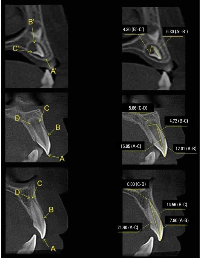
soft-ware). The reference distances used were as follows:
»
AB - maximum width between the incisal edge
or cusp tip and cementoenamel junction;
»
BC - maximum width between the
cemen-toenamel junction and the most apical point
of the root;
»
AC - maximum width between the incisal
edge or cusp tip and the most apical point
of the root;
»
CD - maximum width of the apical foramen;
»
A’B’ - maximum width between the
inci-sal edge or cusp tip and the end of dental
crown, used in teeth that no root formation
was detected;
»
B’C’ - maximum width of the apical
fora-men, used in teeth where no root formation
was detected.
The calibrated examiners measured all 238
teeth at different development stages using the
CBCT images and assessed the dimensions in the
directions described above. When a consensus was
not reached a third observer made the final
deci-sion. Due to peculiarities of distinct dental groups,
especially for multirooted teeth, measurements
were made specifically for each root. The B’C’
ref-erence for teeth with more than one root used the
mean distance between roots.
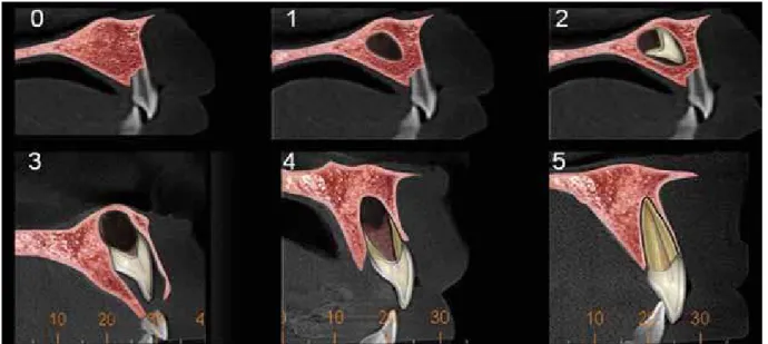
Using these measurements a quantitative
mod-el with five scores was suggested for all dental
groups (with the exception of the third molar):
0 = absence of dental crypt; 1 = presence of
den-tal crypt; 2 = denden-tal crown partially formed; 3 =
dental crown completely formed; 4 = beginning
of root formation – open apex; 5 = end of root
formation – closed apex) (Fig 1).
RESuLtS
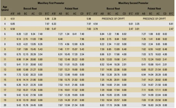
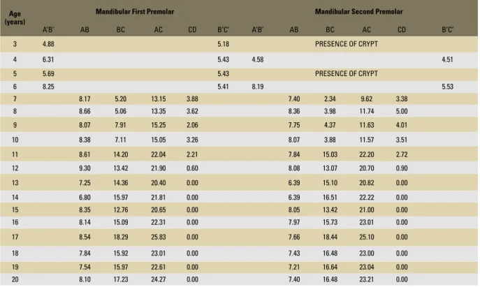
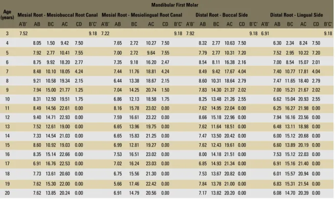
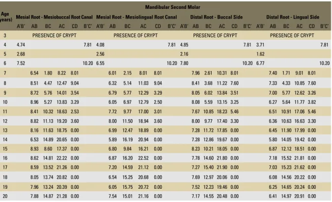
Linear measurements (mm) of the dental
devel-opment stages are shown in Tables 1 to 16. Table 17
presents the mean values (mm) of dental
develop-ment stages on CBCT scans. Figures 2 to 21 show
the images of dental development stages.
diSCuSSiOn
The formation stages of deciduous and
perma-nent teeth are basically the same, differing only in
time periods. The dental lamina of deciduous
denti-tion begins between the sixth and eighth week of
embryonic development. Permanent teeth begin
their development between the twentieth week of
intra-uterine life and the tenth month after birth;
permanent molars, between the twentieth week of
intra-uterine life (first molar) and the fifth year of
life (third molar).
33Dental development starts
dur-ing the intra-uterine life and lasts approximately
until the second decade of life.
TABLE 1 - Linear measurements (mm) of dental development stages of maxillary anterior teeth (Coronal view).
Age (years)
Maxillary Central Incisor Maxillary Lateral Incisor Maxillary Canine
A’B’ AB BC AC CD B’C’ A’B’ AB BC AC CD B’C’ A’B’ AB BC AC CD B’C’
3 9.60 5.79 6.30 4.30 7.13 5.41
4 11.40 6.04 10.06 5.53 9.92 6.74
5 13.23 5.52 10.15 5.53 10.24 6.18
6 12.41 7.70 19.57 4.49 10.04 2.67 12.50 5.83 10.63 1.71 12.20 7.62
7 13.62 9.06 22.07 3.58 12.01 4.12 15.95 5.66 10.44 3.06 13.22 7.30
8 12.43 13.33 24.80 3.23 11.23 9.04 19.50 5.02 13.00 2.91 15.81 8.77
9 10.85 11.01 20.87 0.00 10.72 10.88 20.24 0.00 10.10 10.12 19.68 3.80
10 12.04 15.58 26.44 0.00 10.47 14.49 23.87 1.28 11.77 8.80 20.24 5.02
11 12.04 12.38 23.24 0.00 10.83 13.00 22.75 0.00 11.51 17.77 27.90 0.00
12 12.28 15.15 26.27 0.00 11.61 15.70 26.17 0.00 13.01 14.30 26.76 3.79
13 11.12 14.81 25.05 0.00 9.65 14.85 23.39 0.00 11.61 17.05 27.51 0.00
14 11.09 14.48 24.96 0.00 10.07 14.37 23.74 0.00 10.05 16.75 26.01 0.00
15 11.29 13.18 23.68 0.00 9.48 12.88 21.46 0.00 9.95 18.09 26.97 0.00
16 11.65 13.59 24.56 0.00 9.67 14.78 23.35 0.00 11.29 19.25 29.50 0.00
17 11.26 10.00 20.32 0.00 10.01 11.17 19.78 0.00 10.59 15.25 24.53 0.00
18 12.79 13.10 25.44 0.00 11.20 13.21 23.34 0.00 12.61 16.39 28.24 0.00
19 11.93 15.09 26.42 0.00 9.81 15.33 24.01 0.00 9.65 18.41 27.46 0.00
20 13.06 14.75 26.58 0.00 10.79 16.24 25.37 0.00 11.41 18.09 28.04 0.00 TABLE 2 - Linear measurements (mm) of dental development stages of maxillary anterior teeth (Sagittal view).
Age (years)
Maxillary Central Incisor Maxillary Lateral Incisor Maxillary Canine
A’B’ AB BC AC CD B’C’ A’B’ AB BC AC CD B’C’ A’B’ AB BC AC CD B’C’
3 8.50 4.70 5.24 3.90 7.30 6.36
4 11.03 5.47 9.31 4.20 10.22 6.84
5 11.50 4.50 7.85 3.61 9.77 5.77
6 9.30 8.61 17.57 4.24 7.87 5.60 13.10 3.61 9.02 3.06 11.88 4.80
7 10.90 8.64 18.84 3.22 8.63 5.20 13.72 3.81 10.70 2.81 12.78 5.46
8 11.19 14.02 24.79 2.81 8.55 9.77 18.00 2.81 11.38 4.37 15.42 5.69
9 8.66 12.34 19.85 0.00 7.28 11.79 18.43 0.00 8.35 11.22 19.00 2.01
10 9.85 16.12 25.08 0.00 7.53 14.84 21.65 0.00 9.93 10.32 19.67 2.81
11 8.74 12.76 21.01 0.00 7.84 13.97 21.01 0.00 9.04 17.03 25.02 0.00
12 11.06 13.49 24.00 0.00 8.40 14.23 21.93 0.00 10.44 15.69 25.40 2.09
13 9.18 14.49 22.83 0.00 7.47 15.56 22.17 0.00 9.07 18.05 26.46 0.00
14 9.63 12.53 21.78 0.00 7.22 15.45 22.17 0.00 7.62 18.58 25.55 0.00
15 10.33 14.36 24.01 0.00 7.47 13.34 20.50 0.00 8.48 18.75 26.61 0.00
16 8.83 14.05 21.78 0.00 7.50 13.68 20.53 0.00 8.35 19.50 27.34 0.00 17 9.33 12.17 20.80 0.00 7.95 13.10 20.54 0.00 8.92 15.18 23.41 0.00
18 9.57 15.23 23.77 0.00 7.80 14.56 21.40 0.00 9.51 19.94 28.22 0.00
19 10.31 16.32 25.80 0.00 8.06 15.09 22.15 0.00 7.97 18.87 26.06 0.00
Age (years)
Maxillary First Premolar Maxillary Second Premolar
Buccal Root Palatal Root Buccal Root Palatal Root
A’B’ AB BC AC CD B’C’ A’B’ AB BC AC CD B’C’ A’B’ AB BC AC CD B’C’ A’B’ AB BC AC CD B’C’
3 4.51 5.98 2.28 5.98 PRESENCE OF CRYPT PRESENCE OF CRYPT
4 6.85 7.97 6.33 7.97 4.54 6.61 3.35 6.61
5 6.58 7.60 5.47 7.60 3.79 2.47 3.01 2.47
6 8.20 1.22 9.34 7.40 7.27 1.34 8.41 7.40 6.84 1.22 7.96 8.02 7.27 1.08 8.02 8.02
7 9.14 2.15 11.03 7.96 8.40 7.96 6.99 2.15 8.84 9.02 7.35 1.41 8.60 9.02
8 9.22 4.22 13.05 8.26 7.73 4.56 12.00 8.26 9.22 2.34 11.02 9.00 7.62 2.34 9.65 9.00
9 7.97 7.89 15.45 5.43 7.40 7.77 15.07 5.43 7.59 6.65 13.89 6.48 7.82 6.55 14.02 6.48
10 8.75 12.15 20.39 3.54 6.91 10.45 17.20 3.54 8.66 9.31 17.66 4.00 7.03 9.73 16.63 4.00
11 8.98 11.94 20.60 0.80 7.82 12.48 20.22 0.00 8.29 12.03 19.90 2.41 7.73 12.24 19.63 2.41
12 9.41 11.51 20.60 0.82 7.62 11.81 19.25 0.80 8.52 10.44 18.38 2.01 8.41 10.88 18.41 2.01
13 8.80 12.66 21.27 0.00 7.07 13.21 19.90 0.00 7.57 14.95 22.06 0.00 6.68 15.37 21.54 0.00
14 7.73 12.83 20.22 0.00 7.22 12.86 19.80 0.00 7.86 13.30 20.76 0.00 6.94 14.04 20.39 0.00
15 8.16 14.14 21.80 0.00 7.50 13.75 21.02 0.00 7.25 14.26 20.91 0.00 7.47 14.21 20.82 0.00
16 8.26 14.31 22.35 0.00 7.53 14.41 21.66 0.00 7.86 14.60 21.84 0.00 7.33 14.87 21.50 0.00
17 7.52 10.31 17.26 0.00 7.16 10.61 17.32 0.00 7.29 10.68 17.64 0.00 7.11 10.85 17.11 0.00
18 9.42 12.42 21.56 0.00 7.67 12.28 19.89 0.00 8.46 15.05 22.95 0.00 7.67 14.99 22.04 0.00
19 8.10 13.19 20.62 0.00 7.23 14.20 21.01 0.00 7.53 16.54 23.57 0.00 7.03 17.28 23.50 0.00
20 9.43 15.76 24.45 0.00 7.67 13.94 21.54 0.00 8.63 17.15 24.96 0.00 7.84 16.83 24.39 0.00 TABLE 4 - Linear measurements (mm) of dental development stages of maxillary premolars teeth (Sagittal view).
TABLE 3 - Linear measurements (mm) of dental development stages of maxillary premolars teeth (Coronal view).
Age (years)
Maxillary First Premolar Maxillary Second Premolar
Buccal Root Palatal Root Buccal Root Palatal Root
A’B’ AB BC AC CD B’C’ A’B’ AB BC AC CD B’C’ A’B’ AB BC AC CD B’C’ A’B’ AB BC AC CD B’C’
3 4.30 4.88 3.31 4.88 PRESENCE OF CRYPT PRESENCE OF CRYPT
4 6.85 4.24 5.47 4.24 4.24 4.58 3.66 4.58
5 6.85 5.11 5.77 5.11 3.66 3.66 2.77 2.77
6 7.98 1.81 9.62 4.20 7.40 1.40 8.74 4.20 7.56 1.40 8.82 4.18 7.38 1.22 8.51 4.18
7 8.54 2.43 10.72 4.44 8.59 4.44 7.78 1.02 8.74 4.60 7.81 1.02 8.75 4.60
8 8.40 6.07 14.00 3.26 7.07 4.68 11.42 3.26 7.52 3.81 11.02 4.02 7.33 3.41 10.44 4.02
9 7.97 8.12 15.63 2.21 6.84 7.69 14.21 2.01 7.78 6.80 14.44 3.21 7.40 6.80 14.04 3.61
10 7.86 11.69 19.01 1.41 6.85 11.61 18.25 1.41 7.53 11.29 18.42 2.01 6.90 10.65 17.46 2.40
11 8.73 12.91 20.80 1.22 7.67 13.10 20.22 0.00 7.84 13.12 20.42 1.79 7.53 12.70 19.81 1.22
12 8.85 12.81 20.60 1.26 7.81 12.37 19.64 0.82 7.52 11.51 18.27 0.63 7.97 11.71 19.22 0.63
13 7.15 14.76 21.40 0.00 7.15 12.73 19.40 0.00 6.77 15.89 22.01 0.00 6.32 15.85 21.61 0.00
14 6.96 14.16 20.45 0.00 6.84 14.32 20.63 0.00 6.77 15.16 21.40 0.00 6.32 14.80 20.60 0.00
15 7.66 15.67 22.43 0.00 7.38 14.96 22.01 0.00 7.40 14.12 21.01 0.00 7.15 14.12 20.80 0.00
16 7.72 14.63 21.80 0.00 7.18 14.56 21.26 0.00 7.72 16.36 23.43 0.00 6.99 16.06 22.51 0.00
17 7.35 12.33 18.81 0.00 7.16 11.07 17.61 0.00 6.49 11.47 17.51 0.00 6.41 11.04 16.71 0.00
18 8.03 14.12 21.40 0.00 7.38 13.14 20.52 0.00 7.78 15.07 22.42 0.00 7.17 15.25 21.86 0.00
19 7.47 14.37 21.20 0.00 7.15 14.01 20.60 0.00 7.15 15.82 22.40 0.00 7.40 16.51 23.21 0.00
Age (years)
Maxillary First Molar
Mesiobuccal Root Distalbuccal Root Palatal Root
A’B’ AB BC AC CD B’C’ A’B’ AB BC AC CD B’C’ A’B’ AB BC AC CD B’C’
3 6.63 11.16 6.60 11.16 7.74 11.16
4 9.60 1.90 11.24 10.01 7.50 10.01 8.30 2.72 10.90 10.01
5 7.79 2.10 9.83 10.80 7.71 2.12 9.72 10.80 8.36 2.18 10.19 10.80
6 6.71 9.23 15.81 2.00 7.34 9.93 16.41 1.65 8.54 9.70 17.84 2.72
7 7.92 9.63 17.41 4.42 7.62 9.34 16.84 3.49 8.35 10.40 18.58 2.67
8 7.96 10.41 18.01 4.08 7.47 10.80 17.56 2.34 6.84 11.00 17.69 2.61
9 7.21 12.08 18.83 0.00 7.23 11.74 18.43 0.00 7.42 13.92 21.00 0.00
10 7.42 14.08 21.31 0.00 7.80 12.48 20.25 0.00 8.14 13.88 21.95 0.00
11 7.10 12.23 18.91 0.00 7.73 12.36 19.81 0.00 8.06 13.08 20.91 0.00 12 7.96 13.35 20.72 0.00 7.42 13.65 20.22 0.00 8.93 15.07 23.62 0.00
13 6.71 12.66 19.20 0.00 6.48 12.04 18.40 0.00 7.43 13.67 20.46 0.00
14 6.85 12.13 18.71 0.00 6.71 10.95 17.41 0.00 7.79 11.57 19.10 0.00
15 7.28 13.25 20.22 0.00 7.38 12.06 19.40 0.00 8.03 12.61 20.42 0.00
16 7.30 13.22 20.24 0.00 6.87 14.31 21.02 0.00 7.52 15.03 22.37 0.00
17 7.29 10.85 17.25 0.00 7.04 9.71 16.25 0.00 7.76 11.03 18.54 0.00
18 8.86 12.03 20.52 0.00 8.24 11.30 19.28 0.00 7.07 13.81 20.63 0.00
19 7.81 13.45 20.82 0.00 7.28 14.12 21.00 0.00 8.22 14.81 22.69 0.00
20 8.93 12.18 20.41 0.00 7.86 14.14 21.60 0.00 9.14 15.42 23.99 0.00 TABLE 6 - Linear measurements (mm) of dental development stages of maxillary first molar tooth (Sagittal view).
TABLE 5 - Linear measurements (mm) of dental development stages of maxillary first molar tooth (Coronal view).
Age (years)
Maxillary First Molar
Mesiobuccal Root Distalbuccal Root Palatal Root
A’B’ AB BC AC CD B’C’ A’B’ AB BC AC CD B’C’ A’B’ AB BC AC CD B’C’
3 7.50 7.22 7.50 7.22 10.06 7.85
4 7.59 2.72 10.26 6.64 7.52 2.28 9.72 6.64 9.02 1.90 10.92 6.64
5 7.71 3.06 10.57 6.63 7.35 3.06 10.24 6.63 8.88 2.85 11.44 6.63 6 6.85 8.91 15.61 2.20 8.77 8.66 16.80 2.01 6.79 8.68 15.01 3.35
7 7.86 9.85 17.80 1.08 7.96 9.42 17.23 1.00 8.29 11.18 18.42 3.01 8 6.94 11.74 18.40 1.61 7.53 11.64 18.84 1.41 8.44 10.96 18.82 3.22 9 6.84 12.36 18.80 0.00 7.03 11.91 18.83 0.00 8.22 13.49 20.82 0.00
10 6.36 14.74 20.81 0.00 7.64 13.80 21.42 0.00 8.35 15.77 23.27 0.00
11 6.60 14.31 20.32 0.00 7.57 12.06 19.40 0.00 8.22 14.95 22.03 0.00 12 7.81 13.18 20.60 0.00 8.01 13.10 20.94 0.00 8.30 16.02 23.50 0.00
13 6.36 12.99 19.02 0.00 6.48 12.53 18.82 0.00 7.23 14.60 21.26 0.00
14 6.26 12.03 18.03 0.00 6.68 11.44 18.00 0.00 7.47 13.22 20.00 0.00
15 6.99 14.04 20.62 0.00 7.81 12.21 20.02 0.00 7.47 13.82 20.42 0.00
16 6.79 13.85 20.22 0.00 7.24 13.64 20.80 0.00 7.98 16.07 22.86 0.00
17 6.32 11.47 17.05 0.00 6.91 9.58 16.25 0.00 7.60 11.96 18.54 0.00
18 7.03 14.04 20.62 0.00 7.30 12.56 19.63 0.00 7.54 13.74 20.22 0.00
TABLE 8 - Linear measurements (mm) of dental development stages of maxillary second molar tooth (Sagittal view). TABLE 7 - Linear measurements (mm) of dental development stages of maxillary second molar tooth (Coronal view).
Age (years)
Maxillary Second Molar
Mesiobuccal Root Distalbuccal Root Palatal Root
A’B’ AB BC AC CD B’C’ A’B’ AB BC AC CD B’C’ A’B’ AB BC AC CD B’C’
3 ABSENCE OF CRYPT ABSENCE OF CRYPT ABSENCE OF CRYPT
4 5.32 7.81 3.06 7.81 5.18 7.81
5 4.54 3.00 4.74
6 7.80 10.19 7.02 10.19 9.06 10.19
7 8.42 9.87 8.40 9.87 9.22 9.87
8 8.23 1.26 9.18 12.01 7.15 1.71 8.55 12.01 7.88 1.22 8.92 12.01 9 7.78 5.88 13.01 8.24 7.28 5.53 12.43 8.24 7.28 5.41 12.50 8.24
10 7.34 7.57 14.41 2.81 7.28 6.23 13.27 2.83 7.77 6.03 13.64 4.08
11 8.49 9.01 16.43 2.01 7.50 7.00 14.82 1.08 7.66 8.55 16.16 1.97
12 8.03 8.66 16.28 2.72 7.78 6.85 14.56 3.68 8.16 9.43 17.50 2.24
13 6.99 11.60 18.19 0.00 6.58 10.25 16.71 0.00 7.47 12.50 19.60 0.00
14 6.21 11.61 17.41 0.00 6.48 10.82 17.27 0.00 7.97 13.62 21.26 0.00
15 7.67 11.64 18.49 0.00 7.62 10.01 17.20 0.00 8.41 12.13 19.68 0.00
16 7.62 12.81 20.24 0.00 7.03 14.01 20.94 0.00 8.12 14.41 22.42 0.00
17 6.80 11.95 18.04 0.00 6.88 10.85 17.01 0.00 8.31 12.12 19.06 0.00
18 9.67 11.68 21.20 0.00 7.33 12.50 19.60 0.00 8.51 13.22 21.42 0.00
19 7.47 13.06 20.01 0.00 6.60 13.60 19.40 0.00 7.86 13.80 21.49 0.00
20 8.54 13.16 20.85 0.00 7.54 12.46 19.33 0.00 7.78 15.93 23.43 0.00 Age
(years)
Maxillary Second Molar
Mesiobuccal Root Distalbuccal Root Palatal Root
A’B’ AB BC AC CD B’C’ A’B’ AB BC AC CD B’C’ A’B’ AB BC AC CD B’C’
3 ABSENCE OF CRYPT ABSENCE OF CRYPT ABSENCE OF CRYPT
4 5.11 7.00 3.01 7.00 4.84 7.00
5 4.26 3.31 4.26
6 7.57 6.85 7.22 6.85 7.98 6.85
7 8.66 7.07 8.04 7.07 8.79 7.07
8 7.09 2.43 9.26 7.10 6.81 1.65 8.40 7.10 7.42 3.03 10.06 7.10
9 7.47 6.21 13.21 4.40 7.22 4.90 12.01 4.40 7.84 5.10 12.50 3.80
10 6.91 8.22 14.67 2.04 6.65 6.60 13.21 2.47 7.53 8.29 15.01 3.49
11 7.25 9.41 16.21 1.02 7.60 6.71 14.14 1.00 7.72 9.63 16.51 3.21
12 7.47 10.31 17.34 2.21 6.99 7.86 14.62 2.04 8.20 9.49 17.04 2.61
13 6.46 11.61 17.60 0.00 6.45 11.30 17.29 0.00 7.28 13.03 19.25 0.00
14 6.14 12.32 17.82 0.00 6.36 11.69 17.64 0.00 7.07 14.99 21.00 0.00
15 7.23 11.76 18.29 0.00 7.44 10.25 17.60 0.00 7.40 13.27 20.22 0.00
16 7.28 14.52 20.72 0.00 7.03 13.42 20.45 0.00 7.84 16.02 22.87 0.00
17 6.43 13.46 19.07 0.00 6.33 12.21 18.29 0.00 6.91 11.94 18.17 0.00
18 7.78 13.98 20.72 0.00 7.60 11.64 19.22 0.00 8.14 14.95 22.00 0.00
19 7.21 13.26 19.80 0.00 7.21 13.06 20.12 0.00 7.43 14.71 21.95 0.00
Age (years)
Mandibular Central Incisor Mandibular Lateral Incisor Mandibular Canine
A’B’ AB BC AC CD B’C’ A’B’ AB BC AC CD B’C’ A’B’ AB BC AC CD B’C’
3 8.40 4.85 8.19 4.85 7.11 4.80
4 10.90 5.53 10.55 6.63 9.53 6.41
5 11.89 5.43 11.16 5.18 9.31 5.11
6 10.44 8.24 18.09 5.13 10.26 5.41 15.49 6.36 11.80 8.54
7 10.14 12.03 21.41 0.00 10.63 11.88 21.65 2.34 11.74 6.91 18.27 6.02
8 11.07 12.52 22.62 0.00 11.32 13.15 23.84 2.60 11.76 6.03 17.46 7.42
9 10.59 14.98 24.50 0.00 11.00 13.73 23.80 1.75 12.18 10.69 22.09 6.50
10 10.36 15.10 24.50 0.00 10.64 13.61 23.36 1.82 12.10 10.44 21.82 6.17
11 10.45 14.14 23.53 0.00 10.72 15.03 24.56 0.00 13.05 14.45 26.08 2.83
12 10.34 13.07 22.82 0.00 10.66 14.52 24.05 0.00 11.79 13.59 24.09 0.00
13 9.43 10.58 19.46 0.00 8.60 13.24 21.02 0.00 9.77 14.23 22.60 0.00
14 9.46 12.97 21.40 0.00 9.75 14.48 23.27 0.00 11.61 15.21 25.55 0.00
15 10.00 10.88 20.06 0.00 10.80 12.26 22.01 0.00 11.84 13.09 24.11 0.00
16 9.80 13.72 22.29 0.00 10.33 15.12 23.94 0.00 12.28 15.29 26.65 0.00
17 9.85 13.61 22.67 0.00 11.29 12.70 22.99 0.00 13.44 17.27 29.47 0.00
18 9.57 14.45 23.19 0.00 9.40 15.98 24.56 0.00 11.85 15.58 26.19 0.00
19 8.99 13.58 21.60 0.00 9.49 14.81 23.22 0.00 9.95 16.83 25.81 0.00
20 8.55 13.74 21.47 0.00 9.51 13.91 22.49 0.00 11.22 18.72 28.60 0.00 TABLE 10 - Linear measurements (mm) of dental development stages of mandibular anterior teeth (Sagittal view).
TABLE 9 - Linear measurements (mm) of dental development stages of mandibular anterior teeth (Coronal view).
Age (years)
Mandibular Central Incisor Mandibular Lateral Incisor Mandibular Canine
A’B’ AB BC AC CD B’C’ A’B’ AB BC AC CD B’C’ A’B’ AB BC AC CD B’C’
3 8.45 3.35 7.50 3.35 7.31 4.80
4 9.97 3.31 10.36 3.91 9.90 5.71
5 10.90 3.00 10.65 3.61 9.30 5.32
6 8.19 8.72 16.81 2.18 8.16 7.26 15.07 2.77 9.02 2.42 11.21 6.00
7 8.63 12.64 21.02 0.00 7.87 13.65 21.15 0.00 8.88 9.43 18.05 3.21
8 9.37 13.06 22.60 0.00 9.18 14.51 23.40 0.60 9.95 8.09 17.66 3.61
9 9.12 14.52 23.51 0.00 8.73 15.57 24.01 1.50 9.10 12.25 21.12 3.50
10 8.10 15.85 23.76 0.00 9.10 16.02 24.81 1.03 8.46 13.06 21.05 3.25
11 8.59 12.53 20.80 0.00 8.60 15.26 23.43 0.00 9.49 17.47 26.44 1.60
12 8.88 13.66 22.20 0.00 8.74 15.00 23.52 0.00 8.92 15.93 24.39 0.00
13 6.71 12.68 19.01 0.00 6.84 14.47 20.82 0.00 7.53 14.26 21.05 0.00
14 7.92 13.67 21.40 0.00 7.42 15.42 22.41 0.00 8.54 14.85 23.03 0.00
15 8.74 9.81 18.31 0.00 8.91 10.72 19.40 0.00 8.79 13.39 21.65 0.00
16 8.59 12.68 21.00 0.00 8.83 14.40 22.61 0.00 9.62 16.16 25.41 0.00
17 8.20 13.50 21.40 0.00 8.94 15.47 23.90 0.00 9.67 20.52 28.81 0.00
18 7.23 14.60 21.61 0.00 7.53 15.06 22.01 0.00 7.86 18.68 25.89 0.00
19 7.28 14.14 21.00 0.00 7.78 14.71 22.20 0.00 7.66 18.95 26.00 0.00
Age (years)
Mandibular First Premolar Mandibular Second Premolar
A’B’ AB BC AC CD B’C’ A’B’ AB BC AC CD B’C’
3 4.37 4.81 PRESENCE OF CRYPT
4 6.93 5.73 4.69 5.60
5 5.41 4.81 PRESENCE OF CRYPT
6 8.47 8.11 7.82 8.53
7 9.28 3.42 12.63 5.46 7.07 2.67 9.31 6.01
8 9.39 4.00 13.16 6.91 8.84 3.22 11.73 5.44
9 9.30 6.50 15.26 6.50 8.40 3.04 10.91 6.58
10 8.94 6.79 15.10 6.29 7.50 3.78 10.96 6.96
11 9.21 13.61 21.67 1.79 9.30 12.41 21.11 4.08
12 9.14 13.28 22.00 1.20 9.26 11.94 20.16 0.90
13 8.33 13.93 21.00 0.00 7.62 13.97 20.60 0.00
14 8.44 15.05 22.60 0.00 6.58 16.75 22.80 0.00
15 8.94 12.29 20.42 0.00 9.11 12.96 21.42 0.00
16 9.11 14.76 22.91 0.00 8.80 15.69 23.24 0.00
17 10.63 17.46 27.22 0.00 9.12 16.49 24.85 0.00
18 8.99 14.95 23.00 0.00 8.43 15.92 23.40 0.00
19 7.69 15.78 22.43 0.00 7.53 16.12 23.02 0.00
20 9.22 16.83 25.02 0.00 7.96 16.88 23.90 0.00
TABLE 12 - Linear measurements (mm) of dental development stages of mandibular premolars teeth (Sagittal view). Age
(years)
Mandibular First Premolar Mandibular Second Premolar
A’B’ AB BC AC CD B’C’ A’B’ AB BC AC CD B’C’
3 4.88 5.18 PRESENCE OF CRYPT
4 6.31 5.43 4.58 4.51
5 5.69 5.43 PRESENCE OF CRYPT
6 8.25 5.41 8.19 5.53
7 8.17 5.20 13.15 3.88 7.40 2.34 9.62 3.38
8 8.66 5.06 13.35 3.62 8.36 3.98 11.74 5.00
9 8.07 7.91 15.25 2.06 7.75 4.37 11.63 4.01
10 8.38 7.11 15.05 3.26 8.07 3.88 11.57 3.51
11 8.61 14.20 22.04 2.21 7.84 15.03 22.20 2.72
12 9.30 13.42 21.90 0.60 8.08 13.07 20.70 0.90
13 7.25 14.36 20.40 0.00 6.39 15.10 20.82 0.00
14 6.80 15.97 21.81 0.00 6.39 16.51 22.22 0.00
15 8.35 12.76 20.65 0.00 8.05 13.42 21.00 0.00
16 8.14 15.09 22.31 0.00 7.97 15.73 23.01 0.00
17 8.54 18.29 25.83 0.00 7.66 18.44 25.10 0.00
18 7.84 15.92 23.01 0.00 7.43 16.48 23.00 0.00
19 7.54 15.97 22.61 0.00 7.21 16.64 23.04 0.00
20 8.10 17.23 24.27 0.00 7.40 16.48 23.21 0.00
TABLE 14 - Linear measurements (mm) of dental development stages of mandibular first molar tooth (Sagittal view). TABLE 13 - Linear measurements (mm) of dental development stages of mandibular first molar tooth (Coronal view).
Age (years)
Mandibular First Molar
Mesial Root - Mesiobuccal Root Canal Mesial Root - Mesiolingual Root Canal Distal Root - Buccal Side Distal Root - Lingual Side
A’B’ AB BC AC CD B’C’ A’B’ AB BC AC CD B’C’ A’B’ AB BC AC CD B’C’ A’B’ AB BC AC CD B’C’
3 7.52 9.18 7.22 9.18 7.92 9.18 6.91 9.18
4 8.05 1.50 9.42 7.50 7.65 2.72 10.27 7.50 8.32 2.77 10.63 7.50 6.30 2.34 8.24 7.50
5 7.92 2.77 10.41 7.55 7.00 2.72 9.64 7.55 7.79 2.77 10.31 7.20 7.52 2.95 10.22 7.20
6 8.75 9.92 18.20 2.77 7.35 9.18 16.20 2.47 8.54 8.11 16.38 2.16 7.00 8.54 15.07 2.01
7 8.48 10.10 18.05 4.24 7.44 11.76 18.81 4.24 8.49 9.42 17.67 4.04 7.40 10.77 17.81 4.04
8 9.21 10.58 19.34 2.15 6.44 13.38 18.67 2.15 8.60 10.31 18.64 2.79 7.47 11.65 18.40 2.79 9 7.94 15.00 21.77 1.25 7.04 14.25 20.74 1.50 7.83 14.30 21.37 2.02 7.00 15.21 21.67 2.02
10 8.31 12.50 19.51 1.75 6.86 12.13 18.58 1.75 8.25 13.48 21.26 2.55 6.62 15.04 20.93 2.55
11 8.49 14.56 22.61 0.00 8.16 15.78 23.02 0.00 7.62 14.95 22.04 0.00 6.25 16.27 21.98 0.00 12 9.40 14.71 22.93 0.00 7.59 16.61 23.22 0.00 8.66 15.18 22.96 0.00 7.94 16.16 23.56 0.00
13 7.52 12.61 19.00 0.00 6.65 13.96 19.75 0.00 7.62 11.64 18.51 0.00 6.48 13.11 18.98 0.00
14 7.33 14.54 21.03 0.00 6.65 15.83 21.25 0.00 7.47 13.50 20.42 0.00 6.00 15.12 20.68 0.00
15 8.60 10.92 19.03 0.00 6.99 12.81 19.27 0.00 7.62 12.43 19.61 0.00 6.60 13.89 20.19 0.00
16 8.35 15.14 22.66 0.00 7.53 16.51 23.02 0.00 8.00 14.18 21.51 0.00 7.53 15.12 22.03 0.00
17 6.91 16.76 22.53 0.00 7.02 16.24 23.03 0.00 6.85 14.93 21.34 0.00 6.91 15.16 21.40 0.00
18 7.73 13.61 20.60 0.00 6.75 15.56 21.30 0.00 7.53 13.67 20.82 0.00 6.01 15.57 20.94 0.00
19 7.62 15.30 22.00 0.00 5.66 17.46 22.42 0.00 7.84 13.78 21.00 0.00 6.83 15.31 21.54 0.00
20 7.62 13.85 20.24 0.00 6.91 14.79 20.56 0.00 7.17 13.82 20.20 0.00 6.08 14.70 20.39 0.00 Age
(years)
Mandibular First Molar
Mesial Root - Mesiobuccal Root Canal Mesial Root - Mesiolingual Root Canal Distal Root - Buccal Side Distal Root - Lingual Side
A’B’ AB BC AC CD B’C’ A’B’ AB BC AC CD B’C’ A’B’ AB BC AC CD B’C’ A’B’ AB BC AC CD B’C’
3 8.11 8.24 7.26 8.24 8.08 8.24 6.98 8.24
4 7.85 2.01 9.60 7.52 8.08 2.42 10.26 7.52 7.20 1.82 9.00 7.52 7.06 1.82 8.88 7.52
5 8.11 3.13 11.12 8.83 7.51 3.42 10.87 8.83 8.90 2.42 11.14 8.83 8.08 3.00 10.75 8.83
6 7.79 8.40 16.00 3.01 8.00 8.72 16.54 2.70 8.45 7.35 16.24 3.31 7.79 9.04 16.77 3.35
7 7.92 10.49 18.20 1.41 7.80 11.41 19.10 1.52 8.49 9.48 17.96 2.21 7.57 10.32 17.89 1.90
8 8.71 10.79 19.21 2.15 7.57 12.04 19.21 1.34 8.62 9.95 18.53 2.79 8.25 10.75 18.87 1.34
9 7.20 14.86 21.26 0.79 7.22 15.65 22.32 1.06 7.76 13.75 21.32 0.79 6.91 14.01 20.77 1.03
10 7.67 14.87 21.40 1.60 7.04 14.94 21.15 0.71 7.67 13.76 21.10 1.52 6.80 14.39 20.75 0.75
11 6.65 16.48 22.61 0.00 7.09 16.50 23.20 0.00 7.67 14.27 21.61 0.00 7.21 14.89 22.03 0.00
12 7.21 15.89 22.52 0.00 7.71 15.89 23.13 0.00 8.66 13.88 22.49 0.00 7.59 14.78 22.24 0.00
13 6.55 12.37 18.47 0.00 6.68 12.71 19.10 0.00 7.69 11.80 18.91 0.00 6.99 12.48 19.20 0.00
14 6.32 14.74 20.60 0.00 6.71 15.90 22.20 0.00 7.35 13.81 20.74 0.00 6.75 15.01 21.61 0.00
15 6.99 12.76 19.25 0.00 7.64 12.52 19.90 0.00 7.67 11.21 18.76 0.00 6.32 12.76 17.63 0.00
16 7.15 15.75 21.95 0.00 7.03 16.26 22.57 0.00 7.86 13.62 21.38 0.00 7.40 14.60 21.61 0.00
17 6.32 17.26 23.02 0.00 6.79 16.08 22.44 0.00 6.16 15.61 21.62 0.00 6.99 15.02 21.81 0.00
18 6.91 14.25 20.65 0.00 6.96 14.85 21.42 0.00 7.62 12.63 20.02 0.00 6.87 13.06 19.80 0.00
19 7.28 14.14 20.81 0.00 6.90 15.37 21.63 0.00 6.71 14.81 21.26 0.00 6.90 15.34 21.63 0.00
TABLE 16 - Linear measurements (mm) of dental development stages of mandibular second molar tooth (Sagittal view). TABLE 15 - Linear measurements (mm) of dental development stages of mandibular second molar tooth (Coronal view).
Age (years)
Mandibular Second Molar
Mesial Root - Mesiobuccal Root Canal Mesial Root - Mesiolingual Root Canal Distal Root - Buccal Side Distal Root - Lingual Side
A’B’ AB BC AC CD B’C’ A’B’ AB BC AC CD B’C’ A’B’ AB BC AC CD B’C’ A’B’ AB BC AC CD B’C’
3 PRESENCE OF CRYPT PRESENCE OF CRYPT PRESENCE OF CRYPT PRESENCE OF CRYPT
4 4.74 7.81 4.08 7.81 4.85 7.81 3.71 7.81
5 2.68 2.56 2.16 1.62
6 7.52 10.20 6.55 10.20 7.80 10.20 6.77 10.20
7 6.54 1.80 8.22 8.01 6.01 2.15 8.01 8.01 7.96 2.61 10.31 8.01 7.40 1.71 9.01 8.01
8 8.51 4.47 12.47 9.04 6.32 5.14 11.03 9.04 8.41 3.68 11.22 7.60 7.33 4.33 10.85 7.60
9 8.72 5.76 14.01 3.54 6.79 5.77 12.29 3.29 8.05 6.02 13.84 3.51 7.00 5.77 12.62 3.26 10 8.96 5.27 13.83 3.29 6.05 6.97 12.79 2.50 8.08 5.59 13.15 3.25 6.27 5.64 11.77 3.82
11 8.41 10.32 18.63 2.53 7.72 9.77 17.00 3.01 7.67 10.85 18.23 5.46 6.51 10.91 17.06 5.46
12 8.82 11.13 19.20 3.60 8.00 11.50 18.94 3.60 8.00 9.77 17.40 3.30 6.36 10.63 16.63 3.30
13 8.16 11.63 18.75 0.00 6.99 12.47 18.89 0.00 7.28 11.72 17.85 0.00 6.45 11.90 17.99 0.00
14 6.53 14.89 20.65 0.00 5.89 16.19 20.94 0.00 7.28 12.86 19.67 0.00 5.80 14.05 19.42 0.00
15 8.93 8.60 17.37 0.00 6.80 9.84 16.21 0.00 8.23 10.21 18.05 0.00 6.87 12.12 18.51 0.00 16 8.62 14.81 22.22 0.00 6.87 16.20 22.52 0.00 7.78 14.60 21.80 0.00 7.18 15.52 21.81 0.00
17 8.59 13.52 21.26 0.00 7.20 14.59 21.12 0.00 7.27 15.40 21.90 0.00 7.03 15.23 21.62 0.00
18 8.05 13.74 20.82 0.00 6.54 15.25 20.68 0.00 7.69 12.97 20.06 0.00 6.08 14.56 20.22 0.00
19 7.96 13.24 20.39 0.00 6.05 15.75 20.72 0.00 7.52 12.23 19.46 0.00 6.25 14.65 20.24 0.00 20 7.88 14.87 21.28 0.00 7.54 15.01 21.16 0.00 7.17 14.55 20.48 0.00 6.41 14.97 20.91 0.00 Age
(years)
Mandibular Second Molar
Mesial Root - Mesiobuccal Root Canal Mesial Root - Mesiolingual Root Canal Distal Root - Buccal Side Distal Root - Lingual Side
A’B’ AB BC AC CD B’C’ A’B’ AB BC AC CD B’C’ A’B’ AB BC AC CD B’C’ A’B’ AB BC AC CD B’C’
3 PRESENCE OF CRYPT PRESENCE OF CRYPT PRESENCE OF CRYPT PRESENCE OF CRYPT
4 4.80 8.45 4.51 8.45 4.58 8.45 4.08 8.45
5 3.31 3.31 2.42 4.20
6 8.53 10.36 7.30 10.36 8.47 10.36 7.13 10.36
7 8.24 1.84 9.65 7.59 7.05 1.98 8.68 7.59 7.66 1.61 9.13 7.59 6.88 1.60 8.24 7.59
8 8.10 5.10 12.86 2.61 7.86 5.50 12.80 3.41 8.14 4.22 11.98 3.31 7.25 3.80 10.82 3.50
9 8.14 6.64 14.27 2.55 7.04 6.32 13.06 3.01 7.62 5.88 13.44 3.16 7.27 5.02 12.10 3.02
10 8.14 6.41 14.04 2.61 7.02 6.41 13.12 2.55 7.52 5.40 12.89 3.35 7.38 5.64 12.91 3.16
11 7.98 11.22 18.42 2.28 6.96 11.85 18.20 2.68 7.73 10.00 17.64 2.60 6.94 10.01 16.83 2.83
12 7.87 12.26 19.02 1.27 7.42 12.43 19.05 1.50 8.37 9.64 17.74 1.80 7.13 10.52 17.44 1.80
13 7.53 12.29 18.78 0.00 6.99 12.46 18.73 0.00 7.42 11.25 18.05 0.00 7.03 11.45 18.01 0.00
14 6.83 14.67 20.22 0.00 6.51 12.71 18.09 0.00 7.21 13.00 19.74 0.00 6.99 13.72 20.40 0.00
15 8.29 11.98 18.98 0.00 8.43 10.28 18.66 0.00 7.69 12.09 18.47 0.00 8.03 10.25 18.16 0.00
16 7.60 16.64 22.53 0.00 7.89 13.60 21.45 0.00 7.87 16.32 22.47 0.00 6.75 14.40 21.05 0.00
17 7.67 16.20 22.88 0.00 7.96 15.52 22.59 0.00 7.09 16.12 22.61 0.00 7.78 15.13 22.40 0.00
18 7.80 14.46 21.21 0.00 7.10 14.82 21.22 0.00 7.86 12.66 20.22 0.00 8.22 11.80 19.80 0.00
19 7.35 13.45 20.00 0.00 7.17 13.89 20.20 0.00 7.50 12.24 19.60 0.00 7.47 13.02 20.24 0.00
TABLE 17 - Dimensions (mm) of dental development stages measured with CBCT.
0 – Absence of dental crypt; 1 – Presence of dental crypt; 2 – Dental crown partially formed; 3 – Dental crown totally formed; 4 – Beginning of root forma-tion – open apex; 5 – End of root formaforma-tion – closed apex.
Considering this research is a preliminary essay,
the determination of the anatomical landmarks of
human teeth with clinical importance may be an
ini-tial reference for a dental anatomy study based on
the CBCT imaging method.
Growth and development may be estimated
us-ing parameters of chronological and biological age.
The indicators of biological age are: stature, weight,
mental, sexual, skeletal and dental ages.
23Dental
age may be determined by eruptive chronology and
by dental mineralization stages. A high correlation
is observed between dental age and
chronologi-cal age. The measurements obtained in the present
study corresponding to different stages of dental
Score MAXILLARY TEETH
Central Incisor Lateral Incisor Canine First Premolar Second Premolar First Molar Second Molar
0
1
2 >9.60-11.41 >6.30-8.84 >7.13-9.10 >2.28-5.34 >3.01-3.67 >6.60-6.99 >3.00-4.31 3 >10.85-11.99 >9.48-10.51 >9.65-11.15 >6.91-8.04 >6.68-7.66 >6.48-7.69 >6.21-7.71 4 >7.70-10.03 >2.67-7.58 >1.71-6.82 >1.22-7.25 >1.08-6.31 >1.90-7.25 >1.22-6.02
5 >10-13.59 >10.88-13.86 >15.25-17.45 >10.31-13.14 >10.68-14.69 >9.71-12.90 >10.01-12.47
Score MANDIBULAR TEETH
Central Incisor Lateral Incisor Canine First Premolar Second Premolar First Molar Second Molar
0 1
2 >8.40-10.40 >8.19-9.97 >7.11-9.44 >4.47-6.30 >4.69-7.82 >6.91-7.39 >1.62-4.59
3 >8.55-9.94 >8.60-10.29 >9.77-11.76 >7.69-9.04 >7.07-8.25 >5.66-7.47 >5.89-7.34 4 >8.24-10.88 >5.41-11.56 >6.03-9.70 >3.42-7.93 >2.67-6.18 >1.50-9.15 >1.71-6.57 5 >10.58-13.24 >12.26-14.21 >13.09-15.53 >12.29-15.13 >12.96-15.60 >10.92-14.65 >8.60-13.69
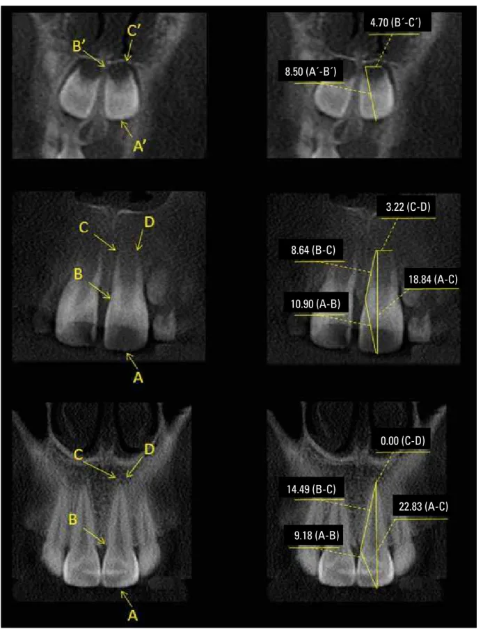
FIGURE 2 - Linear measurements of dental development stages of maxillary central incisor using CBCT (Coronal view).
4.70 (B´-C´)
8.50 (A´-B´)
3.22 (C-D)
18.84 (A-C)
10.90 (A-B)
0.00 (C-D)
14.49 (B-C)
22.83 (A-C)
FIGURE 3 - Linear measurements of dental development stages of maxillary central incisor using CBCT (Sagittal view).
6.04 (B´-C´)
11.40 (A´-B´)
3.58 (C-D)
9.06 (B-C)
22.07 (A-C)
13.62 (A-B)
0.00 (C-D)
15.58 (B-C)
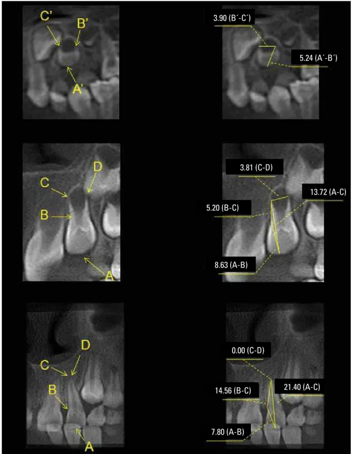
FIGURE 4 - Linear measurements of dental development stages of maxillary lateral incisor using CBCT (Coronal view).
3.90 (B´-C´)
5.24 (A´-B´)
3.81 (C-D)
13.72 (A-C)
5.20 (B-C)
8.63 (A-B)
0.00 (C-D)
14.56 (B-C)
7.80 (A-B)
FIGURE 5 - Linear measurements of dental development stages of maxillary lateral incisor using CBCT (Sagittal view).
4.30 (B´-C´)
5.66 (C-D)
4.72 (B-C)
12.01 (A-B)
15.95 (A-C)
0.00 (C-D)
14.56 (B-C)
21.40 (A-C)
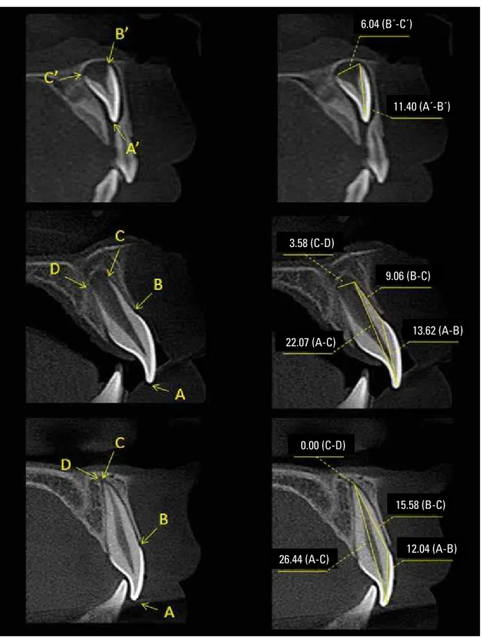
FIGURE 6 - Linear measurements of dental development stages of maxillary canine using CBCT (Coronal view).
6.36 (B´-C´)
7.30 (A´-B´)
4.80 (C-D)
3.06 (B-C)
9.02 (A-B)
11.88 (A-C)
0.00 (C-D)
18.58 (B-C)
7.62 (A-B)
FIGURE 7 - Linear measurements of dental development stages of maxillary canine using CBCT (Sagittal view).
5.41 (B´-C´)
7.13 (A´-B´)
3.80 (C-D)
10.12 (B-C)
15.25 (B-C)
10.59 (A-B)
0.00 (C-D)
24.53 (A-C)
FIGURE 8 - Linear measurements of dental development stages of maxillary first premolar using CBCT (Coronal view).
5.18 (B´-C´)
4.88 (A´-B´)
3.26 (C-D)
7.11 (B-C)
8.38 (A-B)
15.97 (B-C)
0.00 (C-D)
21.81 (A-C)
FIGURE 9 - Linear measurements of dental development stages of maxillary first premolar using CBCT (Sagittal view).
7.97 (B´-C´)
6.85 (A´-B´)
5.43 (C-D)
7.89 (B-C)
15.45 (A-C)
7.97 (A-B)
0.00 (C-D)
12.66 (B-C)
21.27 (A-C)
FIGURE 10 - Linear measurements of dental development stages of maxillary first molar using CBCT (Coronal view).
7.22 (B´-C´)
7.50 (A´-B´)
2.20 (C-D)
9.91 (B-C)
6.85 (A-B)
0.00 (C-D)
20.22 (A-C)
13.85 (B-C)
FIGURE 11 - Linear measurements of dental development stages of maxillary first molar using CBCT (Sagittal view).
7.74 (A´-B´)
2.67 (C-D)
18.58 (A-C)
0.00 (C-D)
15.03 (B-C)
22.37 (A-C)
7.52 (A-B)
10.40 (B-C)
8.35 (A-B)
FIGURE 12 - Linear measurements of dental development stages of mandibular central incisor using CBCT (Coronal view).
8.45 (A´-B´)
3.35 (B´-C´)
8.19 (A-B)
16.81 (A-C)
2.18 (C-D)
9.46 (A-B)
21.40 (A-C)
12.97 (B-C)
FIGURE 13 - Linear measurements of dental development stages of mandibular central incisor using CBCT (Sagittal view).
10.90 (A´-B´)
5.53 (B´-C´)
18.09 (A-C)
10.44 (A-B)
8.24 (B-C)
10.36 (A-B)
15.10 (B-C)
24.50 (A-C)
FIGURE 14 - Linear measurements of dental development stages of maxillary lateral incisor using CBCT (Coronal view).
7.50 (A´-B´)
3.35 (B´-C´)
8.16 (A-B)
7.26 (B-C)
15.07 (A-C)
2.77 (C-D)
7.53 (A-B)
15.06 (B-C)
22.01 (A-C)
FIGURE 15 - Linear measurements of dental development stages of maxillary lateral incisor using CBCT (Sagittal view).
8.19 (A´-B´)
4.85 (B´-C´)
15.49 (A-C)
10.26 (A-B)
5.51 (B-C)
11.29 (A-B)
12.70 (B-C)
22.99 (A-C)
FIGURE 16 - Linear measurements of dental development stages of mandibular canine using CBCT (Coronal view).
7.31 (A´-B´)
4.80 (B´-C´)
9.95 (A-B)
8.09 (B-C)
3.61 (C-D)
8.54 (A-B)
14.85 (B-C)
23.03 (A-C)
FIGURE 17 - Linear measurements of dental development stages of mandibular canine using CBCT (Sagittal view).
9.31 (A´-B´)
5.11 (B´-C´)
11.76 (A-B)
17.46 (A-C)
7.46 (C-D)
24.11 (A-C)
11.84 (A-B)
13.06 (B-C)
FIGURE 18 - Linear measurements of dental development stages of mandibular first premolar using CBCT (Coronal view).
5.69 (A´-B´)
5.43 (B´-C´)
8.07 (A-B)
15.25 (A-C)
2.06 (C-D)
7.54 (A-B)
22.61 (A-C)
15.97 (B-C)
0.00 (C-D)
FIGURE 19 - Linear measurements of dental development stages of mandibular first premolar using CBCT (Sagittal view).
5.41 (A´-B´)
4.81 (B´-C´)
15.26 (A-C)
6.50 (C-D)
9.30 (A-B)
6.50 (B-C)
8.44 (A-B)
15.05 (B-C)
22.60 (A-C)
FIGURE 20 - Linear measurements of dental development stages of mandibular first molar using CBCT (Coronal view).
7.92 (A´-B´)
9.18 (B´-C´)
21.37 (A-C)
7.83 (A-B)
14.30 (B-C)
8.00 (A-B)
21.51 (A-C)
14.18 (B-C)
FIGURE 21 - Linear measurements of dental development stages of mandibular first molar using CBCT (Sagittal view).
8.08 (A´-B´)
8.24 (B´-C´)
8.45 (A-B)
7.35 (B-C)
7.86 (A-B)
13.62 (B-C)
21.28 (A-C)
0.00 (C-D)
16.24 (A-C)
development (3 to 20 years of age) represent a
ref-erence value of length, which should be associated
with caution to maturation stage or skeletal age.
The present study was conducted using databases
from private radiology clinics, in subjects whose
ge-netic, nutritional, physiologic, pathologic,
socioeco-nomic, and housing patterns were not standardized.
The measurements acquired on dental groups are
in accordance with estimates from previously
pub-lished investigations.
9,36,50However, this tool
consti-tutes a noninvasive technique which permits in vivo
studies. Investigations with observation methods
using conventional radiographs to evaluate the
de-velopment of human permanent teeth, chronology
and sequence of eruption represent the most widely
employed study models.
20,21,34,35,44,49A classical study by Nolla
35reported that
ev-ery dentist treating children must have a good
understanding of the development of the
denti-tion. The variability in tooth development may
indicate differences between mean values. The
author used serial oral radiographs of twenty-five
boys and twenty-five girls, and suggested stages of
development of human permanent teeth, which
were graded on a scale from 0 to 10 (0- absence of
crypt; 1- presence of crypt; 2- start of calcification;
3- one-third of crown completed; 4- two thirds
of crown completed; 5- crown almost completed;
6- crown completed; 7- one-third of root
com-pleted; 8- two-thirds of root comcom-pleted; 9- root
almost completed - open apex; 10- apical end of
root completed). Mean differences in the general
sequence of development were not apparent
be-tween genders and few development differences
were found between right and left teeth.
The possibility of obtaining information on
three-dimensional anatomic structures in vivo with
image handling has great potential and constitutes
an achievement for all dental areas.
6Liu et al
25de-termined the accuracy of volumetric analysis of
teeth in vivo using CBCT. The volume of 24
bicus-pid teeth extracted for orthodontic purposes were
determined. The measurements slightly deviated
from the volumes within -4% to 7%. Smoothing
operations reduce volume measurements.
Cur-rently, no requirements for accuracy of volumetric
determinations of tooth volume have been
estab-lished. Baumgaertel et al
4investigated the
reliabil-ity and accuracy of dental measurements made on
CBCT reconstructions. Thirty human skulls were
scanned with dental CBCT, and 3-dimensional
reconstructions of the dentitions were generated.
Ten measurements (overbite, overjet, maxillary
and mandibular intermolar and intercanine widths,
available arch length, and required arch length)
were made directly on the dentitions of the skulls
with a high-precision digital caliper and on the
digital reconstructions with commercially
avail-able software. Dental measurements from CBCT
volumes can be used for quantitative analysis. A
small systematic error was found, which became
statistically significant only when combining
sev-eral measurements. An adjustment for this error
allowed improved accuracy.
Several studies have used the CBCT
mea-surement tool to determine distances between
maxillofacial anatomical structures.
1,4,7,19,25,29-31,45CBCT measurements have more important
ap-plications and reliability than conventional
imag-ing methods.
5,11-13,15,45COnCLuSiOnS
Under the tested conditions and within the
limi-tations of this preliminary study, one can conclude
that CBCT images of different development stages
may contribute to treatment diagnosis, planning
and outcome. The dimensions of dental crowns and
roots may have good clinical and research
applica-tion. However, further studies are recommended to
minimize variables in the methodology.
ACKnOwLEdgMEntS
1. Al-Rawi B, Hassan B, Vandenberge B, Jacobs R. Accuracy assessment of three-dimensional surface reconstructions of teeth from cone beam computed tomography scans. J Oral Rehabil. 2010 May 1;37(5):352-8.
2. Ambrose J. Computerized transverse axial scanning (tomography). II. Clinical application. Br J Radiol. 1973;46:1023-47.
3. Arai Y, Tammisalo E, Iwai K, Hashimoto K, Shinoda K.
Development of a compact computed tomographic apparatus for dental use. Dentomaxillofac Radiol. 1999 Jul;28(4):245-8. 4. Baumgaertel S, Palomo JM, Palomo L, Hans MG. Reliability
and accuracy of cone-beam computed tomography dental measurements. Am J Orthod Dentofacial Orthop. 2009 Jul;136(1):19-25.
5. Bender IB. Factors inluencing the radiographic appearance of
bone lesions. J Endod. 1982 Apr;8(4):161-70.
6. Bueno MR, Estrela C. Cone beam computed tomography in endodontic diagnosis. In: Estrela C. Endodontic Science. 2nd ed.
São Paulo: Artes Médicas; 2009. p. 119-54.
7. Cavalcanti MG, Vannier MW. Measurement of the volume of oral tumors by three-dimensional spiral computed tomography. Dentomaxillofac Radiol. 2000 Jan;29(1):35-40.
8. Cotti E, Campisi G. Advanced radiographic techniques for the detection of lesions in bone. Endodontic Topics. 2004;7:52-72.
9. De Deus QD. Topograia da cavidade pulpar e do periápice. 5ª
ed. Medsi: Rio de Janeiro; 1992. p. 11-56.
10. Dudic A, Giannopoulou C, Leuzinger M, Kiliaridis S. Detection of apical root resorption after orthodontic treatment by using panoramic radiography and cone-beam computed tomography of super-high resolution. Am J Orthod Dentofacial Orthop. 2009 Apr;135(4):434-7.
11. Estrela C, Bueno MR, Leles CR, Azevedo B, Azevedo JR. Accuracy of cone beam computed tomography and panoramic and periapical radiography for detection of apical periodontitis. J Endod. 2008 Mar;34(3):273-9.
12. Estrela C, Bueno MR, Azevedo BC, Azevedo JR, Pécora JD. A new periapical index based on cone beam computed tomography. J Endod. 2008 Nov;34(11):1325-31. 13. Estrela C, Bueno MR, De Alencar AH, Mattar R, Valladares J
Neto, Azevedo BC, et al. Method to evaluate inlammatory
root resorption by using Cone Beam Computed Tomography. J Endod. 2009 Nov;35(11):1491-7.
14. Garib DG, Raymundo R Junior, Raymundo MV, Raymundo
DV, Ferreira SN. Tomograia computadorizada de feixe cônico
(Cone Beam): entendendo este novo método de diagnóstico por imagem com promissora aplicabilidade na Ortodontia. Rev Dental Press Ortod Ortop Facial. 2007 mar-abr;12(2):139-56. 15. Grimard BA, Hoidal MJ, Mills MP, Mellonig JT, Nummikoski PV,
Mealey BL. Comparison of clinical, periapical radiograph, and cone-beam volume tomography measurement techniques for assessing bone level changes following regenerative periodontal therapy. J Periodontol. 2009 Jan;80(1):48-55.
16. Hägg U, Taranger J. Dental development, dental age and tooth counts. Angle Orthod. 1985 Apr;55(2):93-107.
17. Hounsield GN. Computerized transverse axial scanning
(tomography). I. Description of system. Br J Radiol. 1973 Dec;46(552):1016-22.
18. Huumonen S, Orstavik D. Radiological aspects of apical periodontitis. Endod Topic. 2002;1:3-25.
19. Janson GR, Martins DR, Tavano O, Dainesi EA. Dental maturation in subjects wit extreme vertical facial types. Eur J Orthod. 1998 Feb;20(1):73-8.
20. Kobayashi K, Shimoda S, Nakagawa Y, Yamamoto A. Accuracy in measurement of distance using limited cone-beam computerized tomography. Int J Oral Maxillofac Implants. 2004
Mar-Apr;19(2):228-31.
21. Kochhar R, Richardson A. The chronology and sequence of eruption of human permanent teeth in Northern Ireland. Int J Paediatr Dent. 1998 Dec;8(4):243-52.
REfEREnCES
22. Krailassiri S, Anuwongnukroh N, Dechkunakorn S. Relationships
between dental calciication stages and skeletal maturity indicator
in Thai individuals. Angle Orthod. 2002 Apr;72(2):155-66. 23. Krogman WM. The concept of maturity from a morphological
viewpoint. Child Dev. 1950 Mar;21(1):25-32.
24. Liliequist B, Lundberg M. Skeletal and tooth development. A methodologic investigation. Acta Radiol Diagn (Stockh). 1971 Mar;11(2):97-112.
25. Liu Y, Olszewski R, Alexandroni ES, Enciso R, Xu T, Mah JK. The validity of in vivo tooth volume determinations from cone-beam computed tomography. Angle Orthod. 2010 Jan;80(1):160-6. 26. Liversidge HM, Lyons F, Hector MP. The accuracy of three
methods of age estimation using radiographic measurements of developing teeth. Forensic Sci Int. 2003 Jan 9;131(1):22-9. 27. Liversidge HM, Speechly T, Hector MP. Dental maturation in
British children: are Demirjian’s standards applicable? Int J Paediatr Dent. 1999 Dec;9(4):263-9.
28. Liversidge HM. Crown formation times of human permanent anterior teeth. Arch Oral Biol. 2000 Sep;45(9):713-21. 29. Lund H, Gröndahl K, Gröndahl HG. Accuracy and precision of
linear measurements in cone beam computed tomography Accuitomo® tomograms obtained with different reconstruction.
Dentomaxillofac Radiol. 2009;28:379-86.
30. Lund H, Gröndahl K, Gröndahl HG. Cone beam computed tomography for assessment of root length and marginal bone level during orthodontic treatment. Angle Orthod. 2010 May;80(3):466-73.
31. Misch KA, Yi ES, Sarment DP. Accuracy of cone beam computed tomography for periodontal defect measurements. J Periodontol. 2006 Jul;77(7):1261-6.
32. Mozzo P, Procacci C, Tacconi A, Martini PT, Andreis IA. A new volumetric CT machine for dental imaging based on the cone-beam technique: preliminary results. Eur Radiol. 1998;8(9):1558-64.
33. Nanci A. Ten Cate´s oral histology: development, structure and functions. 7th ed. Montreal: Mosby; 2008. p. 98-9.
34. Nicodemo RA, Moraes LC, Médici E Filho. Tabela cronológica da mineralização dos dentes permanentes entre brasileiros. Rev Fac Odontol São José dos Campos. 1974;3:55-6.
35. Nolla CM. The development of the permanent teeth. J Dent Child. 1960;27:254-66.
36. Pucci FM, Reig R. Condutos radiculares: anatomia, patologia e terapia. Buenos Aires: Ed. Medico – Quirurgico; 1945. p.144-305. 37. Raju TN. The Nobel chronicles. 1979: Allan MacLeod Cormack
(b 1924); and Sir Godfrey Newbold Hounsield (b 1919). Lancet.
1999 Nov 6;354(9190):1653.
38. Rasmussen P, Kotsaki A. Inherited retarded eruption in the permanent dentition. J Clin Pediatr Dent. 1997 Spring;21(3):205-11.
39. Reventlid M, Mörnstad H, Teivens AA. Intra and inter-examiner variation in four dental methods for age estimation of children. Swed Dent J. 1996;20(4):133-9.
40. Rosen AA, Baumwell J. Chronological development of the dentition of medically indigent children: a new perspective. ASDC J Dent Child. 1981 Nov-Dec;48(6):437-42.
41. Sandhu S, Kaur T. Radiographic study of the positional changes and eruption of impacted third molars in young adults of an Asian Indian population. J Oral Maxillofac Surg. 2008 Aug;66(8):1617-24.
42. Scarfe WC, Farman AG, Sukovic P. Clinical applications of cone-beam computed tomography in dental practice. J Can Dent Assoc. 2006 Feb;72(1):75-80.
43. Sherrard JF, Rossouw PE, Benson BW, Carrillo R, Buschang PH. Accuracy and reliability of tooth and root lengths measured on cone-beam computed tomographs. Am J Orthod Dentofacial Orthop. 2010 Apr;137(4 Suppl):S100-8.
44. Silva SRP, Nouer PRA, Garbui IU, Ramalho AS. Deinição da época para o início do tratamento ortodôntico. Rev Gaúcha Odontol.
Contact address
Carlos Estrela
Rua C-245, Quadra 546, Lote 9, Jardim América CEP: 74.290-200 - Goiânia / GO, Brazil E-mail: [email protected] Submitted: July 2010
Revised and accepted: August 2010
45. Simonton JD, Azevedo B, Schindler WG, Hargreaves KM. Age- and gender-related differences in the position of the inferior alveolar nerve by using cone beam computed tomography. J Endod. 2009 Jul;35(7):944-9.
46. Staaf V, Mörnstad H, Welander U. Age estimation based on tooth development: a test to reliability and validity. Scand J Dent Res. 1991 Aug;99(4):281-6.
47. Teivens A, Mörnstad H. A modiication of the Demirjian method
for age estimation in children. J Forensic Odontostomatol. 2001 Dec;19(2):26-30.
48. Togashi K, Kitaura H, Yonetsu K, Yoshida N, Nakamura T. Three-dimensional cephalometric using helical computer tomography: measurement error caused by head inclination. Angle Orthod. 2002 Dec;72(6):513-20.
49. Vieira CL, Oliveira AEF, Ribeiro CCC, Lima AASJ. Relação entre os índices de maturação das vértebras cervicais e os estágios de
calciicação dentária. Rev Dental Press Ortod Ortop Facial. 2009
mar-abr;14(2):45-53.
50. Woelfel JB, Scheid RC. Anatomia dental: sua relevância para a