The article was published by Academy of Chemistry of Globe Publications www.acgpubs.org/RNP © Published 05/15/2016 EISSN:1307-6167
Rec. Nat. Prod
. 10:6 (2016) 735-743
New Chemical Constituents from the Endophytic Fungus
Xylaria papulis Cultivated on Taiwanese Lepidagathis stenophylla
Yi-Shuan Chen
1, Hsun-Shuo Chang
1,2*, Ming-Jen Cheng
3*, Hing-Yuen Chan
3,
Ming-Der Wu
3, Sung-Yuan Hsieh
3, Hui-Chun Wang
1and Ih-Sheng Chen
1,2*1
Graduate Institute of Natural Products, College of Pharmacy, Kaohsiung Medical University,
Kaohsiung 807, Taiwan
2
School of Pharmacy, College of Pharmacy, Kaohsiung Medical University, Kaohsiung 807,Taiwan
3
Bioresource Collection and Research Center, Food Industry Research and Development Institute,
Hsinchu 300, Taiwan
(Received December 02, 2015; Revised January 26, 2016; Accepted February 06, 2016)
Abstract: The endophytic strain Xylaria papulis was originated from the stem of Taiwanese Lepidagathis stenophylla C. B. Clarke ex Hayata (Acanthaceae) growing in the Hengchun Peninsula of southern Taiwan. Bioassay-guided fractionation of an EtOAc-soluble fraction of the liquid fermentate of this strain resulted in the isolation of two new isopimarane-type diterpene glycosides compounds, i.e., xylapapusides A & B (1 & 2), along with five previously identified compounds, 3–7. The structures of 1 and 2 were elucidated by detailed spectroscopic analysis including 1H- and 13C-NMR, COSY, HSQC, HMBC, and HR-ESI-MS, while the structures of the known compounds 3–7 were deduced from comparison of their spectral data with those in the literature. The effects of some isolates on the inhibition of NO production in lipopolysaccharide-activated RAW 264.7 murine macrophages were evaluated. Xylapapuside A (1) showed most potent of NO inhibition with Emax value of 34.3 μM.
Keywords:Xylaria papulis; Lepidagathis stenophylla; endophytic fungus;isopimarane-type diterpene glycoside.
© 2016 ACG Publications. All rights reserved.
1. Introduction
Endophytes were microorganisms that reside in the internal tissues of living plants without causing any immediate overt negative effects or apparent symptoms [1–3]. The research on endophytes showed that they are rich sources of diverse and bioactive secondary metabolites [2–5]. During the long periods of co-evolution, endophytes kept a state of balanced antagonism with the host plant [3], which regulated the metabolism of the endophytes and endowed them with limitless potential for the production of bioactive secondary metabolites. During a program of investigation on potentially bioactive secondary metabolites from Formosan endemic plant endophytes, we had isolated
*
and identified over 100 endophytic fungal strains from Formosan endemic plants and the crude EtOAc and BuOH extracts from these endophytic strains were screened for their inhibitory activity on lipopolysaccharide (LPS)-induced nitric oxide (NO) release in RAW 264.7 murine macrophages. An EtOAc-soluble fraction was obtained from the whole broth of Xylaria papulis, which showed inhibitory activity on LPS-induced NO release in RAW 264.7 murine macrophages, as determined by our primary screening (approximately 85% inhibition at a concentration of 10 μg/ml). The genus
Xylaria (family Xylariaceace) is very diverse with respect to their chemical constituents including terpenoids [6–9], cyclopeptides [10][11], polyketides [12][13], cytochalasins [14], xanthones [15][16], and unique unclassified xyloketals [17]. However, many species in this genus still remain chemically and biologically unexplored. One previous work revealed one new and several known metabolites produced by the BuOH-soluble fraction of the 95% ethanolic extract of rice fermented with the fungus
X.papulis [18]. Since different culture conditions are known to afford different metabolites, and in an attempt to maximize chemical diversity of the metabolites produced by this fungus, we used malt extract agar (MEA) as culture media. Chemical investigation of the EtOAc extract from the culture filtrate of fermentations on MEA medium led to the isolation and characterization of two new compounds, ie. xylapapusides A & B (1 & 2) as well as five known constituents (3–7) (Fig. 1). Herein we describe the structure identification of the new compounds and the inhibitory activity of the main isolates on NO production in macrophage RAW 264.7 cells.
2. Materials and Methods
2.1. Microorganism Material
The fungal strain 09F0222 was isolated from the stem of Lepidagathsis stenophylla C. B. Clarke ex Hayata which was collected in Mt. Kaoshih, Pingtung County, Taiwan, during December of 2008. Fungal identification was performed based on sequencing of the ITS regions. The fungal strain grew slowly on MEA (malt extract agar) medium at 25 ºC. Color of the colony was white for the first 7 d, and then gradually became gray with black areas and eventually turned to black. Spores were generated after more than 14 days, and only a few spores can be observed by naked eyes. The fungal strain was identified as X. papulis (family Xylariaceae) by Dr. Sung-Yuan Hsieh, basing on cultural and anamorphic data. The identification was further confirmed by sequence analysis of the rDNA-ITS (internal transcribed spacer) region. A nucleotide-to-nucleotide BLAST (nucleotide sequence comparison program) to search the GenBank database, it has been shown to have 100% similarity to X. papulis (GenBank Accession No. GU300100)
2.2. Cultivation and Preparation of the Fungal Strain
Two-weeks-old colonies of the X. papulis strain on malt extract agar (MEA) medium in 9-cm Petri dish were cut into the bottle and blended for 30 s with 100 ml of dist. H2O to prepare the fungal
inoculum for liquid fermentation. To each 500-ml flask containing 150 ml of liquid cultural media (ingredients: corn starch, 30 g; corn steep liquor, 10 g; yeast extract, 5 g; and sea salt, 2 g in 1 l of dist.
H2O, pH 6) were added 10 ml of fungal inocula and incubated at 25° for 2 weeks on a rotary shaker at
the speed of 100 circles/min without illuminated. A total of 14 l of fungal fermented broth were harvested and then filtered to remove fungal mycelium. The strain is preserved with the Bioresource Collection and Research Center (BCRC) of the Food Industry Research and Development Institute (FIRDI), under the ID No. 09F0222.
The liquid fermentation broth (H2O) of X. papulis 09F0222 (14 l) was extracted with EtOAc, and
the EtOAc-layer (9.2 g) was separated. The H2O-layer was further extracted with BuOH, and the
BuOH-layer (26.4 g) was separated. The EtOAc-layer was subjected to C.C. (silica gel; CH2Cl2/MeOH gradient) to get 13 fractions (Frs. 1-13). Fr. 3 was subjected to MPLC (Silica gel;
CH2Cl2/acetone 10:1) to produce 11 fractions, Frs. 3.1-3.11. Frs. 3-4 and 3-5 were combined together
after TLC analysis and were subjected to MPLC (RP-18; MeOH/H2O 1:1) to produce 6 fractions, Frs. 3.4.1-3.4.6. Fr. 3.4.1 is 4 (122.2 mg). Fr. 5 was subjected to MPLC (Silica gel; CH2Cl2/MeOH 15:1)
to produce 11 fractions, Frs. 5.1-5.11. Fr. 5.7 was subjected to MPLC (Silica gel; CH2Cl2/MeOH
10:1) to produce 6 fractions, Frs. 5.7.1-5.7.6. Fr. 5.7.3 was purified with TLC (RP-18; acetone/H2O
6:5) to obtain 7 (Rf 0.26; 3.0 mg). Fr. 5.7.4 was purified with TLC (RP-18; acetone/H2O 6:5) to obtain
6 (Rf 0.29; 1.8 mg). Fr. 5.8 was purified with TLC (RP-18; acetone/H2O 1:1) to obtain 5 (Rf 0.29; 2.2
mg). Fr. 7 was subjected to MPLC (silica gel; CH2Cl2/MeOH 8:1) to produce 7 fractions, Frs. 7.1-7.7. Fr. 7.4 was subjected to MPLC (RP-18; acetone/H2O 6:5) to produce 10 fractions, Fr.7.4.1-7.4.10. Fr. 7.4.4 was subjected to MPLC (RP-18; MeOH/H2O 2:1) to produce 7 fractions, Fr. 7.4.4.1-7.4.4.7. Fr. 7.4.4.6 was purified with prep. TLC (Silica gel; CH2Cl2/MeOH 5:1) to obtain 1 (Rf 0.43; 14.7 mg). Fr. 7.4.6 was purified with prep. TLC (Silica gel; CH2Cl2/MeOH 8:1) to obtain 2 (Rf 0.55; 16.2 mg)
and 3 (Rf 0.48; 12.0 mg).
2.4. Xylapapuside A (=16-
(α
-glucopyranosyloxy)norisopimar-7-en-
4α
-ol;
1
).
Whitish powder. [α]25 D= +26.2 (c 1.5, MeOH). IR υmax (ATR): 3344 (OH), 1643 (C=C). 1H- and 13C-NMR: Table 1. ESI-MS: 477 ([M+Na]+). HR-ESI-MS:477.2825 ([M+Na]+, C25H42NaO+
7; calc. 477.2823).
2.5. Xylapapuside B (=16-(
α
-mannopyranosyloxy)norisopimar-7-en-4
β
-ol;
2
).
Amorphous solid. [α]25 D = +22.2 (c 0.55, MeOH). IR υmax (ATR): 3345 (OH), 1645 (C=C). 1H- and 13C-NMR: Table 1. ESI-MS: 477 ([M+Na]+). HR-ESI-MS:477.2823 ([M+Na]+, C25H42NaO+
7; calc. 477.2823).
2.6. Determination of NO Production and Cell Viability Assay.
Mouse macrophage cell line (RAW 264.7) was obtained from Bioresource Collection and Research Center (BCRC 60001) and cultured at 37ºC in Dulbecco's Modified Eagle's Medium (DMEM) supplemented with 10% fetal bovine serum (FBS; Gibco), 4.5 g/L glucose, 4 mm glutamine, penicillin (100 units/ml), and streptomycin (100 mg/ml) in a humidified atmosphere in a 5% CO2
incubator. The cells were treated with 10, 25, and 50 mm natural products in the presence of 1 mg/ml Whitish LPS (Sigma-Aldrich) for 20 h. The concentration of NO in culture supernatants was determined as nitrite, a major stable product of NO, by Griess reagent assay [19], and cell viabilities were determined using the MTT assay as described in [20].
3. Results and Discussion
3.1. Structure Elucidation
Compound 1 was obtained as optically active white powder with [α]25 D= +26.2 (c = 1.5, MeOH), and its
molecular formula was deduced as C25H42O7 from HR-ESI-MS data, implying five degrees of
unsaturation. Its IR spectrum showed bands corresponding to the absorptions of OH and C=C groups at 3344 and 1643 cm-1, respectively. The 1H- (Table 1), 13C-NMR (Table 1), and HSQC spectra revealed that 1 contained three Me singlets, an olefinic proton corroborated by 13C-NMR signals at δ(C) 122.2
quaternary C-atoms, one olefinic C-atom, one O-bearing quaternary C-atom, and also bearing an O-α-glucosyl moiety.
Table 1. 1H- and 13C-NMR Data of1and2a) (600 and 150 MHz, resp.; in CD3OD). δ in ppm, J in Hz.
Position 1 2
δ(H) δ(C) δ(H) δ(C)
1 1.07 (br. dd, J = 13.7, 3.0) 40.4 1.00–1.05 (m) 40.5
1.81 (br. d, J = 13.7) 1.83–1.86 (m)
2 1.34–1.47 (m) 21.3 1.38–1.41 (m) 19.1
1.50–1.61 (m) 1.78–1.81 (m)
3 1.34–1.47 (m) 43.8 1.36–1.38 (m) 42.0
1.75–1.77 (m) 1.68–1.72 (m)
4 – 73.1 – 72.0
5 1.43 (m) 53.2 1.15–1.17 m 50.5
6 1.89–1.92 (m) 23.5 1.97–2.02 m 23.5
2.11–2.15 (m) 2.05–2.11 m
7 5.37 (d, J = 5.4) 122.2 5.36 (dd, J = 6.0, 1.8) 122.2
8 – 137.2 – 137.1
9 1.74 (br. dd, J = 12.0, 1.8) 53.7 1.63–1.65 (m) 52.7
10 – 37.4 – 36.4
11 1.34–1.47 (m) 21.7 1.33–1.35 (m) 21.3
1.50–1.61 (m) 1.51–1.55 (m)
12 1.25–1.30 (m) 38.2 1.24–1.29 (m) 38.3
1.50–1.61 (m) 1.51–1.55 (m)
13 – 33.9 – 34.1
14 1.92–1.96 (m) 48.5 1.91–1.94 (m) 48.6
15 1.50–1.61 (m) 45.1 1.49–1.52 (m) 45.3
16 3.51 (ddd, J = 15.6, 6.0, 3.0) 65.6 3.49 (ddd, J = 15.9, 7.7, 2.3) 65.2 3.84 (ddd, J = 15.6, 6.6, 3.0) 3.84 (ddd, J = 15.9, 7.2, 2.6)
17 0.81 (s) 22.2 0.81 (s) 22.2
18 – – 1.15 (s) 30.8
19 1.20 (s) 23.5 – –
20 0.85 (s) 15.1 1.03 (s) 15.0
1' 4.76 (d, J = 3.6) 100.2 4.72 (d, J = 1.8) 101.7
2' 3.38 (dd, J = 9.5, 3.6) 73.6 3.77 (dd, J = 3.3, 1.8) 72.3 3' 3.62 (dd, J = 9.5, 9.2) 75.2 3.68 (dd, J = 9.6, 3.3) 72.7 4' 3.27 (dd, J = 9.9, 9.2) 71.9 3.60 (dd, J = 9.6, 9.6) 68.7 5' 3.57 (ddd, J = 9.9, 6.0, 2.4) 73.8 3.53 (ddd, J = 9.6, 5.7, 2.7) 74.7 6' 3.67 (dd, J = 12.0, 6.0) 62.8 3.71 (dd, J = 12.0, 6.0) 63.0
The glycosidic bond was α-oriented according to the small coupling constant (J = 3.6) of the anomeric H-atom (δ(H) 4.76). The above findings accounted for two of the five degrees of unsaturation, which indicated that 1 contains three rings. Comparing the 1H- and 13C-NMR data and unsaturation degrees with those of a published compound elaeicolaside B (3) from literature [22], compound 1 was tentatively classified as an isopimarane-type diterpene analogue.
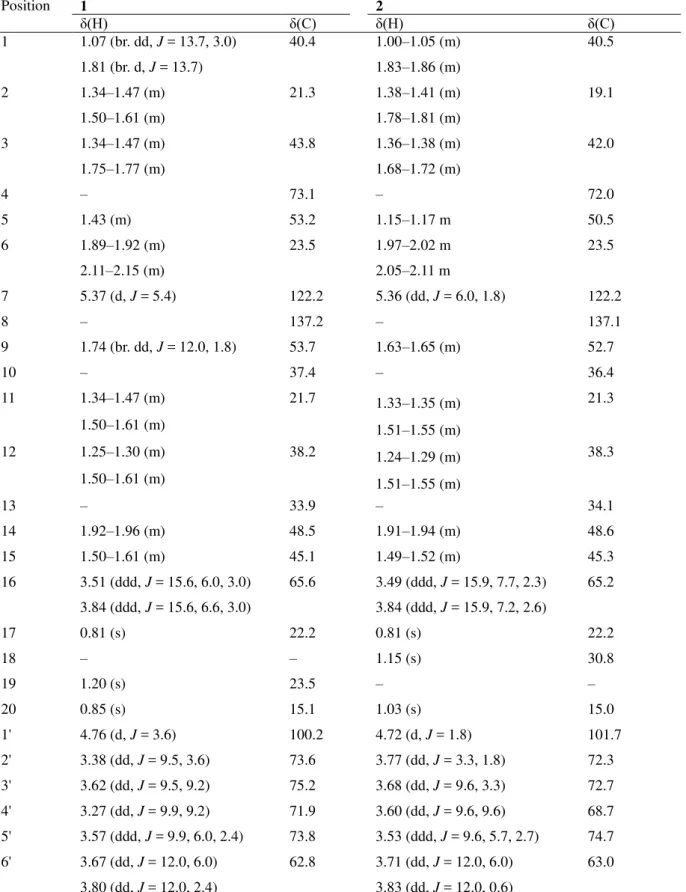
Figure 1. Chemical structures of the isolated 1–7 from X. papulis.
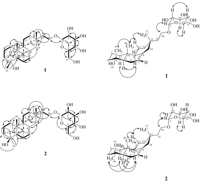
In the HMBC spectrum of 1 (Fig. 2), key cross-peaks H–C(17)/C(12), C(13), C(14), and C(15); H–C(19)/C(3), C(4), and C(5); and H–C(20)/C(1), C(5), C(9), and C(10) established the locations of all the functionalities. In addition, a COSY spectrum (Fig. 2) and detailed analysis of the long-range 1H–13C correlations (Fig. 2) from H-C(1) to C(2), C(3), from H-C(5) to C(9), from H-C(6) to C(8), C(10), from H-C(7) to C(5), C(14), from H-C(14) to C(9), C(12), and from H-C(16) to C(13), C(15) confirmed the skeleton of 1. The HMBC cross peaks from H–C(1') to C(16) and from CH2(16) to C(1') positioned the
glucosyl moiety is connected to C(16) with α-orientation O-linkage. So, the xylapapuside A (1) is an isopimarane-type diterpene glycoside.
The relative configuration of 1 was proposed on the basis of the NOESY experiments (Fig. 3) and
1
H,1H coupling constants. The β-orientation of the Me(17), Me(19) and Me(20) were deduced from the observation of the quasi-1,3-diaxial interactions of Me(19) and Me(20), Me(20) and Hax–C(11a), and
Hax–C(11a) and Me–C(17). Thus, the OH group at C(4) is on the α-face of this molecule. The NMR
data of the sugar moieties were very similar to those of α-D-glucopyranose by comparing the NMR data with those in the literature [21], which suggested that 1 was a glucopyranose. Furthermore, the NOESY (Fig. 3) displayed the key correlation from H–C(1') to H–C(16) to confirm that the glucosyl moiety was located at C(16) with α-orientation O-linkage once again. Based on above spectral evidence, the structure of 1 was elucidated as 16-(α-D-glucopyranosyloxy)norisopimar-7-en-4α-ol, named xylapapuside A.
Compound 2 was obtained as an optically active amorphous solid with [α]25
D= +22.2 (c = 0.55,
MeOH) and assigned the molecular formula C25H42O7, as deduced from ESI-MS [m/z 477 [M+Na] +
and HR-ESI-MS data, which implies five degrees of unsaturation. The IR spectrum revealed the presence of an OH group (3345 cm-1) and a C=C bond (1645 cm-1). Based on the 1H- and 13C-NMR (Table 1), HSQC, COSY (Fig. 2), HMBC (Fig. 2), and NOESY (Fig. 3) experiments, and the structure of 2 was elucidated as 16-(α-D-mannopyranosyloxy)norisopimar-7-en-4β-ol, named xylapapuside B.
The 1H-NMR and 13C-NMR spectra (Table 1) of 2 were similar to those of elaeicolaside B (3) [22], also isolated in this study, except that Me(18) and OH–C(19) of 2 replaced Me(19) and OH–C(18) of elaeicolaside B (3) [22], suggesting that the two compounds most likely are stereoisomers at C(4).
Comparison of the 1H- and 13C-NMR signals (Table 1) with those of elaeicolaside B (3) [22], the downfield shift of Me(20) [δ(H) 1.03 vs. 0.84], H–C(2a) [δ(H) 1.78–1.81 (m) vs. 1.53–1.59 (m)] and Me(18) [δ(C) 30.8 vs. 23.6] and the upfield shift of H–C(5) signal [δ(H) 1.15–1.17 (m) vs. 1.40–1.44 (m)], C(2) [δ(C) 19.1 vs. 21.3] and C(5) [δ(C) 50.5 vs. 53.3] were also observed.
Figure 2. Key COSY (bold line) and HMBCs (H→C) correlations for compounds 1 & 2
Figure 3. Key NOESY correlations for compounds 1 & 2
H–C(11b)/H–C(17) indicated that Me(17), Me(20), and OH-C(4) were β-oriented, and Me(18), and H-C(5) were α-oriented. The sugar moiety was identified as α-D-mannopyranose by comparing the
1
H- and 13C-NMR data with those in the literature [18]. Furthermore, the HMBC (Fig. 2) displayed the key correlation from H–C(1') to C(16) and the coupling constant of [δ(H) 4.72 (d, J = 1.8, H–C(1'));
δ(C) 101.7 (C(1'))] to confirm that the mannosyl moiety was located at C(16) with α-orientation O-linkage.
Additionally, five known compounds were assigned as following: one diterpenoid, elaeicolaside B (3) [22], one benzenoid, tyrosol (4) [23], one amide, N-acetyltyramine (5) [24], and two sesquiterpenoids, hypoxylonol A (6) [24], xylaranol B (7) [25] by comparing their spectroscopic data with published literature values.
3.2. Anti-inflammatory Activity
Nitrite production induced (%) and cell viability (%) of iNOS inhibitory activity of compounds
1–3 and 5 were obtained at the concentration range of 100 μM, and the results were shown in Table 2.
The most active compounds 1–3 had nitrite production induced values of 65.71, 88.74 and 78.53 %, respectively. The increase in cell viability of compound 5 indicates these compounds might have some activities other than lipopolysaccharide (LPS)-induced nitrite production, which is worthy to be explored by other evaluation models.
a) All experiments were repeated three times. Results are shown as the mean ± standard error of means (SE) from three independent experiments. aEmax indicates mean maximum inhibitory effect of nitrite production, at a concentration of 100 μM, expressed as a percentage inhibition induced by LPS (200 ng/mL) in the presence of vehicle. The Emax of iNOS inhibitors aminoguanidine (AG) was evaluated at a concentration of 100 μM. b) positive control (a selective iNOS inhibitor)
Acknowledgment
This work kindly supported by the Ministry of Economic Affairs. Taiwan, ROC to Food Industry Reaserch & Development Institute (FIRDI).
Supporting Information
Supporting Information accompanies this paper on http://www.acgpubs.org/RNP
References
[1] C. W. Bacon, J. F. White, 'Microbial endophytes', Marcel Dekker Inc., New York, 2000.
[2] J. W. Blunt, B. R. Copp, M. H. G. Munro, P. T. Northcote, and M. R. Prinsep (2011). Marine natural products, Nat. Prod. Rep. 28, 196-268.
Table 2. Inhibitory Effects of the four isolates (1–3, and 5) from X. papulis on LPS-activated NO productions in RAW 264.7 macrophages.a)
compounds Nitrite Production (%) Emax (%) Cell Viability (%) Xylapapuside A (1) 65.71 ± 1.51 34.29 ± 1.51 97.07 ± 5.09 Xylapapuside B (2) 88.74 ± 0.73 11.26 ± 0.73 67.84 ± 4.42 Elaeicolaside B (3) 78.53 ± 0.45 21.47 ± 0.45 96.95 ± 4.18
Xylaranol B (5) 105.24 ± 0.37 -5.24 ± 0.37 102.45 ± 2.77
[3] J. W. Blunt, B. R. Copp, R. A. Keyzers, M. H. G. Munro and M. R. Prinsep (2012). Marine natural products,
Nat. Prod. Rep. 29, 144-222.
[4] A. A. Leslie Gulatilaka (2006). Natural products from plant-associated microorganisms: distribution, structural diversity, J. Nat. Prod. 69, 509-526.
[5] J. W. Blunt, B. R. Copp, R. A. Keyzers, M. H. G. Munro and M. R. Prinsep (2013). Marine natural products,
Nat. Prod. Rep. 30, 237-323.
[6] Z. Y. Hu, Y. Y. Li, Y. J. Huang, W. J. Su and Y. M. Shen (2008). Three new sesquiterpenoids from Xylaria sp. NCY2, Helv. Chim. Acta91, 46-52.
[7] S. T. Deyrup, J. B. Gloer, K. O’Donnell and D. T. Wicklow (2007). Kolokosides A−D: Triterpenoid glycosides from a Hawaiian isolate of Xylaria sp., J. Nat. Prod. 70, 378-382.
[8] S. J. Coval, M. S. Puar, D.W. Phife, J. S. Terracciano and M. Patel (1995). SCH57404, an antifungal agent possessing the rare sordaricin skeleton and tricyclin moiety, J. Antibiot. 48, 1171-1172.
[9] C. J. Smith, N. R. Morin, G. F. Bills, A.W. Dombrowski, G. M. Salituro, S. K. Smith, A. Zhao and D. J. MacNeil (2002). Novel sesquiterpenoids from the fermentation of Xylariapersicaria are selective ligands for the NPY Y5 receptor, J. Org. Chem. 67, 5001-5004.
[10] Y. Lin, X. Wu, S. Feng, G. Jiang, S. Zhou, L. L. P. Vrijmoed, and E. B. G. Jones (2001). A novel
N-cinnamoylcyclopeptide containing an allenic ether from the fungus Xylaria sp. (strain #2508) from the South China Sea, TetrahedronLett. 42, 449-451.
[11] H. Huang, Z. She, Y. Lin, L. L. P. Vrijmoed, and W. Lin (2007). Cyclic peptides from an endophytic fungus obtained from a mangrove leaf (Kandeliacandel), J. Nat. Prod. 70, 1696-1699.
[12] S. Boonphong, P. Kittakoop, M. Isaka, D. Pittayakhajonwut, M. Tanticharoen and Y. Thebtaranonth (2001). Multiplolides A and B, new antifungal 10-membered lactones from Xylariamultiplex, J. Nat. Prod. 64, 965-967.
[13] D. O’ Hagana, S. V. Rogers, G. R. Duffin and R. L. Edwards (1992). Biosynthesis of the fungal polyketide, cubensic acid from Xylariacubensis, TetrahedronLett. 33, 5585-5588.
[14] H. Jayasuriya, K. B. Herath, J. G. Ondeyka, J. D. Polishook, G. F. Bills, A.W. Dombrowski, M. S. Springer, S. Siciliano, L. Malkowitz, M. Sanchez, Z. Guan, S. Tiwari, D.W. Stevenson, R. P. Borris and S. B. Singh (2004). Isolation and structure of antagonists of chemokine receptor (CCR5), J. Nat. Prod. 67, 1036-1038. [15] R. A. Davis and G. K. Pierens (2006). 1H and 13C NMR assignments for two new xanthones from the
endophytic fungus Xylaria sp. FRR 5657, Magn. Reson. Chem. 44, 966-968.
[16] P. C. Healy, A. Hocking, N. Tran-Dinh, J. I. Pitt, R. G. Shivas, J. K. Mitchell, M. Kotiw and R. A. Davis (2004). Xanthones from a microfungus of the genus Xylaria, Phytochemistry65, 2373-2378.
[17] Y. Lin, X. Wu, S. Feng, G. Jiang, J. Luo, S. Zhou, L. L. P. Vrijmoed, E. B. G. Jones, K. Krohn, K.
Steingröver and F. Zsila (2001). Five unique compounds: xyloketals from mangrove fungus Xylaria sp. from the South China Sea coast, J Org Chem.66, 6252-6256.
[18] Y. C. Ting, H. H. Ko, H. C. Wang, C. F. Peng, H. S. Chang, P. C. Hsieh and I. S. Chen (2015). A New Pyrrole Metabolite from the Endophytic Fungus of Xylariapapulis, Chem. Nat. Compd. 51, 515-518. [19] Y. C. Ting, H. H. Ko, H. C. Wang, C. F. Peng, H. S. Chang, P. C. Hsieh and I. S. Chen (2014). Biological
evaluation of secondary metabolites from the roots of Myricaadenophora, Phytochemistry103, 89-98. [20] H. C. Wang, C. C. Wu, T. S. Cheng, C. Y. Kuo, Y. C. Tsai, S. Y. Chiang, T. S. Wong, Y. C. Wu and F. R.
Chang (2013). Active Constituents from Liriope platyphylla root against cancer growth in vitro,
Evidence-BasedComplementAltern. Med. 857929, 1.
[21] K. H. Son, J. O. Park, K. C. Chung, H.W. Chang, H. P. Kim, J. S. Kim, S. S. Kang (1992). Flavonoids from aerial parts of Lonicerajaponica, Arch. Pharmacal. Res. 15, 365-370.
[23] V. Rukachaisirikul, N. Khamthong, Y. Sukpondma, S. Phongpaichit, N. Hutadilok-Towatana, P. Graidist, J. Sakayaroj and K. Kirtikara (2010). Cyclohexene, diketopiperazine, lactone and phenol derivatives from the sea fan-derived fungi Nigrospora sp. PSU-F11 and PSU-F12, Arch. Pharm. Res. 33, 375-380.
[24] C. W. Chang, H. S. Chang, M. J. Cheng, T. W. Liu, S. Y. Hsieh, G. F. Yuan and I. S. Chen (2014). Inhibitory effects of constituents of an endophytic fungus Hypoxylon investiens on nitric oxide and interleukin-6 production in RAW264.7 macrophages, Chem. Biodivers. 11, 949-961.
[25] Y. Y. Li, Z. Y. Hu, C. H. Lu and Y. M. Shen (2010). Four new terpenoids from Xylaria sp. 101, Helv. Chim.
Acta93, 796-802.