Mastopexy with breast implants and the pectoralis
major muscle lap: a technique adopted by the
Department of Plastic Surgery of Unicamp
Mastopexia com uso de implantes associados a retalho de músculo peitoral maior:
técnica utilizada na Disciplina de Cirurgia Plástica da Unicamp
ABSTRACT
Background: The correction of breast ptosis associated with skin sagging and low projection is still a subject of controversy in the literature. This study aims to describe the experience of the Plastic Surgery Department of Universidade Estadual de Campinas (Unicamp) with the technique of mastopexy with breast implants in a double plane and the pectoralis major muscle lap. Methods: A retrospective study of 20 patients with grade II or III mammary ptosis, who underwent surgery between June 2008 and September 2010, was performed.
Results: A 9- and 12-month follow-up of patients showed neither breast or chest deformities nor recurrence of ptosis. All patients presented with good breast projection and adequate upper pole ill, with long-lasting and satisfactory results. Conclusions: Mastopexy with breast implants and the pectoralis major muscle lap technique is easy to perform, with a relatively short learning curve, good reproducibility, and satisfactory long-lasting results.
Keywords: Mammaplasty. Breast/surgery. Breast implantation. Surgical laps.
RESUMO
Introdução: A correção da ptose mamária associada à lacidez de pele e baixa projeção é
ainda tema de discussões e controvérsias na literatura. O objetivo deste estudo é descrever a experiência da Disciplina de Cirurgia Plástica da Universidade Estadual de Campinas (Unicamp) com a técnica de mastopexia com implante mamário associado a retalho de sustentação do músculo peitoral maior. Método: Foi realizado estudo retrospectivo de
20 pacientes com ptose mamária graus II ou III, operadas no período de junho de 2008 a setembro de 2010. Resultados: Após seguimento pós-operatório entre 9 meses e 12 meses, não foram observados casos de deformidades mamárias ou torácicas e nenhuma paciente evoluiu com recidiva da ptose. Foram observados, em todas as pacientes, manutenção de boa projeção da mama e adequado preenchimento do polo superior mamário, gerando resultados duradouros e satisfatórios. Conclusões: A técnica de mastopexia com uso de implantes
associados a retalho de músculo peitoral maior é de fácil realização, com curva de apren -dizado relativamente curta, boa reprodutibilidade, e resultados duradouros e satisfatórios.
Descritores: Mamoplastia. Mama/cirurgia. Implante mamário. Retalhos cirúrgicos. This study was conducted at the
School of Medical Sciences of Universidade Estadual de Campinas (Unicamp), Campinas, SP, Brazil.
Submitted to SGP (Sistema de Gestão de Publicações/Manager Publications System) of RBCP (Revista Brasileira de Cirurgia Plástica/Brazilian Journal of Plastic Surgery).
Paper received: July 15, 2011 Paper accepted: October 31, 2011
1. Resident Physician of Plastic Surgery at Universidade Estadual de Campinas (Unicamp), Campinas, SP, Brazil.
2. Specialist Member of the Brazilian Society of Plastic Surgery (SBCP), Collaborator Physician of the Discipline of Plastic Surgery at Unicamp, Campinas, SP, Brazil.
3. Specialist Member of the Brazilian Society of Plastic Surgery (SBCP), Assistant Physician of the Discipline of Plastic Surgery at Unicamp, Campinas, SP, Brazil.
4. Specialist Member of the Brazilian Society of Plastic Surgery (SBCP), Associate Professor of the Discipline of Plastic Surgery at Unicamp, Campinas, SP, Brazil.
5. Full-time Member of the Brazilian Society of Plastic Surgery (SBCP), Professor Doctor and Head of the Discipline of Plastic Surgery at Unicamp, Campinas, SP, Brazil.
AndreA Boldrin SoAreS1
FernAndo FABrício FrAnco2
endrigo TorezAn roSim1
BrendA ArTuzi renó1
JuSSArA olivo Pinheiro
AlveS hAchmAnn3
mArcelode cAmPoS guidi3
mArco AnToniode
cAmArgo Bueno4
INTRODUCTION
Breast ptosis is caused by an imbalance between the breast skin and its content. There are several degrees of breast pto sis according to the relationship between the nipple and the in framammary crease as classiied by Regnault in 1976 (Chart 1)1,2.
The correction of breast ptosis associated with skin sa g ging and low projection of the breast is a subject of con tro -versy. A critical review of the aesthetic results and the de gree of satisfaction of patients and surgeons is not well es tablished in the medical literature2-5.
Several studies have described the correction of breast pto sis by an increase in breast volume, removal of excess skin, or a combination of both2-5. In cases of massive weight loss, fat catabolism and connective tissue alterations lead to reduction in breast projection and increased sagging, ge -nera ting an unsightly appearance6-10. The correction of these de fects is very difficult for surgeons under training, and even for ex perienced surgeons, and different techniques have been des cribed in the literature.
Techniques showing good reproducibility and a low rate of complications have been investigated to achieve sa tisfactory long-lasting results. The use of the pectoralis major muscle type I was described by Caldeira & Lucas11, in 2000. A
mastopexy procedure that includes the placement of textured silicone implants in a double plane for the treatment of breast ptosis, and the use of a portion of the pectoralis major muscle to support the implant in its lower quadrant is used at the Plas tic Surgery Department of Universidade Estadual de Cam pinas (Unicamp).
The present study describes the experience of the Plastic Surgery Department of Unicamp with the technique of masto-pexy with breast implants in a double plane and a pectoralis major muscle lap.
METHODS
A retrospective study of patients with grade II or III breast ptosis, diagnosed according to the classiication of Reg -nault1, who underwent surgery from June 2008 to September
2010, was performed.
Twenty patients who were followed up for 9–12 months after the surgery were included in the study. The average age of the patients was 32.2 years (range, 26–53 years). With re gard to ethnicity, 16 patients were Caucasian, 3 were of mi xed race, and 1 was of African descent.
Preoperative examination revealed an average body mass index of 23.2 kg/m². On physical examination of the breast, al most all patients were found to have poor quality skin, and on ly 1 had no stretch marks on the breast.
All patients underwent mastopexy with breast implants and a supporting lap of the pectoralis major muscle in a dou ble plane, according to the technique described below.
Skin Markings
The skin markings were performed with the patient in an upright position, deining the midline, meridians, and ma m-mary creases (Figure 1). Point A was marked as a projection of the mammary crease in both breasts. Points B and C were marked by digital clamping, and point D was marked 2 cm above the breast crease. These points were connected in a si milar manner to the marking proposed by Peixoto6.
Anesthesia and Surgical Technique
General anesthesia was used in all cases. With the patient in a supine position and a slight elevation of the dorsum (30 degrees), a vertical incision was made below the areolarpa -pillary complex, followed by the generation of a subglandular cavity for the implant.
An incision was then made in the pectoralis major muscle in the direction of its ibers, in the transition between the mid and lower thirds of the muscle (Figure 2). The lower portion
Chart 1 – Regnault’s1 classification of breast ptosis.
Breast ptosis
True ptosis
Grade I
Areola at the level of the mammary crease and above the contour of the gland
Grade II
Areola below the level of the ma m-mary crease and above the contour of the gland
Grade III
Areola below the level of the ma m-mary crease and below the contour of the gland
Partial ptosis Areola above the crease and gland ptosis
Pseudoptosis Areola above the mammary crease.
of the muscle was detached, and after rigorous hemostasis, the implants were positioned in a double plane. The upper part of the implant was positioned in the subglandular region and the lower part was submuscular, with the pectoralis muscle pro viding greater support to the implant (Figure 3).
The prepared muscular girdle was attached to the glan-dular tissue with nonabsorbable sutures. The need for closed drainage was assessed during surgery.
Using a bi-digital maneuver, the excess skin was marked and resected, resulting in a periareolar scar and a vertical or in verted T, depending on the amount of excised skin.
Implants
Round, high proile, textured implants with a cohesive gel (Winner and Perthese brands), obtained by donation, were used.
RESULTS
The volume of the implants ranged between 160 and 300 cc (average, 246.2 cc). Closed drainage (aspiration) with a por -to vac drain was used in 1 patient and maintained in place until 1 day after surgery.
No complications, including hematomas, seromas, infec-tions, or capsular contractures, were reported in the patients
included in this study. There were 3 cases of partial dehiscen ce and 2 cases reporting enlarged scars.
At the 9 and 12-month follow-up examinations, there we re no cases of breast or chest deformities, and none of the pa tients showed recurrence of ptosis. All patients presented with good breast projection and maintenance of adequate up per pole ill, with long-lasting and satisfactory results.
In the late postoperative period, the aesthetic results were considered satisfactory by the surgical staff and the patients (Table 1).
Figures 4 to 6 illustrate some cases of patients included in the study.
DISCUSSION
Mastopexy with breast implants is a procedure associated with a high degree of dificulty, which is even greater in pa -ti ents with signiicant weight loss. This is because the skin of such patients is often of poor quality, characterized by stre tch marks, excessive sagging, and decreased elasticity. This procedure is the subject of ongoing debate in the ield of plastic surgery mainly because of the associated poten tial for complications, variation in results, and possibility of early recurrence of ptosis2-10.
The development of a mastopexy method capable of pro -ducing satisfactory and long-lasting results with good re pro-ducibility has been the subject of research for many years. In the present study, we performed mastopexy using a technique that involves breast implants and the pectoralis major mus cle lap. In this technique, an incision is made in the middle third of the pectoralis major muscle in the di rection of its ibers, and the lower portion of the implant is supported by the pec toralis muscle lap while the upper por tion is positioned above the muscle.
The use of muscular girdles in mastopexies has been reported in the literature for the reduction of early recurrence of breast ptosis, and this is achieved by providing better su pport to the implant and the parenchyma5,7,8,10-12. Moreover, the decrease in tension could potentially reduce the formation of scars11,12, resulting in a low incidence of dehiscences and
enlarged or hypertrophic scars, such as the one presented in this study.
Figure 2 – Marking of the pectoralis major muscle lap.
Figure 3 – Breast implant in double plane.
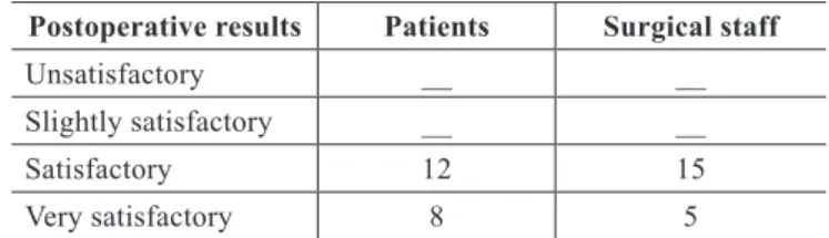
Table 1 – Assessment of the results obtained according to the patients and surgical staff.
Postoperative results Patients Surgical staff
Unsatisfactory __ __
Slightly satisfactory __ __
Satisfactory 12 15
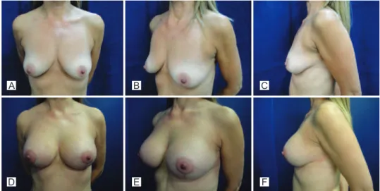
Figure 6 – Case 3. A to C, preoperative period. D to F, postoperative period of 9 months.
A
D
B
E
C
F A
D
B
E
C
F
Figure 4 – Case 1. A to C, preoperative period. D to F, postoperative period of 10 months.
A
D
B
E
C
F
However, some disadvantages associated with the use of the pectoralis major muscle flap in mastopexies have been described in the literature, such as the possibility of thoracic deformities, muscular contractures, and potential need for the use of drains to avoid hematomas11. In the present study,
aspirative drainage was only necessary in 1 patient, and no postoperative complications were reported.
CONCLUSIONS
Mastopexy with the insertion of implants and the use of pec toralis major muscle lap is a technique that can be easily im plemented; it has a relatively short learning curve, good re producibility, and satisfactory short- and long-term results.
REFERENCES
1. Regnault P. Breast ptosis. Deinition and treatment. Clin Plast Surg. 1976;3(2):193-203.
2. Spear SL, Pelletiere CV, Menon N. One-stage augmentation combined with mastopexy: aesthetic results and patient satisfaction. Aesthetic Plast Surg. 2004;28(5):259-67.
3. Spear SL, Low M, Ducic I. Revision augmentation mastopexy: indica -tions, opera-tions, and outcomes. Ann Plast Surg. 2003;51(6):540-6. 4. Spear SL, Giese SY. Simultaneous breast augmentation and mastopexy.
Aesthetic Plast Surg. 2001;107:1294-9.
5. Hurwitz DJ, Agha-Mohammadi S. Postbariatric surgery breast resha-ping: the spiral lap. Ann Plast Surg. 2006;56(5):481-6.
6. Peixoto G. Reduction mammaplasty: a personal view. In: Goldwin RM, ed. Reduction mammaplasty. London: Little, Brown;1990. p. 337-62. 7. Colwell AS, Driscoll D, Breuing KH. Mastopexy techniques after mas
-sive weight loss: an algorithmic approach and review of the literature. Ann Plast Surg. 2009;63(1):28-33.
8. Hamdi M, Van Landuyt K, Blondeel P, Hijjawi JB, Roche N, Monstrey S. Autologous breast augmentation with the lateral intercostal artery perforator lap in massive weight loss patients. J Plast Reconstr Aesthet Surg. 2009;62(1):65-70.
9. Thornton DJ, Fourie R. Autologous augmentation-mastopexy after bariatric surgery: waste not want not! Aesthetic Plast Surg. 2010; 34(4): 519-24.
10. Graf RM, Mansur AE, Tenius FP, Ono MC, Romano GG, Cruz GA. Mastopexy after massive weight loss: extended chest wall-based lap associated with a loop of pectoralis muscle. Aesthetic Plast Surg. 2008; 32(2):371-4.
11. Caldeira AM, Lucas A. Pectoralis major muscle lap: a new support approach to mammaplasty, personal technique. Aesthetic Plast Surg. 2000;24(1):58-70.
12. Auersvald A, Auersvald LA. Breast augmentation and mastopexy using a pectoral muscle loop. Aesthetic Plast Surg. 2010;35(3):333-40.
Correspondence to: Andrea Boldrin Soares