Vol.53, n. 2: pp. 361-368, March-April 2010
ISSN 1516-8913 Printed in Brazil BRAZILIAN ARCHIVES OF
BIOLOGY AND TECHNOLOGY
A N I N T E R N A T I O N A L J O U R N A L
Cytochrome P450 Detection in Liver of the Catfish Ancistrus
multispinis (Osteichthyes, Loricariidae)
Claudio Klemz
1, Ligia Maria Salvo
1, Jayme da Cunha Bastos Neto
2, Afonso Celso Dias
Bainy
3and Helena Cristina da Silva de Assis
1*1Departamento de Farmacologia; Universidade Federal do Paraná; C. P.: 19031; 81531-990; Curitiba - PR -
Brasil. 2Departamento de Bioquímica; Universidade do Estado do Rio de Janeiro; Rio de Janeiro - RJ - Brasil.
3
Departamento de Bioquímica; Universidade Federal de Santa Catarina; Florianópolis - SC - Brasil
ABSTRACT
Sensitive biological responses to environmental contaminants are useful as early warning signals to predict the damages by long-term exposure. Protocols standardization to quantify biochemical parameters in different fish species is required to validate its use as biomarkers. Comparative studies from different fish species and its interpretation are a challenge for the validation of its use as general biomarkers, representative of environmental impact. In this study, the protocol for liver cytochrome P450 (CYP) analysis from the native Brazilian fish Ancistrus multispinis was established. The microsome contamination by hemoglobin during the analysis of CYP in liver was detected, leading to misinterpretation of the results. The spectrophotometric method for CYP analysis was adapted in order to diminish the hemoglobin interference. Additionally, the western blotting method for CYP1A analysis was tested with success for this fish species.
Key words: cytochrome P450, fish, Ancistrus multispinis, biomarker
*Author for correspondence:[email protected]
INTRODUCTION
Contamination of the environment with anthropogenic compounds is a widespread phenomenon and occurs both from fixed and diffused sources. Ecotoxicological implications, however, often remain obscure. Although a deterioration of environmental health can be observed, in every case it cannot be ascribed to contaminants, as the synergistic mode of action of a complex mixture of pollutants present is not understood. The use of biomarkers may bridge the gap between the cause and effect (Bucheli and Fent, 1995). Biomarkers so called biological responses to xenobiotics have been studied as
isoenzyme cytochrome P4501A (Bucheli and Fent, 1995). However, in Brazilian fish species, these biomarkers standardization need to be carried out for subsequent utilization as possible biomarkers in each indicator species. It is particularly necessary to use the biological indicators in the aquatic environment, especially because it is the fate for a variety of pollutants from focal or non-point sources. Fish species are useful as bioindicators, since they are promptly exposed to diverse anthropogenic water contaminants (Sturm et al, 2000). The mixed function oxygenases (MFO) system plays an essential role in the metabolism of xenobiotics thanks to the function of cytochromes P450 (CYP) that mediate the NADPH-dependent oxidation of endogenous compounds and xenobiotics. CYP proteins are proposed as biomarkers because their transcriptional activation cytochromes P450 (CYP) that mediate (Bainy et al, 1999, Leitão et al, 2000, Stegemann and Hahn, 1994, Bucheli and Fent, 1995). Among the CYP forms, CYP1A induction has been characterized and involves the binding of xenobiotics, such as polynuclear
aromatic hydrocarbons (PAH), planar
polychlorinated biphenyls (PCB) and dioxins, to the Aryl hydrocarbon receptor (Ah receptor). This complex associates to the Aril receptor nuclear translocase (ARNT), which bind to the promoter region of CYP1A gene, activating the transcription and subsequently, protein synthesis (Hahn and Chandran, 1996).
The CYP system in the fish is structurally similar to that in mammals, but some differences are found; for example, the enzyme activity of fish liver microsomes is lower than in mammals, and the system could be regulated by exogenous factors (Klotz et al 1984, Stegeman and Hahn, 1994). In recent years, research in fish CYP1A has increased with the attempt to provide data to biomonitoring and pollutant metabolism (Da Silva et al, 2004).
Ancistrus multispinis (Regan, 1912) (cascudo) is a
Brazilian tropical fish belonging to the Loricariidae family that can be found in many Brazilian rivers. It has been chosen due to its detritivorous habit, meaning that the fish is in contact with xenobiotics that interact with algae
Quantification of total CYP in tropical and subtropical fish species have faced analytical difficulties while using protocols already described for total CYP analysis in other species (Cunha Bastos et al, 1989; Leitão et al 2000). To cope with this, the spectrophotometric method was adapted for CYP analysis in Ancistrus multispinis. Additionally, the western blotting method for CYP1A analysis was also tested for this species, improving even more the interpretation of results.
MATERIAL AND METHODS
Fish
Ancistrus multispinis was collected from a
non-contaminated site at the Marumbi River in the city of Morretes, Paraná, Brazil. Male and female fishes weighing approximately 10.7 ± 0.75 g (mean ± standard deviation) were collected and transported to the laboratory when they were kept for at least 20 days under controlled light (12 h dark/12 h light), temperature (25oC) and aeration conditions.
Experimental design
Fishes were randomly taken, measured, weighed and grouped in 30 liters test aquaria (12 fishes in each aquarium). Induced group (positive control) received an intracoelomic (IC) injection of 3-methylcholantrene (3-MC) (30 mg/kg), which induced CYP1A isoforms. Control group received an IC injection of sunflower oil in the same volume as the induced group. Both the groups were kept under the same experimental conditions for 96 h before liver collection. Liver was removed, washed with saline solution and immediately frozen in liquid nitrogen before microsomes isolation.
Microsome isolation
microsomal fraction. All the spectroscopic CYP analyses were carried out with this fraction. Protein concentration was determined using bovine serum albumin (BSA) as a standard (Bradford, 1976).
Total cytochrome P450 (CYP)
Analysis of CYP was based on the methods described before (Omura and Sato, 1964, Johannesen and Depierre, 1978) using differential visible spectroscopy with some adaptations. This protocol compares an oxidized aliquot of microsomal fraction with another reduced aliquot, which is bubbled with carbon monoxide. Liver perfusion was performed with an isotonic saline solution (0.9%) in order to prevent contamination by hemoglobin. Because liver samples of
Ancistrus multispinis were very small, the
perfusion was carried out by injecting the isotonic solution into each fragment of tissue until it showed a yellowish color.
Blood of A. multispinnis was collected by cardiac puncture in order to obtain hemoglobin. The hemoglobin was treated exactly as the microsome samples and UV-vis spectra were recorded. Aiming at reducing the influence of hemoglobin on CYP spectra, concentrations of 20 µM of phenasine methosulphate (PMS) and 2 mM sodium ascorbate were tested, which have been suggested to reduce contaminant hemoglobin without reducing CYP (Johannesen and Depierre, 1978). Analysis was carried out in a Beckman DU-650 spectrophotometer.
Cytochrome P450 1A (CYP1A)
Western blotting (Kloepper-Sams et al, 1987) quantified the CYP1A isoform. Microsomal proteins (40µg) were dissolved in 12% SDS-PAGE gels, electrophoretically transferred to a nitrocellulose sheet, which was blocked in solution containing 5% powdered skim milk in 0.05M TRIS; 0.15M NaCl; pH 7.5. The primary antibodies used were the 1-12-3 antibody (donated by John J. Stegeman). The secondary antibodies used were peroxidase conjugated goat anti-rabbit IgG. Protein bands were detected according to the
Amersham Pharmacia Kit for chemiluminescence
detection of Western blots and the protein bands were visualized by autoradiography on Fuji Medical X-ray film. Autoradiograms were scanned and densitometrically analyzed using NIH image. Relative concentration of CYP1A was determined comparing the integrated density of each sample protein content in µg.
Statistical analysis
Data were expressed as mean ± s.e.m. Statistical analysis was carried out by t-test followed by Welch test. Data were considered different at a significance level of p < 0.05.
RESULTS
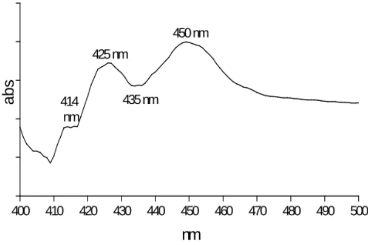
Total cytochrome P450 (CYP): Two spectra were obtained following the classical method (Omura and Sato, 1964). These spectra were composed of the expected 450nm peak, which resulted from the combination of the CYP with carbon monoxide (CO), and of an additional peak near 420 nm that could be caused by the degradation of the CYP into CYP420 or by contamination of the microssomal sample with hemoglobin (Fig. 1). Spectra obtained from microsomes prepared after the liver perfusion showed a peak at 450nm and a peak at 420 nm (Fig. 2). The effect of hemoglobin from blood sample from A. multispinis compared to perfused microsome spectra is shown in Figure 3. There was a coincidence between the spectra valleys around 409 nm and also between the peaks around 415-430nm. Comparison of these spectra led to conclude that the 420 nm peak could have been caused by the hemoglobin contamination.
Cytochrome P450 1A (CYP1A)
Results of the western blotting method are shown in Figure 5.
Evidently, the 3-MC effectively induced A.
multispinis CYP1A isoform. The value obtained
Figure 1 - Differential spectrum of microsomes from liver of A. multispinis after 30 mg/kg of 3-MC administration.
400 410 420 430 440 450 460 470 480 490 500
450 nm
435 nm 425 nm
414 nm
nm
a
b
s
Figure 2 - Differential spectrum of microsomes prepared from liver perfused with 0.9% isotonic
saline solution.
400 410 420 430 440 450 460 470 480 490 500
a
b
s
400 410 420 430 440 450 460 470 480 490 500
421nm 450nm
nm
a
b
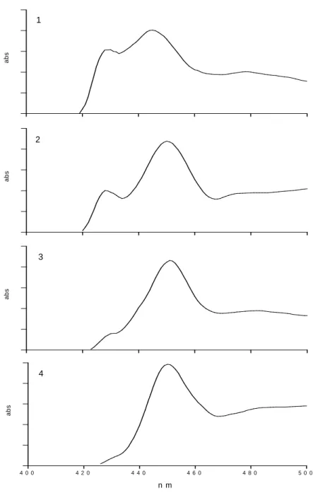
Figure 4 - Sequence of differential spectra after microsome treatment with 20 µM PMS and 2 mM sodium ascorbate: 1. immediately scanned; 2. scanned after 30 s; 3. scanned after 1 min; 4. scanned after 1 min and 30 sec.
a
b
s
1
a
b
s
2
a
b
s
3
4 0 0 4 2 0 4 4 0 4 6 0 4 8 0 5 0 0
n m
a
b
s
a
b
Figure 5 - a) Western blotting results (1- control male, 2- control female, 3- 3-MC). b) CYP1A isoform expression in hepatic microsomes of Ancistrus multispinis (* p<0.001).
DISCUSSION
There are generally marked differences between the species in P450 enzyme content, activity, and susceptibility. It could explain the different physiology, habitats, behavior, and feeding habitats, to uptake, accumulate, distribute, and metabolize contaminants. In addition, strain differences in CYP1A activity could occur (Goksøyr and Förlin, 1992). There are only few
Unusual spectra were obtained following the classical method for 450 detection by Omura and Sato (1964). Studies on Hypostomus punctatus, a fish that belonged to the same Order as A.
multispinis (Siluriformes), also showed similar
uncommon spectra (Cunha Bastos et al, 1989). Assays with rat lung microsomes showed similar results and attributed them to the residues of hemoglobin since tissues could not be adequately perfused. Other study with a Neotropical
control 3MC
0 1 2 3 4 5
*
U
A
.
µ
g
-1
p
ro
te
of microsomes (Leitão et al, 2000). Therefore the perfusion method for A. multispinis was not very efficient or degraded the CYP. The lack of related data in the literature makes it difficult to explain this situation. Aiming at reducing the influence of hemoglobin on CYP spectra, phenasine methosulphate (PMS) and sodium ascorbate were tested. Concentrations of 20 µM PMS and 2 mM sodium ascorbate caused the reduction of the peak at 420nm, which showed the reduction of hemoglobin influence in the spectra without reducing CYP as suggested by Johannesen and Depierre (1978). Adaptations on the classical method for cytochrome P450 (CYP) detection have been described for its application on
Ancistrus multispinis, in order to diminish the
hemoglobin interference on results.
In this study, the CYP of A. multispinnis showed to be inducible to Aryl hydrocarbon receptor (AhR)-ligand as 3-MC. This is in agreement with studies that have been carried out on the induction of hepatic CYP1A in fish by AhR-binding ligands, such as 3-methylcholanthrene (3MC) and β -naphthoflavone (BNF) (Goksøyr and Förlin, 1992; Stegeman and Hahn, 1994). The western blotting method used (Kloepper-Sams et al, 1987) was suitable for CYP1A analysis. Various methods to measure each step of the induction have been established and, to some degree, complement one another. The total concentration of P450 a sum parameter, frequently used and quite easy to determine, could be a screening method of contamination, mainly to polynuclear aromatic hydrocarbons (PAH), and planar polychlorinated biphenyls (PCB). Although some authors have shown that this method was less sensitive to induction than specifically inducible isoenzymes or their respective enzyme activities (Monod et al 1988, Lange et al, 1998).
Standardizing analytical techniques is a crucial step that must be carried out before field application of biomarkers. Further studies must focus the effect of xenobiotics on the induction of cytochrome P450-dependent monooxygenases from this fish to be a useful tool for monitoring programs.
ACKNOWLEDGEMENTS
The authors gratefully acknowledge the financial support by CAPES (Coordination for the Improvement of Higher Education Personnel) and the use of the facilities and technical support from the Department of Biochemistry, Federal University of Santa Catarina.
RESUMO
Respostas biológicas sensíveis aos contaminantes ambientais são úteis para prever efeitos prejudiciais devido a exposições crônicas. Padronização de protocolos para quantificar parâmetros bioquímicos em diferentes espécies de peixes é necessária para validar o uso como biomarcador. Estudos comparativos de diferentes espécies de peixe e sua interpretação são um avanço para a validação do uso de biomarcadores gerais, representativos do impacto ambiental. Neste estudo o protocolo para a análise do citocromo P450 (CYP) do peixe nativo brasileiro Ancistrus multispinis foi estabelecido. Cyp é um biomarcador de exposição principalmente de hidrocarbonetos policíclicos aromáticos (HAP), bifenilas policloradas (PCB) e dioxinas. A contaminação do microssomo pela hemoglobina durante as análises do CYP no fígado foi detectada, levando a uma interpretação errônea dos resultados. O método espectrofotométrico para análise do CYP foi adaptado para diminuir a interferência da hemoglobina. Além disso, o método de western blotting para análise de CYP1A foi testado com sucesso para essa espécie de peixe.
REFERENCES
Bainy, A.C.D., Woodin, B.R., Stegeman, J.J. (1999), Elevated levels of multiple cytochrome P450 forms in Tilapia from the Billings Reservoir-São Paulo, Brazil. Aquat. Toxicol., 44, 289-305.
Bucheli, T.B., Fent, K. (1995), Induction of cytochrome P450 as a biomarker for environmental contamination in aquatic ecosystems. Crit. Rev. Environ. Sci. Technol., 25, 201-268.
Cunha Bastos, J., Cunha Bastos, V.L.F., Burth, P., Harab, R., De Luna, M.G., Rossini, A., Castro Faria, M.V. (1989), Drug metabolism components in liver microsomes from benthic fish (Cascudo). Comp. Biochem. Physiol., 94C (2), 683-689.
Da Silva, M.E.F., Silva, J.A., Marangoni, S., Novello, J.C., Meirelles, N. C (2004), A new method to purify hepatic CYP1A of Prochilodus scrofa, a Brazilian freshwater fish. Comp. Biochem. Physiol., 138C, 67-74.
Goksøyr, A., Förlin, L. (1992), The cytochrome P-450 system in fish, aquatic toxicology and environmental monitoring. Aquat. Toxicol., 22, 287-312.
Hahn, M.E., Chandran, K. (1996), Uroporphyrin accumulation associated with cytochrome P4501A induction in fish hepatoma cells exposed to aryl hydrocarbon receptor agonists, including 2,3,7,8-tetrachlorodibenzo-p-dioxin and planar chlorobiphenyls. Arch. Biochem. Biophys., 329 (2), 163-174.
Johannesen, K.A.M., Depierre, J.W. (1978), Measurement cytochrome P450 in the presence of large amounts of contaminating hemoglobin and methemoglobin. Anal. Biochem., 86, 725-732. Kloepper-Sams. P.J., Park, S.S., Gelboin, H.V.,
Stegeman, J.J. (1987), Specificity and cross-reactivity of monoclonal and polyclonal antibodies against cytochrome P-450E of the marine fish scup. Arch. Biochem. Biophys., 253, 268-278.
Klotz, A.V., Stegeman, J.J., Walsh, C. (1984), An alternative 7-ethoxyresorufin O-deethylase activity assay: a continuous visible spectrophotometric method for measurement of cytochrome P450 monooxygenase activity. Anal. Biochem., 140, 138-145.
Lange, U., Saborowski, R., Siebers, D., Buchholz, F., Karbe, L. (1998), Temperature as a key factor determining the regional variability of the xenobiotic-inducible EROD activity in the liver of dab (Limanda limanda [L.]). Can. J. Fish. Aquat. Sci., 55, 328–338. Leitão, M.A.S., Affonso, E.G., Da Silva, M.F.E., Meirelles, N.C., Rantin, F.T., Vercesi, A.E., Junqueira, V.B.C., Degterev, I.A. (2000), The liver monooxygenase system of Brazilian freshwater fish.
Comp. Biochem.Physiol., 126, 29-38.
Monod, G., Devaux, A., Riviere, J.L. (1988), Effects of chemical pollution on the activities of hepatic xenobiotic metabolizing enzymes in fish from the River Rhone. Sci. Total Environ., 73, 189-201. Omura, T., Sato, R. (1964), The carbon
monoxide-binding pigment of liver microsomes. I. Evidence for its hemoprotein nature. J. Biol. Chem., 239 (7), 2370-2378.
Payne, J.F. (1976), Field evaluation of benzopyrene hydroxylase induction as a monitor for marine pollution. Science, 191, 945-946.
Payne, J.F., Penrose, W.R. (1975), Induction of aryl hydrocarbon (benzo[a]pyrene hydroxylase in fish by petroleum. Bull.Environ.Contamin.Toxicol., 14, 112-115.
Silva de Assis, H.C. (1998), Der Einsatz von Biomarkern zur summarischen Erfassung von Gewässerverschmutzungen. PhD Thesis Technical University of Berlin, Alemanha.
Stegeman, J.J., Binder, R.L., Orren, A. (1979), Hepatic and extrahepatic microssomal electron transport components and mixed-function oxigenases in the marine fish Stenotomus versicolor. Biochem. Pharmacol., 28, 3431-3439.
Stegeman, J.J., Hahn, M.E. (1994), Biochemistry and molecular biology of monooxigenases: current perspectives on forms, functions and regulation of cytochrome P450 in aquatic species. In- Aquatic Toxicology: Molecular, Biochemical and Cellular Perspectives. ed. D.C. Mallins, G.K. Ostrander, CRC Press, Boca Raton, FL, pp. 87-206.
Sturm, A., Wogram, J., Segner, H., Liess, M. (2000), Different sensitivity to organophosphates of acetylcholinesterase and butyrylcholinesterase from three-spined stickleback (Gasterosteus aculeatus) application in biomonitoring. Environ. Toxicol. Chem., 6, 1607-1615.
Walker, C.H., Hopkin, S.P., Sibly, R.M., Peakall, D.B. (1996), Principles of Ecotoxicology. Taylor and Francis, London, pp.321.