(Annals of the Brazilian Academy of Sciences)
Printed version ISSN 0001-3765 / Online version ISSN 1678-2690 www.scielo.br/aabc
Evaluation of wound healing and antimicrobial properties of aqueous extract from Bowdichia virgilioides stem barks in mice
ISABELA K.R. AGRA1, LUANA L.S. PIRES2, PAULO S.M. CARVALHO1, EURÍPEDES A. SILVA-FILHO2, SALETE SMANIOTTO1 and EMILIANO BARRETO1
1Laboratório de Biologia Celular, Instituto de Ciências Biológicas e da Saúde, Universidade Federal de Alagoas,
Campus A.C. Simões, Avenida Lourival Melo Mota, s/n, Tabuleiro dos Martins, 57072-970 Maceió, Al, Brasil
2
Laboratório de Genética de Microorganismos, Instituto de Ciências Biológicas e da Saúde, Universidade Federal de Alagoas, Campus A.C. Simões, Avenida Lourival Melo Mota, s/n, Tabuleiro dos Martins, 57072-970 Maceió, Al, Brasil
Manuscript received on September 2, 2011, accepted for publication on October 19, 2011
ABSTRACT
The decoction of the stem barks from Bowdichia virgilioides KUNTH is a folk remedy used to treat inflammatory disorders in Latin American and Brazil. In the present study, the wound healing activity of aqueous extract of the stem bark from B. virgilioides, called AEBv, was evaluated by the rate of healing by wound contraction and period of epithelization at different days post-wound using the wound excisional model. On day 9, the AEBv-treated animals exhibited significative reduction in the wound area when compared with controls. In wound infected with S. aureus, the AEBv significantly improved the wound contraction when compared to the saline-treated mice. The histological analysis showed that AEBv induced a collagen deposition, increase in the fibroblast count and few inflammatory cells than compared to saline-treated group. The expression of collagen type I was increased in the group treated with AEBv as indicated by immunohistochemical staining. In vitro, the AEBv was effective only against S. aureus but not against P. aeruginosa. Together, the results of this study demonstrate, for the first time, the healing and antimicrobiological effects of aqueous extract of the stem bark from B. virgilioides in the therapy of skin wounds.
Key words: wound healing, antimicrobial effect, Bowdichia virgilioides, medicinal plant.
Correspondence to Emiliano Barreto E-mail: [email protected]
INTRODUCTION
Bowdichia virgilioides Kunth (Fabaceae) is a plant that grows commonly in several South American countries such as Venezuela, Guiana and Brazil (Flores and Rodrigues 2010). Various parts of B. virgilioides are used in the Brazilian traditional medicine for treatment of diseases. The bark is used for wound healing, as anti-ulcer and anti-diabetic
(Bacchi 1986, Oliveira and Saito 1989, Braga
1953). Other plant parts such as seeds are used in
folk medicine to treat rheumatism, arthritis, and skin diseases (Cruz 1965).
The stem bark preparations of the B. virgilioides are reported to have antimalarial (Deharo et al.
2001), analgesic and anti-inflammatory activities
(Silva et al. 2010, Thomazzi et al. 2010, Barros et al. 2010). The stem bark contains several chemical substances such as lupeol, lupeol acetate, sitosterol
and stigmasterol (Melo et al. 2001). Others
and podopetaline (Barbosa-Filho et al. 2004). The antimicrobial property from B. virgilioides has been attributed to its essential oil of seeds (Almeida et al. 2006) and leaves (J.G.R. Feitosa et al., unpublished data). However, there are no published reports on the antimicrobial activity of the stem barks from B. virgilioides.
Open wounds are particularly prone to
infection, especially by bacteria, and also provide an entry point for systemic infections. Infected wounds heal less rapidly and also often result in the formation of unpleasant exudates and toxins that will be produced with concomitant killing of regenerating cells. Consequently, there is a need to stimulate healing and restore the normal functions of the affected part of the body to ease the discomfort and pain associated with wounds, preventing infection, and activating tissue repair processes. Antibacterial and healing compounds in a traditional remedy can induce this occurrence
and may be beneficial in treating wounds (Reddy
et al. 2008). In spite of recorded uses of the B. virgilioides, there is no scientific evidence that
confirms the healing effect and antibacterial activity
of stem bark of B. virgilioides. Thus, this work was undertaken to explore the antimicrobial and wound healing effects of B. virgilioides stem bark extract.
MATERIALS AND METHODS
PLANT MATERIAL AND PREPARATIONS OF AQuEOuS EXTRACT
Stem bark from B. virgilioides Kunth (Family Fabaceae) was collected in Maceió, Alagoas State, Brazil (9°33'12''S and 35°46'9''W). The plant was
identified by Prof. Rosângela Lemos, Instituto
do Meio Ambiente, Maceió, Brazil, and the voucher specimen (number MAC29914) has been deposited at the Herbarium MAC of the Instituto do Meio Ambiente.
After collection, the stem barks were dried at ambient temperature and triturated. The aqueous extract of B. virgilioides, called AEBv, was prepared
by infusing 50 g of powdered plant material for 20 minutes using 300 mL of boiling water. The
extract was filtered and lyophilized. The yield of the infusion was 17.2% (wt=wt). At the time
of use, extract was reconstituted in water (sterile endotoxin-free) at the required concentration (10 mg.kg-1) according as previous results (J.P. Silva, unpublished data).
ANIMALS
Swiss mice of either sex weighing 18–22 g were obtained from the Universidade Federal de Alagoas (UFAL) breeding unit. The animals were maintained with free access to food and water and kept at 22-28°C with a controlled 12-hour light/ dark cycle at the Instituto de Ciências Biológicas e da Saúde, UFAL. Experiments were performed during the light phase of the cycle. The animals were allowed to adapt to the laboratory for at least 2 hours before testing and were used only once. All experiments were carried out in accordance with institutional guidelines and ethics (License Number 23065.12614/2006-89).
ExCISION WOuNd MOdEL
The animals were anesthetized with anesthetic ether and shaved at the predetermined site before
wounding. A circular wound was inflicted by
cutting away approximately 1.6 cm of diameter of the predetermined area on the anterior-dorsal side of each mice using sterile surgical blade (Morton and Malone 1972). The animals were then placed in separate cages to avoid any disturbance. The bedding was changed daily. After skin excision, the wound was left open to the environment.
In other set of experiments, the wound was
inoculated (10 µL) with Staphylococcus aureus (ATCC 25923) at 108 Colony Forming Unit (CFU).
All animals received topical application (200 µL)
standard treatment was used fibrinolysin (Fibrase
SA®) on non-infected wounds or 1% silver sulfadiazine (Dermazine®) on infected wounds. The animals of the Fibrase SA® or Dermazine ® groups were topically treated once a day with 0.5 g of each ointment. Wound contraction was calculated as percentage reduction in wound area. The progressive changes in wound area were monitored by a camera (Sony Cyber Shot, Dsc w80) on wounding day, followed by measurements on 3th, 6th and 9th day. Later on, wound area was evaluated by using ImageJ program (Nicoli et al. 2008). A specimen sample of tissue was isolated from the healed skin of each group of mice for the histopathological examination.
WOuNd HEALING RATE
The wound area of each animal was measured on days 3, 6, and 9 post-surgery. The wound size measurements taken at the time of surgery and at the time of biopsy were used to calculate the percent wound contraction, using equation:
[(A0 – At)/A0] x 100 = % of wound closure
where A0 is the original wound area, and At is the area of wound at the time of biopsy.
ANTIMICROBIAL SENSITIVITY TEST
Antimicrobial activities of extract was evaluated against Staphylococcus aureus (ATCC 25923), Pseudomonas aeruginosa (ATCC 27853) and Staphylococcus aureus (MRSA) which was supplied by Dr. Euripedes A. Silva-Fillho, UFAL, Maceió-AL, Brazil. The MRSA-strain was based on their resistance to methicillin (DMPPC) and oxacillin (MPIPC) according to the guidelines of the National Committee for Clinical Laboratory
Standards (2003). A modified diffusion test was
used to determine the antimicrobial activity (Joung et al. 2010). The colonies were taken directly from the plate and were suspended in 5 mL of sterile 0.85% saline. The turbidity of the initial suspension
was adjusted by comparing with 0.5 McFarland’s standard. When adjusted to the turbidity of the 0.5 McFarland’s standard, the bacteria suspension contains about 108 colony forming unites (CFU). mL-1. In different petridish the bacterial strains were grown to exponential phase in Mueller-Hinton broth
at 37 °C for 18 h and adjusted to a final density of 108 CFU/ml by diluting fresh cultures and comparing with McFarland density (Murray et al. 1995). The blank control was performed with distillated water. Chloramphenicol was used as the positive control. In each petridish were made bores (4 mm) where each
bore was loaded with 40 μL of water, AEBv (1, 2 and
4 mg.mL-1) or chloramphenicol (1 mg.mL-1). After incubation at 37°C for 24 h the inhibition zones around the bores were measured. The tests were performed in triplicate and the results were expressed in mm as the arithmetic media of diameters of the inhibition zones. After incubation, the result of antimicrobial activity test was reported as the average diameter of the inhibition zone surrounding the wells containing the test solution.
HISTOPAThOLOGICAL ANALYSIS
The skin specimens from each group were collected at 9 days after beginning of the experiment to evaluate the histopathological alterations in accordance with Tumen et al. (2012), being the analysis performed blindly by a pathologist.
Samples were fixed in 10% buffered formalin, processed and blocked with paraffin. Then, sample were sectioned into 5 μm-thick sections and stained
with hematoxylin and eosin (HE) and Masson’s trichrome (MT). The tissues were examined by
light microscope (Olympus Bx51 attached dP70
Digital Camera System) and graded subjectively as mild (+), moderate (++) and severe (+++) for epidermal or dermal remodeling, reepithelization;
fibroblast proliferation, mononuclear and/or
IMMuNOhISTOChEMICAL STAINING
Six μm thick skin cryostat sections were used
for detecting type I collagen expression by immunoperoxidase staining. The sections were then treated with 0.3% hydrogen peroxide (H2O2) in phosphate buffered saline (PBS) for 10 min to quench any endogenous peroxidase activity
within the tissue. The nonspecific binding sites
were blocked with 0.5% bovine serum albumin (BSA) for 10 min at room temperature. After washing with PBS, specimens were incubated
with purified rabbit anti-mouse type I collagen
antibody (Novotec - lot 338i) diluted 1:80 in PBS for 1 h at room temperature. After washing, the specimens were incubated with peroxidase-conjugated goat anti-rabbit IgG second antibody (Sigma) diluted 1:200 in PBS for 45 min at room temperature. Immunoreactivity was visualized with a diaminobenzidine (DAB) (Sigma) containing 0.02% H2O2 for 10 min. The control sections were incubated directly with the secondary antibody in the absence of the primary antibody and processed as above. The specimens were observed using light microscope (Nikon Eclipse 50i).
STATISTICAL ANALYSIS
Data are mean ± SEM values. The statistical analysis involving two groups was done using Student’s t test. Analysis of variance followed by the
Student-Neuman-Keuls test was used to compare three or
more groups. values of P < 0.05 were considered as indicative of significance.
RESULTS
WOuNd HEALING ACTIVITY
The wound healing activity of the aqueous extract prepared from the stem barks of Bowdichia virgilioides was evaluated on mice in the excision
wound models to confirm the folkloric usage of the
plant. The histopathological changes induced by this extract and its antimicrobial activity in vitro were also assessed. The area of the wound was measurement on the days 3, 6 and 9 days post surgery in all groups. The measurements of the progress of wound healing induced by the extract, reference drug and saline treated-groups in the excision of non-infected wounds are shown in Table I.
Table I shows the measured values of the closure progression of non-infected wound in different groups. After application of AEBv topically onto non-infected wounds the area of wound reduced 25% of their original size (2 cm2) on day 3, 62.5% on day 6 and 91% on day 9, and complete closure around day 10. In saline-treated animals, the area was reduced to 16.5% (day 3), 26% cm2 (day 6) and 44.5% (day 9). The wound closure in animals treated with reference drug, Fibrase, were 20% (day 3), 30% and 74% (day 9) (Table I). Treatment with
Treatment
Parameter
Wound area (cm2) on day Period of epithelization (days)
3 6 9
Saline 1.67 ± 0.15 1.48 ± 0.11 1.11 ± 0.08 13.00 ± 0.00
Fibrase® 1.60 ± 0.06 1.40 ± 0.09 0.52 ± 0.08 *** 10.75 ± 0.5 ** AEBv 1.50 ± 0.12 c 0.75 ± 0.06 *** a 0.18 ± 0.03 *** b 9.60 ± 0.33 *** c
TABLE I
Effect of aqueous extract of stem barks from Bowdichia virgilioides on non-infected wounds.
values represent the mean ± S.E.M., (n=4). Statistical differences were determined by ANOvA followed Student-Newman-Keuls test. ***P<0.001, **P<0.01, *P<0.05 as compared to respective saline treatment.
AEBv in non-infected wounds was able to reduce to 9 days the period of epithelialization when compared with saline-treated group and Fibrase group, which were, respectively, 13 and 10 days.
Table II shows the measured values of the closure progression of infected wound in different groups. After application of AEBv topically onto infected wounds the area of wound reduced 43% of their original size (2 cm2) on day 3, 84% on day 6 and 95.5% on day 9, and complete closure on day 10
(Table II). On the other hand, in saline-treated animals,
the area was reduced to 16.8% (day 3), 26.3% (day 6) and 44.7% (day 9). The wound closure in animals treated with reference drug, Dermazine®, were 23.8% (day 3), 41.2% and 64.1% (day 9) (Table II). Treatment with AEBv in infected wounds was able to reduce to 10 days the period of epithelialization when compared with saline-treated group and Dermazine® group, which were, respectively, 17.5 and 15 days.
hISTOPAThOLOGICAL ANALYSIS
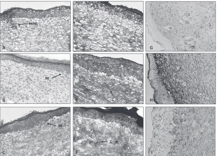
In order to confirm the experimental results,
histopa thological analysis was also performed. Figure 1 shows the histology of saline, AEBv and Fibrase-treated groups at 9 days of analysis in non-infected wound. The AEBv and Fibrase-treated groups shows faster wound healing processes if compared with saline-treated animals. There was
attenuation in the infiltration of inflammatory cells
and enhanced proliferation of fibroblasts as a result of treatment with our extract and the reference drug. There was full thickness reepithelialization, in which epidermis was thin and well organized, comparable to the normal adjacent skin which was not involved in the wound generation and healing process. AEBv-treated wounds were associated with enhanced formation of epidermis and deposition of connective tissue when compared to that of control group animals. Less epithelialization and less collagen formation in saline-treated animals indicated incomplete healing.
The expression of type I collagen was detected by the immunohistochemistry method (Figure 1). In contrast to the group treated with saline, a considerable expression of collagen type I was detected in the tissue after 9 days of treatment with AEBv (Figure 1G - 1H). This increase in collagen type I expression was the most effective in AEBv-treated group than compared to Fibrase-treated group (Figure 1H - 1I).
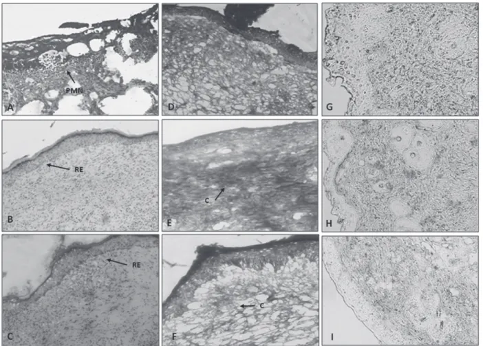
Figure 2 shows the histology of saline, AEBv and Dermazine-treated groups at 9 days of analysis in infected wound. The saline-treated group demonstrated delayed wound healing processes compared to the other groups. The epidermis in infected wounds was thick and disorganized, especially when compared with the adjacent normal skin. Clumps of degenerating tissue, necrotic changes, and the persistence of
Treatment
Parameter
Wound area (cm2) on day Period of epithelization (days)
3 6 9
Saline 1.53 ± 0.03 1.25 ± 0.03 1.04 ± 0.03 17.50 ± 0.50
Dermazine® 1.53 ± 0.05 1.18 ± 0.04 0.72 ± 0.04 *** 15.00 ± 0.00 ** AEBv 1.14 ± 0.12 ** 0.32 ± 0.08 *** a 0.09 ± 0.04 *** a 10.00 ± 0.00 *** a
TABLE II
Effect of aqueous extract of stem barks from Bowdichia virgilioides on infected wounds.
inflammatory exudates in the upper dermis with
loss of epidermis were observed in infected wounds on day 9. AEBv and Dermazine-treated mice showed marked epithelialization and moderate amount of connective tissue synthesis. Following histopathological examination to both infected and non-infected wound, the scored results were combined, summarized and presented in Table III.
The expression of collagen type I was detected in 9 days after injury in infected wounds (Figure 2). The AEBv-treated group demonstrated clusters of slight increase in expression of collagen type I compared with the saline-treated group (Figure 2G
- 2H). Dermazine treatment had less effect on the collagen type I expression in comparison with the AEBv-treated group (Figure 2H- 2I).
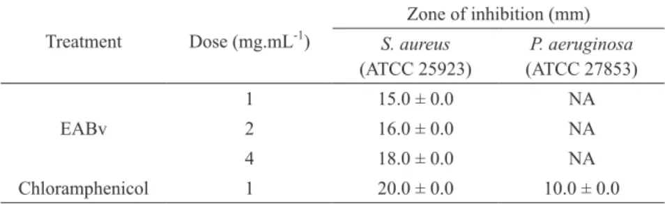
ANTIMICROBIAL SENSITIVITY TEST
Table IV shows the antibacterial activity of aqueous extracts of the stem bark of Bowdichia virgilioides against two bacterial strains, S. aureus and P. aeruginosa. The AEBv showed the highest antibacterial activity against S. aureus while had no effect against P. aeruginosa. Chloramphenicol,
a standard antibiotic, showed a significantly
anti-bacterial activity against the test organisms.
Figure 2 - Histopathological view of epidermal/dermal remodeling in infected wounds. In A, B and C show skin sections stained with hematoxylin and eosin. In D, E and F show skin sections stained with Masson’s trichrome. In G, H and I show immunohistological staining to expression of collagen type I. The original magnification was 100x. data are representative of 4 animals per group. A, D and G Saline-treated group (9-day-old wound tissue treated with only saline); B, E and H AEBv group (9-day-old wound tissue treated with B. virgilioides extract); C, F and I Dermazine-treated group (9-day-old wound tissue treated with Dermazine). Arrows pointing events during wound healing: RE: reepithelization; C: collagen; PMN: polymorphonuclear cells.
Wound healing processes
Groups
Non-infected wounds Infected wounds
Saline Fibrase AEBv Saline Dermazine AEBv
RE +/++ ++ +++ -/+ ++ +++
FP + +++ +++ + +/++ ++
CD + +/++ +++ -/+ -/+ ++/+++
PMN ++ -/+ -/+ ++ + -/+
TABLE III
Wound healing processes of aqueous extract of stem barks from Bowdichia virgilioides.
Due to the high antimicrobial activity of the AEBv against S. aureus ATCC 25923, the antimi-crobial activity against S. aureus MRSA was assessed. Results demonstrated that the AEBv (4 mg.mL-1) induced an inhibition zone of 18 mm for strain MRSA, value similar to that presented by the standard strain.
DISCUSSION
In the present paper we report the wound healing potential of the aqueous extract of the stem bark of Bowdichia virgilioides, AEBv, applied on infected and non-infected wounds in mice. The extract of Bowdichia virgilioides showed antimicrobial activity (J.G.R. Feitosa et al., unpublished data, Almeida et al. 2006), analgesic and
anti-inflammatory effects (Silva et al. 2010, Thomazzi
et al. 2010, Barros et al. 2010). So, if any plant material presents antimicrobial, analgesic and
anti-inflammatory activities together, it can be supposed
that this material also may help to promote wound healing and contribute skin regeneration.
We observed that the topical application of AEBv enhances cutaneous healing, which appeared
completed in 9 days. The histological findings showed
that the original tissue regeneration is much greater in skin wounds treated with the extract than in wounds saline-treated. The wound contraction is mediated by
specialized myofibroblasts found in the granulated
tissue (Moulin et al. 2000). So, the increase in wound contraction in AEBv-treated mice might be a result of
the enhanced activity of fibroblasts.
Indeed, the response to injury involves the
migration and proliferation of cells such as
fibro-blasts, endothelial and epithelial cells, and depo-sition of connective tissue and contraction of the wound. Collagen not only confers strength and integrity to the tissue matrix but also plays an important role in homeostasis and in epithelia-lization at the later phase of healing (Clark 1996).
here, our finding revealed that treatment with
AEBv caused an increased in the deposition of the bands of collagen, a phenomenon that appears to contribute with the increase in wound contraction.
Collagen type I is the most common protein in animals and provides the tensile strength of healing in wounds. Besides contributing to the skin strength, collagen type I is also important to guide keratinocytes
and dermal fibroblasts migration in the wounded
area (Bennett and Schultz 1993). Considering this, our results suggest that topical treatment with AEBv
could be beneficial to wounds skin repair in both
conditions infected and non-infected.
Skin wound healing starts immediately after
injury and consists of three phases: inflammation, proliferation, and maturation. The first response is inflammation, acting as a defense mechanism of the
tissue, able to provide a resistance to the microbial contaminations (Kondo 2007). But, a long duration
in the inflammatory phase causes a delay in healing process. Anti-inflammatory activity is necessary
for shorten the healing period (Shimizu et al.
2000). Therefore, the significant wound healing TABLE IV
Antibacterial activity of the aqueous stem bark extracts of B. virgilioides.
Treatment Dose (mg.mL-1)
zone of inhibition (mm)
S. aureus
(ATCC 25923)
P. aeruginosa
(ATCC 27853)
EABv
1 15.0 ± 0.0 NA
2 16.0 ± 0.0 NA
4 18.0 ± 0.0 NA
Chloramphenicol 1 20.0 ± 0.0 10.0 ± 0.0
activity of AEBv may be related to its remarkable
anti-inflammatory effect as presented in previous
reports (Silva et al. 2010, Thomazzi et al. 2010). Antimicrobial activity is important for the wound healing period, because the wound exposed to external environment is more prone to microbial attacks, which usually results in a delay in the healing process. So, risk factors such as infections may compromise the repair process. S. aureus and P. aeruginosa are the most common pathogens respon-sible for infection in skin wounds (Arora and Kaur 2007). Topical applications of drugs are effective both as microbicide and increasing wound healing rate because of its greater availability at the infected wound site. In this study, the slow rate of wound closure in control mice may be attributable to the presence of microorganisms and their metabolites, which inhibits wound contraction and impair healing. In this study, even in infected wounds where the period of epithelialization is greater, when the animals were treated with AEBv there was a better wound healing if compared to animals treated with saline.
In vitro analysis of the antimicrobial effect of AEBv showed a potential inhibitory effect against Gram-positive bacteria S. aureus, but not against Gram-negative bacteria such as P. aeruginosa. In line with this observation, previous results from J.G.R. Feitosa et al. (unpublished data) showed that essential oil of seeds from B. virgilioides possess an antimicrobial activity against Gram-positive B. subtilis, B. vulgaris, E. faecalis and S. aureus and had low activity in vitro against Gram-negative P. aeruginosa, S. enteritidis and E. coli. This antibacterial effectiveness may be attributed to the fact that cell wall in Gram-positive bacteria consists of a single layer, whereas Gram-negative cell wall is a multilayered structure bounded by an outer cell membrane (Mahomoodally et al. 2010). Moreover,
findings from the present study showed that AEBv
was effective against a methicillin-resistant strain of S. aureus (MRSA). These MRSA are difficult to treat because they are also multiresistant and up to
now there are no satisfactory antimicrobial drugs (Joung et al. 2010). Therefore, regarding to the present result, extract from B. virgilioides seem to be a potential tool to combat the problem of MRSA.
The results of our study indicate, for the first time,
that B. virgilioides may be a potential candidate for
dermal wound healing because of its positive influence
on phases of the healing process and particularly effective in view of it antimicrobial properties. Therefore, there is, the need for further studies into
the stability of the extract to ensure an efficacious
formulation of products for wound healing.
ACKNOWLEDGMENTS
This work was supported by grants from the
Conselho Nacional de desenvolvimento Científico
e Tecnológico (CNPq), the Programa de Cooperação Acadêmica/Coordenação de Aperfeiçoamento de
Pessoal de Nível Superior (PROCAd/CAPES) and
the Fundação de Amparo à Pesquisa do Estado de Alagoas (FAPEAL) (Brazil).
RESUMO
imuno-histoquímica. In vitro, o AEBv foi eficaz apenas contra S. aureus, mas não contra P. aeruginosa. Juntos, os resultados deste estudo demonstram, pela primeira vez, a cura e os efeitos antimicrobianos do extrato aquoso da casca do caule de B. virgilioides na terapia de feridas cutâneas.
Palavras-chave: Cicatrização de feridas, efeito
antimi-crobiano, Bowdichia virgilioides, planta medicinal.
REFERENCES
ALMEIDA JRGS, SILVA-FILhO M, NUNES XP, DIAS CS, PEREIRA FO AND LIMA EO. 2006. Antimicrobial activity of the essential oil of Bowdichia virgilioides Kunt. Rev Bras Farmacogn 16: 638-641.
ARORA DS AND KAUR GJ. 2007. Antibacterial activity of some Indian medicinal plants. J Nat Med 61: 313-317. BACCHI EM. 1986. Ação antiúlcera e cicatrizante de algumas
plantas brasileiras, Rev Bras Farmacog 1: 93-100. BARBOSA-FILhO JM, DA-SILVA ALMEIDA JR, DE-OLIVEIRA
COSTA VC, DA-CUNHA EV, DA-SILVA MS AND B RAz-FILhO R. 2004. Bowdichine, a new diaza-adamantane alkaloid from Bowdichia virgilioides. J Asian Nat Prod Res 6: 11-17.
BARROS CWM, RAO VS, SILVA RM, LIMA JC AND MARTINS DT. 2010. Anti-inflammatory effect of the ethanolic extract from Bowdichia virgilioides H.B.K stem bark. An Acad Bras Cienc 82: 609-616.
BENNETT NT AND SCHULTz GS. 1993. Growth factors and wound healing: biochemical properties of growth factors and their receptors. Am J Surg 165: 728-737.
BRAGA R. 1953. Plantas do nordeste, especialmente do Ceará. Coleção Mossoroense. Ed. Universitária UFRN. 4ª edição, Fortaleza, CE, Brasil, p. 449-453.
CLARK RAF. 1996. Wound repair: overview and general considerations. In: CLARK RA ANd hENSON PM. The molecular and cellular biology of wound repair, 2nd ed., New York: Plenum, p. 3-50.
CRUz GL. 1965. Livro Verde das Plantas Medicinais e Industriais do Brasil. Ed. Helmus, Belo Horizonte, MG, Brasil, 779 p. DEhARO E, BOuRdY G, QuENEvO C, MuNOz V, RUIz G AND SAUVAIN M. 2001. A search for natural bioactive com-pounds in Bolivia through a multidisciplinary approach. Part V. Evaluation of the antimalarial activity of plants used by the Tacana Indians. J Ethnopharmacol 77: 91-98. FLORES S AND ROdRIGuES RS. 2010. Diversidade de
Leguminosae em uma área de savana do estado de Roraima, Brasil. Acta Bot Bras 24: 175-183.
JOuNG H, KWON DY, ChOI JP, SHIN DY, CHUN SS, YU YB AND SHIN DW. 2010. Antibacterial and synergistic effects of Smallanthus sonchifolius leaf extracts against methicillin-resistant Staphylococcus aureus under light intensity. J Nat Med 64: 212-215.
KONdO T. 2007. Timing of skin wounds. Leg Med 9: 109-114.
MAhOMOOdALLY MF, GURIB-FAKIM A AND SUBRATTY AH. 2010. Screening for Alternative Antibiotics: An investigation into the antimicrobial activities of medicinal food plants of Mauritius. J Food Sci 75: 173-177. MELO FN, NAvARRO VR, SILVA MS, DA-CUNHA EV,
BARBOSA-FILhO JM AND BRAz-FILhO R. 2001. Bowdenol, a new 2,3-dihydrobenzofuran constituent from Bowdichia virgilioides. Nat Prod Lett 15: 261-266.
MORTON JJ AND MALONE MH. 1972. Evaluation of vulnerary activity by an open wound procedure in rats. Arch Int Pharmacody Thér 196: 117-126.
MOuLIN V, AUGER FA, GAREL D AND GERMAIN L. 2000. Role of wound healing myofibroblasts on re-epithelization of human skin. Burns 26: 3.
MURRAY PR, BARON EJ, PFALLER MA, TENOvER FC AND YOLKEN RH. 1995. Manual of clinical microbiology. 6th ed., Washington, U.S.A. American Society for Microbiology, 165 p.
NATIONAL COMMITTEE FOR CLINICAL LABORATORY STANDARDS. 2003. Methods for Dilution Antimicrobial Susceptibility Tests for Bacteria That Grow Aerobically, 2nd ed., Approved Standard. NCCLS document M2-A8. 940 West Valley Road, Suite 1400, Wayne, Pennsylvania: NCCLS.
NICOLI S, PADULA C, AVERSA V, VIETTI B, WERTz PW, MILLET A, FALSON F, GOvONI P AND SANTI P. 2008. Characterization of rabbit ear skin as a skin model for in vitro transdermal permeation experiments: histology, lipid composition and permeability. Skin Pharmacol Physiol 21: 218-226.
OLIVEIRA O AND SAITO ML. 1989. Alguns vegetais brasileiros empregados no tratamento do diabetes. Rev Bras Farmacogn 2: 170-196.
REDDY S, REDDY RKK, NAIDU VG, MADHUSUDHANA K, AGWANE SB, RAMAKRISHNA S AND DIWAN PV. 2008. Evaluation of antimicrobial, antioxidant and wound-healing potentials of Holoptelea integrifolia. J Ethnopharmacol 115: 249-256.
SHIMIzU N, WATANABE T, ARAKAWA T, FUJIWARA Y, HIGUCHI K AND KuROKI T. 2000. Pentoxifylline accelerates gastric ulcer healing in rats: roles of tumor necrosis factor alpha and neutrophils during the early phase of ulcer healing. Digestion 61: 157-164.
SILVA JP, ROdARTE RS, CALhEIROS AS, SOuzA Cz, AMENdOEIRA FC, MARTINS MA, SILVA PM, FRuTuOSO VS AND BARRETO E. 2010. Antinociceptive activity of aqueous extract of Bowdichia virgilioides in mice. J Med Food 13: 348-351.
ThOMAzzI SM, SILVA CB, SILVEIRA DC, VASCONCELLOS CL, LIRA AF, CAMBUI EV, ESTEVAM CS AND ANTONIOLLI AR. 2010. Antinociceptive and anti-inflammatory activities of
Bowdichia virgilioides (sucupira). J Ethnopharmacol 127: 451-456.