Effect of
Effect of
Effect of
Effect of
Effect of
Carapa guianensis Aublet
Carapa guianensis Aublet
Carapa guianensis Aublet (Andiroba) and
Carapa guianensis Aublet
Carapa guianensis Aublet
(Andiroba) and
(Andiroba) and
(Andiroba) and
(Andiroba) and Orbignya
Orbignya
Orbignya
Orbignya
Orbignya
phalerata
phalerata
phalerata
phalerata
phalerata (Babassu) in colonic healing in rats
(Babassu) in colonic healing in rats
(Babassu) in colonic healing in rats
(Babassu) in colonic healing in rats
(Babassu) in colonic healing in rats
Efeito da
Efeito da
Efeito da
Efeito da
Efeito da Carapa guianensis Aublet
Carapa guianensis Aublet
Carapa guianensis Aublet
Carapa guianensis Aublet (A
Carapa guianensis Aublet
(A
(A
(Andiroba
(A
ndiroba)
ndiroba
ndiroba
ndiroba
)
)
)
) e
ee
ee O
O
O
O
Orbignya
rbignya
rbignya
rbignya
rbignya phalerata
phalerata
phalerata
phalerata
phalerata (((((Babaçu
Babaçu)
Babaçu
Babaçu
Babaçu
)
)
)
) na
na
na
na
na
cicatrização de colorrafias em ratos
cicatrização de colorrafias em ratos
cicatrização de colorrafias em ratos
cicatrização de colorrafias em ratos
cicatrização de colorrafias em ratos
CÍCERO EVANDRO SOARES SILVA1,2, ORLANDO JOSEDOS SANTOS2, JURANDIR MARCONDES RIBAS-FILHO1; FERNANDO ISSAMU TABUSHI1; MARCIO
HIROAKI KUME1; LEANDRO BRESSIANINI JUKONIS1; IGOR FURLAN CELLA1
A B S T R A C T A B S T R A C T A B S T R A C T A B S T R A C T A B S T R A C T
Objective: Objective: Objective: Objective:
Objective: to evaluate the healing effect of the babassu aqueous extract and andiroba oil on open wounds in the cecum of rats. Methods
Methods Methods Methods
Methods: fifty-four Wistar rats were divided into three groups of 18: 1) babassu group with application of aqueous extract of babassu; 2) andiroba group with application of the oil; and 3) control group, with application of saline solution. All procedures were done by gavage. Each group was divided into three subgroups of six animals according to the observation period of 7, 14 or 21 days. From each animal was removed caecum fragment of 1.5cm² diameter. The areas of the lesions were analyzed macroscopically and resected specimens by light microscopy using hematoxylin-eosin and Masson’s trichrome. ResultsResultsResultsResultsResults: abscess and infection were observed in two aroeira group animals, and in one only hematoma. In relationship to adhesions degree, babassu group had higher incidence of grade II while in the control and aroeira groups predominated adhesions grade I. On microscopic examination on day 7 fibroblast proliferation was greater in aroeira and lower in babassu group (p=0.028). On the 14th day polymorphonuclear were less pronounced in babassu (p=0.007). As for the resistance test of air insufflation, it was
observed that in all andiroba group in all tested days showed be higher. As for collagen, on the 7th day it was present in 100% of
animals of aroeira group. On the 14th day was more pronounced in the control group and at day 21 similar results were found in
the control and aroeira groups. ConclusionConclusionConclusionConclusionConclusion: animals in babassu and andiroba groups showed better cecum healing compared to the control group.
Key words Key words Key words Key words
Key words: Rats. Healing. Phytotherapy.
1. Programa de Pós-Graduação em Princípios da Cirurgia da Faculdade Evangélica do Paraná / Hospital Universitário Evangélico de Curitiba / Instituto de Pesquisas Médicas, Curitiba, PR, Brasil; 2. Hospital São Domingos, São Luis, MA, Brasil.
INTRODUCTION
INTRODUCTION
INTRODUCTION
INTRODUCTION
INTRODUCTION
T
he healing process is similar in all wounds and is based on a complex sequence of events ranging from trauma to injured tissue repair. It consists on a perfect and coordinated cascade of cellular, molecular and biochemical events that are interrelated in order to occur tissue reconstitution1. Such process can be divided into threephases that temporally and continuously overlap: inflammatory, proliferative or granulation, and remodeling or maturation phases1,2.
The interest in wound healing began in antiquity, with records dating back to 3000-2500 b. C. Among them, honey-based, grease, and flax strings are mentioned as medicine that constituted Egyptian pharmacopoeia3,4.
The record of plant use to cure diseases is also reported in 5.000 years old historical records, in which ancient people used herbs for medicinal purposes. Nowadays, there is a growing interest in herbal medicine, which consists of plant use techniques in the treatment of diseases and health recovery5.
Herbal medicines are considered as those obtained with exclusive use of plant active raw materials, whose efficacy and safety are validated through ethnopharmacological surveys, techno-scientific documentation or clinical evidence6. The use of plants as
therapeutic resources is still underutilized. From 300,000 plants currently known, only 2,000 are used in medicine5.
In Brazil, internationally known as having the largest diversified forest reserve in the world, the state of Maranhão is highlighted, not only for having a dense diverse flora, but also by the tradition in research of medicinal plants, having its own herbarium with artisanal production of various products7.
The use of herbal medicines in wound healing has been stimulated by the need to find new substances that play an effective role in surgical repair because, although it is a systemic process, favorable local conditions, through appropriate topical therapy stimulating the physiological process, are necessary8.
properties, Orbignya phalerata and Carapa guianensis Aublet have been widely mentioned in experiments.
Orbignya phalerata (babassu) is found more frequently in the North, Northeast and Midwest regions of Brazil, with more than 50% being concentrated in Maranhão. It has anti-inflammatory and analgesic properties proven in studies9. Martins et al., in an attempt to verify its
healing action in surgical skin wounds, also noted satisfactory effects on the healing process, through microscopic analysis, in mononuclear variables and collagen fibers10.
The species Carapa guianensis Aublet belongs to the family Meliaceae, commonly known as “andiroba”. It is mainly found in wetlands and flooded areas, but it can be grown on land. It has physicochemical properties that gives it anti-inflammatory action11-13.However, Brito et al.,
when assessing the effect of andiroba oil in open wounds in rats, observed a delay in contraction and epithelialization of wounds, suggesting a possible anti-inflammatory action resulting in a loss during the healing process14.
It is then necessary to understand the therapeutic effects of Orbignya phalerata and Carapa guianensis Aublet today in order to expand their use in wound healing.
This study with rats aimed to use both substances in colon wound healing to verify their healing effect.
METHODS
METHODS
METHODS
METHODS
METHODS
This study was performed at the Experimental Surgery Laboratory of the Universidade Federal do Maranhão, São Luís, MA, Brazil, and complied with ani-mal testing requirements of the Sergio Arouca Law (no
11.794/2008). It was approved by the Ethics and Animal Experimentation Committee of the Medical School of Veterinary of the Universidade Estadual do Maranhão, protocol no 039/2012.
Fifty-four Wistar rats, male, aged between 50 to 60 days, with an average weight of 275.64g, were used. The animals were weighed and kept under observation for a seven-day adaptation period prior to the operation. The animals were placed in nine polypropylene cages with a stainless metal grill cover, with six animals per cage, receiving water and species’ standard ration ad libitum at 23±2°C, in a noise-free environment, with a light/dark cycle of 12h. They were randomly distributed in three groups with 18 animals: CG (control group), BG (babassu group), AG (andiroba group), which in turn were divided into six subgroups according to the day of death (7th, 14th and 21st).
Preparation of phytotherapics Preparation of phytotherapics Preparation of phytotherapics Preparation of phytotherapics Preparation of phytotherapics
Aqueous extract of Orbignya phalerata (Babassu) The exsiccate specimen of babassu was cataloged in the Herbarium Ático Seabra of the Universidade Federal do Maranhão. The mesocarp was obtained from mature coconut, which was considered as mature when falling
naturally from bunches. To remove the mesocarp, a wood artifact was used, manually beating the top until the rupture of the shells and after it, with the aid of a spatula, the mesocarp was separated. The material was spread on a bench for three days to dry. After dried, it was placed in a drying oven at a temperature of 45-50ºC for 24h in order to complete the total removal of moisture. The mesocarp was subjected to a grinding process in an electric grinder, where a powdery flour was obtained. For the preparation of the aqueous extract, the powder was weighed on an analytical digital balance and diluted in saline solution at 25mg/ml. Chemical analysis has shown it to have starch (68.3%), moisture (14.9%), fiber (2.51%), protein (1.54%), soluble carbohydrates (1.25%), lipids (0.27%) and other substances - amino acids, hemicellulose and pentosans (11.23%)
Carapa guianensis Carapa guianensis Carapa guianensis Carapa guianensis
Carapa guianensis oil (Andiroba) oil (Andiroba) oil (Andiroba) oil (Andiroba) oil (Andiroba)
The oil is a transparent liquid, yellow, with a very bitter taste, which, below 25°C, solidifies as petrolatum (the oil becomes a solid white fat, whose starting point is 22° C and is completed at 28ºC). The seeds contain 70% of insect-repelling substances and medicinal oil15. The
traditional extraction process is complex, lasting about two months, and can be divided into three stages: 1) collection, selection of good seeds and a first storage time (3-5 days); 2) bulk preparing by baking the seeds in water (1-3h) and a second storage time (up to 20 days), finishing with the withdrawal of the peel and the kneading of the seeds; 3) extraction of oil by dripping (30 days), placing the bulk on an inclined surface16.
The sample used in the study was from the city of Axixá, MA, and 50ml were analyzed by the Laboratory of Food and Water Quality Control, at the Department of Chemistry Technology of the Universidade Federal do Maranhão, with the following grease composition: myristic acid (18.1%); oleic acid (58.9%); linoleic acid (9.2%) and palmitic acid (9.3%); and among the non-fatty compounds, triterpenes and tannins stand out, besides two alkaloids, andirobine and carapine.
The andiroba oil is cataloged in the Herbarium Ático Seabra of the Department of Pharmacy of the Biological and Health Science Center of the Universidade Federal do Maranhão, with registration no 01253.
Surgical procedures Surgical procedures Surgical procedures Surgical procedures Surgical procedures
Surgical technique Surgical technique Surgical technique Surgical technique Surgical technique
After anesthetized, each mouse was placed in decubitus position, immobilized on a wooden plank with containment of the fore and hind limbs; cryoepilation of the upper ventral abdomen with a 4.0cm2 area was made;
anti-disinfection with topic polyvinylpyrrolidone-iodine 10%; and placed one fenestrated field on the animal, delimiting the surgical area.
The animals underwent cross laparotomy from 1 cm below the xiphoid process, , , , , extending 5cm caudally; skin and subcutaneous tissue diaresis using a scalpel blade #15 was made, also in musculoaponeurotic and peritoneum plans entering the abdominal cavity, which was inspected. After identification and exteriorization of the colon, a lon-gitudinal incision with 1cm in length in the anterior wall of the cecum, near the anti-mesenteric line, was made, and separated through four points using a 6-0 polypropylene string in a single plane, cecorrhaphy was made. After this procedure had been done in all groups, each subgroup received by gavage different preparations according to determination prior to the surgical procedure. CG received distilled water in the same volume of the largest of the substances (andiroba), AG received andiroba oil at a dose of 5ml/kg/dose and BG was treated with aqueous extract of mesocarp of Orbignya phalerata, in the dose of 50 mg/ kg/body weight, quantifying 0.6 ml. The synthesis of the abdominal wall occurred in two planes with a mononylon 5-0 thread.A
Post-operative Post-operative Post-operative Post-operative Post-operative
After surgical procedure and recovery from anesthesia, each animal was placed in its cage, with free access to water and food after six hours of the procedure, and packed under the same conditions of temperature and brightness of the pre-operative. The animals were submitted to daily clinical assessment where motor activity, food acceptance, surgical wound and death were observed. It was recorded in an individual protocol until the day of death. The animals were induced to death on days previously established (7th, 14th and 21st) by a lethal dose of
anesthetic, which is four times the standard dose. Subsequently, they were weighed and transferred to the wooden plank.
Macroscopic evaluation Macroscopic evaluation Macroscopic evaluation Macroscopic evaluation Macroscopic evaluation
After the death of the animals, the surgical scar was inspected and evaluated for the presence of signs of infection, wall dehiscence, hematoma and fistulas. Then, a laparotomy consisting of two parallel transverse incisions was made, one cranial and one caudal, one in middle-left 1 cm parallel to the median incision and perpendicular to the two transverse incisions, aiming to secure access to the abdominal cavity. Following, the abdominal cavity was examined, looking for findings suggestive of infection, collections, fistulas and adhesions, the latter being classified and reviewed by the Nair score18. The surgical specimen
containing 2cm above and below the suture was removed (cecum, ascending colon and terminal ileum), not undoing the structures and organs attached to the bowel suture in order not to jeopardize insufflation tests.
Resistance to atmospheric air insufflation Resistance to atmospheric air insufflation Resistance to atmospheric air insufflation Resistance to atmospheric air insufflation Resistance to atmospheric air insufflation test
test test test test
It consisted on the introduction of silicone tubes #6 in the terminal ileum and fixing them with cotton thread 2-0; connection to pressure gauge and hemostatic graspers at the beginning of the ascending colon; specimen submersion in water; ambient air insufflation at speed of 0.1ml/s until the occurrence of air bubbles, the pressure being recorded at the time of the rupture of the specimen in mmHg (Figures 1 A and B).
Microscopic evaluation Microscopic evaluation Microscopic evaluation Microscopic evaluation Microscopic evaluation
The resected segment was opened on its dorsal wall and divided into fragments, measuring an area with 1.5cm2. The inner side of the suture line was turned
downwards, fixed in a styrofoam plate with 2cm2 using
pins. The specimen was fixed in 10% formalin for 48h and sent for histological study, preserved in paraffin blocks and cut with microtome set to thickness of 5 mm and stained
with hematoxylin & eosin and Masson trichrome analyzed by a single pathologist. Data obtained from the cecorhaphy area were classified according to the intensity found and transformed into quantitative variables using an index for the histological finding (0-absent, 1-mild, 2-moderate and 3-severe). The presence of vascular congestion, edema and polymorphonuclear were indicative of an acute inflammatory process (acute phase). The presence of monomorphonuclear, angiogenesis, fibroblast proliferation (fibrosis) and collagenation was indicative of a chronic inflammatory process.
Statistical analysis Statistical analysis Statistical analysis Statistical analysis Statistical analysis
Data were evaluated using IBM SPSS Statistics 20.0 statistical software (2011). Initially, numeric variables such as the initial weight, final weight, xylazine, ketamine
A B
and maximum tension underwent the Lilliefors normality test, and it was found that none showed a normal distribution. Therefore, these variables were evaluated in relation to the group effect and in relation to days within each group by Kruskal-Wallis analysis and Dunn’s post hoc test. In histological variables (NAIR, grade, polymorphonuclear, mononuclear, edema, congestion, angiogenesis, fibrosis and collagen), the effect of the group within each day and the effect of the day in each group were evaluated by the nonparametric Kruskal-Wallis test and by the Dunn’s post hoc test. The association of macroscopic evaluation variables (infection, dehiscence, abscess, fistula, and hematoma) and the location of the rupture with the groups were performed using the Pearson’s chi-squared test. In all tests, the significance level (a) was
5%, that is, it was considered significant when p<0.05.
RESULTS
RESULTS
RESULTS
RESULTS
RESULTS
The average weight of the rats in the control group was 279.89±47.01; those of the babassu group, 286.11±47.01; and those of the andiroba group, 260.94±37.12. There were no deaths in any of the animals as a result of anesthesia or surgical procedure, as well as due to the use of herbal medicines.
Macroscopic evaluation Macroscopic evaluation Macroscopic evaluation Macroscopic evaluation Macroscopic evaluation
No animal showed dehiscence or fistulas; however, the presence of abscess and infection in two animals from the andiroba group were verified, and one had hematoma. No complications in animals of the control and babassu groups were observed.
Regarding the grade of adhesion, the animals of the babassu group had a higher incidence of grade II adhesions, while in the control and andiroba groups grade I adhesions predominated. In the 21st day of the
postoperative period, the control and andiroba groups were similar; however, the babassu group had 100% of their animals with grade II adhesions, with a statistically significant difference (Table 1).
Tensiometric evaluation (resistance to Tensiometric evaluation (resistance to Tensiometric evaluation (resistance to Tensiometric evaluation (resistance to Tensiometric evaluation (resistance to atmospheric air insufflation test)
atmospheric air insufflation test)atmospheric air insufflation test) atmospheric air insufflation test)atmospheric air insufflation test)
It was performed in all rats. The rupture of the anastomosis in rats evaluated on the 7th day was 83.3% in
the control group, 33.3% in the babassu group and 66.6% in the andiroba group. On the 14th day, the andiroba group
predominated (83.3%) and the control and babassu groups were equal (63.3%), while on the 21st day the results were
similar. None of the evaluations had statistical significance. It was found that the animals of the control group had the same number of animals with rupture of an organ distant from the suture and inside the suture area. In the babassu group, most animals had organ ruptures outside the suture lines in any of the evaluated days. On the other hand, in the andiroba group, most animals also had disruption of the organ outside the suture, but, on the 14th
day, there was a higher number of animals when compared to other days. None of inter-group evaluations showed a significant difference.
It was observed that the andiroba group, on any of the days evaluated, presented a higher tension, highlighting the 14th day, with a tension of 157.7mmHg. In
the babassu group, there was a higher tension on the 7th
day, and in the next days the average of its pressures were lower than other groups.
Microscopic evaluation Microscopic evaluation Microscopic evaluation Microscopic evaluation Microscopic evaluation
On the 7th day, the angiogenesis, the
mononuclear and polymorphonuclear were more intense in the andiroba, control and babassu group, respectively, a tendency towards significance only considering mononuclear. Regarding fibroblast proliferation, it was
Table 1 -Table 1 -Table 1 Table 1
-Table 1 - Intergroup evaluation of adhesion grade according to NAIR score on the 7th, 14th and 21st postoperative day.
Group / Day Group / DayGroup / Day Group / Day
Group / Day N A I RN A I RN A I RN A I RN A I R C o n t r o lC o n t r o lC o n t r o lC o n t r o lC o n t r o l B a b a s s uB a b a s s uB a b a s s uB a b a s s uB a b a s s u A n d i r o b aA n d i r o b aA n d i r o b aA n d i r o b aA n d i r o b a ppppp
7 I 3 (50.0) 3 (50.0) 3 (50.0)
II 3 (50.0) 2 (33.3) 2 (33.3) 0.590
III 0 (0.0) 1 (16.70) 1 (16.70)
I 1 (16.7) 2 (33.3) 1 (16.7)
14 II 2 (33.3) 4 (66.7) 4 (66.7) 0.308
III 3 (50.0) 0 (0.0) 1 (16.7)
0 1 (16.7) 0 (0.0) 1 (16.7)
21 I 4 (66.7) 0 (0.0) 4 (66.7) 0.024
II 1 (16.7) 6 (100.0) 1 (16.7)
0 1 (5.6) 0 (0.0) 1 (5.6)
GERAL I 8 (44.4) 5 (27.8) 6 (33.3)
II 6 ( 33.3) 12 (66.7) 9 (50.0)
higher in the andiroba group and smaller in the babassu group, with a statistically significant difference (p=0.028) (Table 2). HE staining, 400x, Andiroba Group 7 days
In animals on the 14th day, the variables related
to acute inflammation and the presence of edema were similar among groups. Polymorphonuclear were less pronounced in the babassu group, with a statistically significant difference (p=0.007). The congestion was more pronounced in the control group, followed by the andiroba and babassu groups, with a significant difference between them (p=0.003). Fibroblastic proliferation was more intense in the control group, although with a statistical significance only for the babassu group (p=0.043). When comparing this variable to control and andiroba groups, no statistically significant difference was observed (Table 2). In the evaluation of the 21st day,
none of the variables evaluated in any group showed a statistically significant difference.
Regarding collagenation in the evaluation of the 7th day, it was present in 100% of animals of the andiroba
group. On the 14th day, it was more pronounced in the
control group, while the babassu group had the lowest quantification. On the 21st day, similar results for the control
and andiroba groups were observed.
DISCUSSION
DISCUSSION
DISCUSSION
DISCUSSION
DISCUSSION
The use of phytotherapics in order to verify its action on the healing of organs and tissues has been frequent, especially in experimental research and in those related to digestive system wound healing10,19,20. In the
literature, there are papers related to wound healing in colonic adopting 3, 7 days9 and 14, 21 days20. These two
herbal medicines are compared because they have common use in our society, with a great popular use and widely distribution in Maranhão.
The choice of monofilament synthetic and non-absorbable polypropylene thread was due to the fact that
non-absorbable materials, in the gastrointestinal tract, are better in promoting healing21.
In this study, the healing of the colon was analyzed from four main aspects: macroscopic, determining the mechanical resistance of the scar through atmospheric air insufflation test; histological study of tissue morphology; and the presence of collagen in the wound. Different days of death were used in order to verify changes resulting from the healing process, since each phase has peculiar characteristics.
In the macroscopic evaluation, the NAIR score was used to evaluate the adhesion of intra-abdominal organs, which, although sometimes being difficult and subjective, is a safe, practical and feasible method. In this study, there was no presence of grade IV adhesions in either group. On the 21st day, there was a predominance of
gra-de II adhesions in the animals of the babassu group (100%); the andiroba and control groups had the same results (gra-de 0- 16.7%; gra(gra-de I- 66.7%; gra(gra-de II- 16.7%), with statistical significance. It was observed that the andiroba group showed abscess and infection in 11% of the animals, 5.5% had hematoma, but none showed dehiscence or fistula. However, in the Santos et al. paper, which compared the effect of babassu and andiroba in gastric healing, clinical signs of infection or dehiscence were not observed22.
Regarding the animals of the babassu group, there were no signs of infection or abscesses in any of them, as well as absence of fistulas, dehiscence or hematoma, a result better than that observed for Baldez, who found a mild surgical site infection and superficial skin dehiscence9.
There are two methods of scar tissue evaluation according to mechanical resistance: resistance of air or water insufflation and linear traction23. In this study, was chosen
to use resistance to air insufflation test because the organ under study is a hollow viscera. Such method is the most suited to the proposal as it is physiological when reproducing pressure vectors that usually are transmitted on the bowel wall resembling the real clinical situation; disruption will occur according to the distention, besides exerting pressure over the entire circumference of the intestinal wall,
Table 2 Table 2 Table 2 Table 2
-Table 2 - Analysis of the presence of several variables among groups (inter-groups).
G r o u p G r o u p G r o u p G r o u p
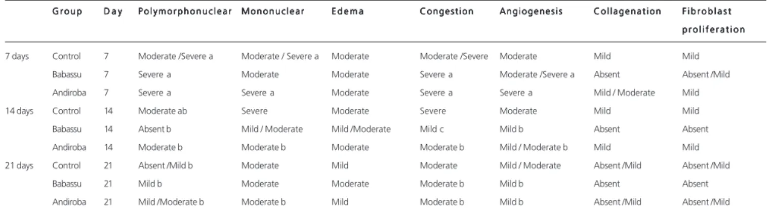
G r o u p D a yD a yD a yD a yD a y P o l y m o r p h o n u c l e a rP o l y m o r p h o n u c l e a rP o l y m o r p h o n u c l e a rP o l y m o r p h o n u c l e a rP o l y m o r p h o n u c l e a r M o n o n u c l e a rM o n o n u c l e a rM o n o n u c l e a rM o n o n u c l e a rM o n o n u c l e a r E d e m aE d e m aE d e m aE d e m aE d e m a C o n g e s t i o nC o n g e s t i o nC o n g e s t i o nC o n g e s t i o nC o n g e s t i o n A n g i o g e n e s i sA n g i o g e n e s i sA n g i o g e n e s i sA n g i o g e n e s i sA n g i o g e n e s i s C o l l a g e n a t i o nC o l l a g e n a t i o nC o l l a g e n a t i o nC o l l a g e n a t i o nC o l l a g e n a t i o n F i b r o b l a s tF i b r o b l a s tF i b r o b l a s tF i b r o b l a s tF i b r o b l a s t p r o l i f e r a t i o n p r o l i f e r a t i o n p r o l i f e r a t i o n p r o l i f e r a t i o n p r o l i f e r a t i o n 7 days Control 7 Moderate /Severe a Moderate / Severe a Moderate Moderate /Severe Moderate Mild Mild
Babassu 7 Severe a Moderate Moderate Severe a Moderate /Severe a Absent Absent /Mild Andiroba 7 Severe a Severe a Moderate Severe a Severe a Mild / Moderate Mild 14 days Control 14 Moderate ab Severe Moderate Severe Moderate Mild Mild Babassu 14 Absent b Mild / Moderate Mild /Moderate Mild c Mild b Absent Absent Andiroba 14 Moderate b Moderate b Moderate Moderate b Mild / Moderate b Mild Mild 21 days Control 21 Absent /Mild b Moderate Mild Moderate Mild / Moderate Absent /Mild Absent /Mild
Babassu 21 Mild b Moderate Moderate Moderate b Mild b Absent Absent Andiroba 21 Mild /Moderate b Moderate b Mild Moderate b Mild b Absent /Mild Absent /Mild
submitting it to seal proof24. It was observed in this
experiment, regarding the average of rupture forces, that in the 7th day there was more pressure on andiroba and
babassu groups when compared with the control group, showing a better healing for babassu and andiroba groups, but without statistical significance. On the 14th day, there
were no significant changes in rupture pressures of surgical wounds. On the 21st day, there was a substantial drop in
pressure averages of the andiroba group, but without statistical significance. The resistance to atmospheric air insufflation test is a good and essential parameter for the evaluation of anastomotic integrity in the first days after surgery, while in the late postoperative period it is best evaluated by linear traction25.
Regarding the site of the organ rupture, it was observed that on the 7th day, in the control group, only one
animal had organ rupture outside the anastomosis; however, in andiroba and babassu groups, four animals had its colon ruptured distant from the anastomosis, a fact different from that reported by Thornton and Barbul26, in which they posit
that only from the second week resistance by anastomosis to the pressure may exceed that of the normal intestinal tissue, and the exhaust gas occurs distant from the anastomosis. On the 14th day, the control and andiroba
groups had similar results, and on the 21st day, the results
were the same for all groups, where four of the animals ruptured the colon outside the suture lines. Santos et al., in their research on the analysis of atmospheric air insufflation test, the rupture of the stomach occurred in all anastomosis22. Likewise, Batista et al. when studying the
effect of aqueous extract of babassu in stomach healing, in no animal, in which it was possible to conduct the tensiometric study, occurred air leakage outside the suture lines27.
Inflammation is essential to healing, characterized by increased vascular permeability, chemotaxis of circulating cells and release of cytokines and growth factors. Neutrophils are the first cells to migrate to the injury, being responsible for the removal of foreign matter and dead tissue. Its maximum action occurs around the second day of healing. Polymorphonuclear, represented by macrophages, reach its highest concentration around the third day. The inflammatory phase extends from the occurrence of lesions to the sixth day of healing. In this study, the animals were put to death on the 7th day, as it
represents the early wound healing phase acute phase -characterized by the presence of edema, congestion and polymorphonuclear. The second phase, known as proliferative, begins around the second or third day, extending up to the 14th day. It is characterized by the
presence of fibroblasts responsible for collagen production, a very important protein for the cell matrix. It is still possible
to find endothelial cells responsible for angiogenesis and myofibroblasts responsible for wound contraction. The third and final phase is characterized by the deposition of collagen in the wound, starting around the 8th day and extending up
to a year and a half.
In attempting to evaluate chronic inflammation parameters (mononuclear cells, angiogenesis, fibroblast proliferation and collagenation), was opted for the death of the other group of animals at the 14th and 21st day after
surgery, as this phase is best evaluated in late periods after postoperative.
When comparing acute inflammatory reaction among the groups, polymorphonuclear was higher in the babassu and andiroba groups in relation to the control group when they are compared on the 7th day, with a
decrease in the last days of evaluation, but without statistical significance. This fact is explained because, in this time, the initial phase of wound healing occurs, also known as inflammatory phase. Andy Petroianu et al., when evaluating the effect of vitamin C and hydrocortisone in the intestinal anastomotic healing, found that in animals subjected only to vitamin and in those who received vitamin and hydrocortisone on the 5th day after surgery there was the presence of
inflammatory infiltrate consisting of polymorphonuclear and vascular congestion28. On the 14th day, the signs of
congestion were more evident in the control group, with a statistical significance.
The signs of chronic inflammation increase over days after injury in detriment of acute signs of inflammation. Mononuclear, representing chronic inflammation, were more pronounced on the 7th day, both in the babassu and
andiroba groups, revealing a possible effect of the aqueous extract of babassu. Baldez, when studying the action of the aqueous extract of babassu in colon healing in rats, showed similar results9.
Experiments show a direct association between the healing efficiency and the number of fibroblasts and collagen fibers, the main structural component of granulation tissue29. It was observed in this study that fibroblast
proliferation, angiogenesis and collagenation were more pronounced on the 7th day after surgery in the babassu and
andiroba groups when compared to the control group; there was a decrease in the 14th and the 21st day. Nunes Jr et
al.30 report that collagen synthesis reaches its peak on the
7th and 14th day, but from the 3rd day it is already possible
to see fibroblasts and collagen in suture areas, fact proved by their experiment babassu using in the healing of the linea alba on the 3rd and 7th days.
R E S U M O R E S U M O R E S U M O R E S U M O R E S U M O
Objetivo: Objetivo: Objetivo: Objetivo:
Objetivo: avaliar o efeito cicatrizante do extrato aquoso do babaçu e do óleo de andiroba em feridas abertas no ceco de ratos. Métodos:
Métodos: Métodos: Métodos:
Métodos: cinquenta e quatro ratos Wistar foram divididos em três grupos de 18: 1) grupo babaçu, com aplicação do extrato aquoso de babaçu; 2) grupo andiroba, com aplicação do óleo; e 3) grupo controle, com aplicação de solução salina. Todos os procedimentos foram feitos por gavagem. Cada grupo foi dividido em três subgrupos de seis animais conforme o período de observação, aos 7, 14 ou 21 dias. De cada animal foi retirado fragmento do ceco com 1,5cm2 de diâmetro. As áreas das lesões
foram analisadas por macroscopia e os segmentos ressecados das feridas por microscopia ótica em colorações de hematoxilina-eosina e tricrômico de Masson. Resultados: Resultados: Resultados: Resultados: foram verificados abscesso e infecção em dois animais do grupo andiroba, e um com Resultados: hematoma. Quanto ao grau de aderências, o grupo babaçu teve maior incidência de aderências grau II enquanto que no grupo controle e andiroba predominaram aderências grau I. Na análise microscópica no sétimo dia a proliferação fibroblástica foi maior no grupo andiroba e menor no grupo babaçu (p=0,028). No 14º dia os polimorfonucleares foram menos acentuados no grupo babaçu (p=0,007). Quanto ao teste de resistência à insuflação de ar atmosférico observou-se que o grupo andiroba em qualquer dos dias avaliados apresentou maior tensão. Quanto à colagenização, no sétimo dia, ela esteve presente em 100% dos animais do grupo andiroba. No 14º dia foi mais acentuada no grupo controle e no 21º dia resultados semelhantes para o grupo controle e andiroba. ConclusãoConclusãoConclusãoConclusãoConclusão: os animais dos grupos babaçu e andiroba apresentaram melhor cicatrização do ceco em comparação ao grupo controle.
Descritores Descritores Descritores Descritores
Descritores: Ratos. Cicatrização. Fitoterapia.
REFERENCES
REFERENCES
REFERENCES
REFERENCES
REFERENCES
1. Campos ACL, Borges-Branco A, Groth AK. Cicatrização de feri-das. ABCD, arq bras cir dig. 2007;20(1):51-8.
2. Isaac C, Ladeira PRS, Rego FMP, Aldunate JCB, Ferreira MC. Pro-cesso de cura das feridas: cicatrização fisiológica. Rev Med. 2010;89(3/4):125-31.
3. Mandelbaum SH, Di Santis EP. Cicatrização: conceitos atuais e recursos auxiliares: parte I. An Bras Dermatol. 2003;78(4):393-410.
4. Silva MI, Ribas Filho JM, Malafaia O, Nassif PAN, Ribas MM, Varaschim M, et al. A utilização da Pfaffia glomerata no processo de cicatrização de feridas da pele. ABCD, arq bras cir dig. 2010;23(4):228-33.
5. Palharin LHDC, Figueiredo Neto E, Camargo-Lopes MP, Bosquê GG. Efeitos fitoterápicos e homeopáticos da babosa. Rev Científ Eletron Agron. 2008;7(14).
6. Brasil. Agência Nacional de Vigilância Sanitária. Resolução - RDC No 14, de 31 de março de 2010. Dispõe sobre os requisitos mínimos
para o registro de medicamentos fitoterápicos. Diário Oficial da União, 5 abr 2010. [Citado 2014 17 nov]. Disponível em: h t t p : / / p o r t a l . a n v i s a . g o v . b r / w p s / w c m / c o n n e c t / e 3 2 1 9 9 0 0 4 2 c f 0 6 e 7 9 b 5 7 d f a f b c 1 8 8 c 8 f / Resolu%C3%A7%C3%A3o+RDC+n%C2%BA+4+de+30+ de+janeiro+de+2014.pdf?MOD=AJPERES.
7. Malafaia O, Campos ACL, Torres O, Goldenberg S. Os fitoterápicos e seu potencial na cicatrização. Acta Cir Bras. 2006;21(3):1. 8. Coelho JM, Antoniolli AB, Silva DN, Carvalho TMMB, Cury-Pontes
ERJ, Odashiro AN. O efeito da sulfadiazina de prata, extrato de ipê roxo e extrato de barbatimão na cicatrização de feridas cutâneas em ratos. Rev Col Bras Cir. 2010;37(1):45-51.
9. Baldez RN. Análise da cicatrização do cólon com uso do extrato aquoso da Orbignya phalerata (Babaçu) em ratos. Acta Cir Bras. 2006;21(2):31-8.
10. Martins NLP, Malafaia O, Ribas Filho JM, Heibel M, Baldez RN, Vasconcelos PRL. Análise comparativa da cicatrização da pele com o uso intraperitoneal de extrato aquoso de Orbignya phalerata (babaçu): estudo controlado em ratos. Acta Cir Bras. 2006;21(3):66-75.
11. Orellana BJP, Kobayashi ES, Lourenço GM. Terapia alternativa através do uso da andiroba. Lato & Sensu. 2004;5(1):136-41. 12. Pereira MRNP, Tonini H. Fenologia da andiroba (Carapaguianensis,
Aubl., meliaceae) no sul do estado de Roraima. Ciênc Florestal. 2012;22(1);47-58.
13. Nayak BS, Kanhai J, Milne DM, Swanston WH, Mayers S, Eversley M. Investigation of the wound healing activity of Carapa guianensis L. (Meliaceae) bark extract in rats using excision, incision and dead space wound models. Larchmont. J Med Food. 2010;13(5). 14. Brito NMB, Silva PRF, Silva GCF, Caselia SFM, Sampaio ARS,
Car-valho RA. Avaliação macroscópica de feridas cutâneas abertas, em ratos, tratadas com óleo de andiroba. Rev para med. 2001;15(2):17-22.
15. Lorenzi H. Árvores brasileiras: manual de identificação e cultivo de plantas arbóreas nativas do Brasil. São Paulo: Instituto Plantarum; 2002.
16. Mendonça AP, Ferraz IDK. Óleo de andiroba: processo tradicional da extração, uso e aspectos sociais no estado do Amazonas, Brasil. Manaus. Rev Acta Amazon. 2007;37(3):353-64.
17. White PF, Johnston RR, Eger EL. Determination of anesthesic requiriment in rats. Anesthesiology. 1974;40(1):52-7.
18. Nair SK, Bhat IK, Aurora AL. Role of proteolytic enzymes in prevention of postoperative intraperitoneal adhesions. Arch Surg. 1974;108:849-53.
19. Garros IC. Campos ACL, Tâmbara EM, Tenório SB, Torres OJM, Agulham MA. Extrato de Passiflora edulis na cicatrização de feri-das cutâneas abertas em ratos: estudo morfológico e histológico. Acta Cir Bras. 2006;21(3):55-65.
20. Santos OJ, Barros Filho AKD, Malafaia O, Ribas Filho JM, Santos RHP, Santos RAP. Schinus Terebinthifolius Raddi (Anacardiaceae) no processo de cicatrização de gastrorrafias em ratos. ABCD, arq bras cir dig. 2012;25(3):140-6.
21. Thornton FJ, Barbul, A. Cicatrização no trato gastrointestinal. In: Barbul A. Cicatrização das feridas. Tradução de Giuseppe Taranto. Rio de Janeiro: Interlivros; 1977.
22. Santos OJ, Malafaia O, Ribas Filho, Marcondes J, Czeczko NG, Santos RHP, et al. Efeito do Schinus Terebinthifolius Raddi (aroeira) e Carapa guianensis aublet (andiroba) na cicatrização de gastrorrafias. ABCD, arq bras cir dig. 2013;26(2):84-91. 23. Nomura LM, Ribas Filho JM, Malafaia O, Dietz UA, Skare TL, Kume
MH. Processo cicatricial de sutura em ceco com os fios polipropilene, poliglecaprone 25 e glicomer 60 em ratos. ABCD, arq bras cir dig. 2009;22(2):82-8.
24. Ballantyne GH. Intestinal suturing: review of the experiment foundations for traditional doctrines. Dis Col Rect. 1983;26(12):836-43.
26. Thornton FJ, Barbul A. Healing in the gastrointestinal tract. Surg Clin of North Am. 1997;77(33):549-73.
27. Batista CP, Torres OJM, Matias JEF, Moreira ATR, Colman D, Lima JHF. Effect of extract of Orbignya phalerata (babassu) in the gastric healing in rats: morphologic and tensiometric study. Acta Cir Bras. 2006;21:26-32.
28. Petroianu A, Rocha CG, Alberti LR, Costa AMC. Rev Col Bras Cir. 2001;28(6):404-7.
29. Nitz AC, Ely JB, d’Acampora AJ, Tames DR, Corrêa BP. Estudo morfométrico no processo de cicatrização de feridas cutâneas em ratos, usando: Coronopu didymus e Calendula officinal. Arq Catarin Med. 2006;35(4):74-9.
30. Nunes Junior JAT, Ribas Filho JM, Malafaia O, Czeczko NG, Inácio CM, Negrão AW. Avaliação do efeito hidroalcoólico de Schinusterebinthifolius Raddi (aroeira) no processo de cicatrização da línea Alba de ratos. Acta Cir Bras. 2006;21(3):8-15.
Received at: 15/03/2015
Accepted for publication: 18/05/2015 Conflict of interest: none.
Source of funding: none.