Ana Isabel Lopes Luís
Stem cells and stroke
2009/20100
Ana Isabel Lopes Luís
Stem cells and stroke
Mestrado Integrado em Medicina
Área: Neurologia
Trabalho efectuado sob a Orientação de:
Prof. Doutor Manuel Joaquim Lopes Vaz da Silva
Acta Neuropathologica
Projecto de Opção do 6º ano - DECLARAÇÃO DE REPRODUÇÃO
Nome: Ana Isabel Lopes Luís
Endereço electrónico: [email protected]
Título da Dissertação/Monografia/Relatório de Estágio: Stem cells and Stroke
Nome completo do Orientador: Prof. Doutor Manuel Joaquim Lopes Vaz da Silva
Nome completo do Co-Orientador: Não aplicável
Ano de conclusão: 2010
Designação da área do projecto de opção: Neurologia
1
Projecto de Opção do 6º ano - DECLARAÇÃO DE INTEGRIDADE
Ana Isabel Lopes Luís
Stem cells and stroke
Faculty of Medicine of the University of Porto (FMUP) Al. Prof. Hernâni Monteiro 4200-319 Porto, Portugal
e-mail: [email protected] / [email protected] Telephone: 00351 964472775
C
ONTENTSAbstract ... 1
Introduction ... 2
Stroke overview, the clinical challenge and rational for cell therapy ... 2
Stem cells: substrate for regeneration ... 6
Definitions and types of stem cells ... 6
Stem cell behavior ... 6
Stem cell applications ... 7
Endogenous stem cells and stroke ... 8
Ischemia-induced neurogenesis ... 10
Neurogenesis in ischemic non-neurogenic brain regions ... 11
Regulation and augmentation of ischemia-induced neurogenesis ... 12
Are there neural stem cells in non-neurogenic regions? ... 14
Effect of age on neuroregeneration ... 14
Is there a role for endogenous bone marrow in neuroregeneration after stoke? ... 15
Putting it briefly… ... 15
Exogenous stem cells ... 16
Introduction to cell transplantation ... 16
Transplantation variables ... 16
Anatomy ... 16
Cell types and transplantation ... 20
Cell transplantation for delivery of trophic molecules ... 24
Clinical trials of stem cells transplantation for stroke patients ... 25
Neuroteratocarcinoma cell trials... 26
Porcine cell trial ... 27
Autologous mesenchymal stem cells... 27
Human fetal cells ... 27
CASE STUDY ... 32
Background ... 32
Methods ... 33
Study population and design ... 33
Main outcome measures ... 34
Statistic analysis ... 34
Ethical considerations ... 35
Results ... 35
Discussion... 46
Conclusion ... 51
Acknowledgements ... 52
References ... 53
F
IGURESL
ISTFigure 1 - Neurogenesis persists in adult mammals throughout life ... 8
Figure 2 - Hypothesis for neural stem-cell development ... 9
Figure 3 - Postulated mechanisms of exogenous stem cell action. ... 23
Figure 4 - Amount of patients admitted to the stroke unit between 1st September and 31st December 2008 ... 36
T
ABLES LIST Table 1. Current and investigational reperfusion strategies in Acute Ischemic Stroke [93] ... 4Table 2 - Synopsis clinical studies involving stem-cells after stroke ... 25
Table 3 - Planned clinical trials using stem cells in stroke patients ... 29
Table 4 - Exclusion criteria for the treatment of acute ischemic stroke with recombinant tissue plasminogen activator (alteplase) within 3 hours of stroke onset [2]... 35
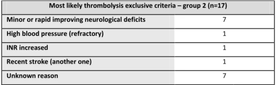
Table 5 - Potential reasons not to perform thrombolysis on group 2 patients ... 36
Table 6 - Demographic and clinical characteristics of Ischemic Stroke Patients ... 37
Table 7 - Demographic and clinical characteristics of Hemorrhagic Stroke Patients ... 38
Table 8 - Baseline Characteristics of the patients ... 39
Table 9 - Classification of subtypes of acute Ischemic Strokes ... 39
Table 10 - Hemorrhagic stroke subtypes and etiology ... 40
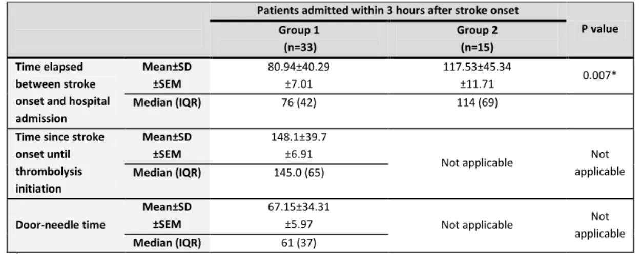
Table 11 - Time since stroke onset until thrombolysis initiation and door-needle time ... 40
Table 12 - Ischemic stroke adverse events... 41
Table 13 - NIHSS and mRS scores after 2-4 months of follow-up among the 4 groups of ischemic stroke patients ... 42
Table 14 - Distribution of scores on the Modified Rankin Scale. ... 42
Table 15 - NIHSS variation at 2-4 months follow-up. ... 43
G
RAPHICSL
IST Graphic 1 - Distribution of scores on the Modified Rankin Scale. ... 43Graphic 2 - Mean±SEM NIHSS reduction/improvement at 2-4 months follow-up among patients who were admitted within 3 hours after stroke onset at the hospital. ... 44
1
A
BBREVIATIONSANOVA One-way analysis of variance
BDNF Brain-derived neurotrophic factor
bFGF Basic fibroblast growth factor
BrdU 5-bromo-29-deoxyuridine-59-monophosphate immunohistochemistry
cDNA Complementary deoxyribonucleic acid
DA Dopaminergic neurons
DCX Doublecortin
DG Dentate gyrus
ECASS III European Cooperative Acute Stroke Study III
EGF Epidermal growth factor
EPE Entidades Públicas Empresariais
EPO Erythropoietin
ESC Embryonic stem cell
ESO European Stroke Organization
ESS European Stroke Scale
EU European Union
FGF-2 Fibroblast growth factor-2
FISH Fluorescent in situ hybridization
G-CSF Granulocyte colony-stimulating factor
GDNF Glia cell line-derived neurotrophic factor
GDNF-hMSCs Glia cell line-derived neurotrophic factor gene modified human marrow stromal cells
GFAP Glial fibrillary acidic protein
GFP Green fluorescent protein
GZ Granular zone
hATSCs Human adipose tissue stromal cells
hMSCs Human marrow stromal cells
IAT Intra-arterial thrombolysis
ICH Intracerebral hemorrhage
i.e. id est
IGF-1 Insulin-like growth factor-1
INR International normalized ratio
iPS Induced pluripotent stem cells
IV Intravenous
LACI Lacunar infarct
LGE Lateral ganglionic eminence
MCA Middle cerebral arterial
MCAO Middle cerebral artery occlusion
mRS Modified Rankin scale
MSCs Mesenchymal stem cells
NIHSS National Institutes of Health Stroke Scale
NINCDS National Institute of Neurological and Communicative Disorders and Stroke
NINDS National Institute of Neurological Disorders and Stroke
NO Nitric oxide
NSCs Neural stem cells
2
OB Olfactory bulb
OCSP Oxfordshire Community Stroke Project
OPCs Oligodendrocyte precursor cells
PACI Partial anterior circulation infarct
PIGF Placental growth factor
PIGF-hMSCs Human marrow stromal cells placental transfected with adenoviral vectors carrying placental growth factor gene
POCI Posterior circulation infarct
PSA-NCAM Poly-sialated neural cell adhesion molecule
RMS Rostral migratory stream
rt-PA Recombinant tissue plasminogen activator
SAM Sistema de Apoio ao Médico/Medical Support System
SCF Stem cell factor
SGZ Subgranular zone
SITS Safe Implementation of Treatments in Stroke
SVZ Subventricular zone
TACI Total anterior circulation infarct
TOAST Trial of Org 10172 in Acute Stroke Treatment
US United States
VEGF Vascular endothelial growth factor
1
A
B S T R A C TStroke is a major cause of morbidity and mortality worldwide, heading the causes of disability and representing
the third leading cause of death in the western world. Yet, apart from thrombolysis in the acute phase of
ischemic stroke, no effective therapy is currently available to promote recovery and reduce the heavy burden
stroke represents. In the background of this imperative clinical need, endogenous and exogenous stem cells
have been studied as a therapeutic option following stroke. This arising strategy is briefly reviewed in this
article, pointing out some of the knowledge acquired in basic research, pre-clinical studies and in the few
developed clinical trials. In order to appraise the potential clinical necessity of alternative therapeutic
strategies, such as stem cell therapy, in a close scenario, a retrospective and observational study was
conducted in a stroke unit of a University Hospital, at Oporto, attempting to identify and characterize stroke
patients admitted within a quadrimester, their baseline characteristics, type of treatment received and
neurological and functional evolution at 2 to 4 months follow-up. A total of 136 patients were studied, 77.2%
with ischemic and 22.8% with hemorrhagic stroke. Among 51 patients admitted within the therapeutic window
(approved at the time), 34 were submitted to thrombolysis. The sooner thrombolysis was given to ischemic
stroke patients the greater seemed to be the benefit.
2
I
N T R O D U C T I O NStroke overview, the clinical challenge and rational for cell therapy
The generic term stroke refers to the sudden impairment of brain function caused by a variety of pathologic
changes involving one (focal) or several (multifocal) cervicocerebral blood vessels [135]. The majority of strokes
are ischemic (87%), with the remaining being result of intracerebral (10%) and subarachnoid hemorrhage (3%)
[75].
Stroke is a major cause of morbidity and mortality worldwide, heading the causes of disability and representing
the third cause of death, in the western world, behind heart disease and cancer [11,23,36,41,66,74-76]. Of all
stroke survivors, 30%–50% do not regain functional independence and 15%–30% are permanently disabled (i.e., not able to walk, talk clearly, or feed themselves with a favored hand), a devastating reality to both
patients and carers [36,66,93]. It is as well the second most common cause of dementia, the most frequent
cause of epilepsy in the elderly, and a frequent cause of depression [104].
Stroke also represents a substantial economical burden, with stroke care estimated to cost more than 5% of
many countries’ healthcare budgets [101]. For instance, in 2006, in the European Union (EU), the overall cost of
stroke was around €38.1 billion [103], and the expected direct and indirect cost of stroke for 2010, in the
United States (US), is $73.7 billion [75].
In the US, each year around 795 000 people experience a new or recurrent stroke. About 610 000 of these are
first attacks, and 185 000 are recurrent attacks. Approximately 1 in 4 people dies within 1 year after having an
initial stroke and mortality data from 2006 revealed that stroke accounted for approximately 1 of every 18
deaths in the US. This means that on average, every 40 seconds, someone in the US has a stroke, and every 4
minutes someone dies of it [75].
Annually, also in the US, around 55 000 more women than men have a stroke. As a matter of fact, the stroke
incidence rate is higher for men compared with women at younger ages, but the scenario changes as they grow
older. The male-to-female incidence ratio was 1.25 in those 55 to 64 years of age, 1.50 in those 65 to 74 years
of age, 1.07 in those 75 to 84 years of age, and 0.76 in those ≥85 years of age [75].
There are also noticeable racial differences in stroke incidence. In fact, blacks have a risk of first-ever stroke
that is almost twice that of whites. The age-adjusted stroke incidence rates in people 45 to 84 years of age are
6.6 per 1000 population in black men, 3.6 in white men, 4.9 in black women, and 2.3 in white women [75].
In Europe, each year, stroke accounts for 1.24 million deaths (among 900 million total estimated population):
over 1 in 6 women (approximately 17%) and over 1 in 10 men (around 11%) dying from the disease [103]. Large
differences in incidence, prevalence and mortality have been reported between Eastern and Western European
countries. This has been attributed to difference in risk factors, with higher levels of hypertension and other
risk factors resulting in more severe stroke in Eastern Europe. But even within Western Europe notable regional
variations have also been found. Yet, stroke in Europe is also the most important cause of morbidity and
serious, long-term disability, and demographic changes will result in an increase in both incidence and
3 In truth, the population in Europe is aging rapidly. In 2004, 13.7% of the European population was aged 65
years or older which was twice the world level (with the higher percentages in Italy, 18.9%, Germany, 18.0%,
Greece, 18.0%, and Sweden, 17.2%) (82). Besides, the percentage of people aged 65 or older is expected to
double to about 30% in 2050 [112]. This trend, also observed in the US, at a shorter term [23], seems to reflect
the longer life expectancy in Western countries [112]. In 2006, the European average life expectancy at birth
was 75.1 years, and 78.6 for the 25 Members States of the European Union. The US life expectancy in 2007 was
77.9 years (80.4 years to women and 75.3 years to men) [86], continuing a long-term increasing trend [46]. Life
expectancy at age 65 was 18.6 years in 2007, an increase of 6 percent since 2000 [86].
With age, the risk of stroke increases steeply. It more than doubles each decade of life over the age 55. So, as
the elderly segment of the population continues to rapidly grow, substantial advances in the prevention and
treatment of stroke are of paramount relevance [23].
Despite more than four decades of intense investigations, no therapy that significantly prevents stroke-induced
brain damage and neurological dysfunction has emerged [74].
Recombinant tissue plasminogen activator (rt-PA), a thrombolytic agent, is the only currently available
intervention to reduce the size of the cerebral infarct. Classically, the thrombolytic therapy with rt-PA has been
approved for use only if administered within 3 hours of the onset of ischemia, being this limit recommended as
a standard in all international stroke treatment guidelines until 2008 [23,126]. However, since January 2009,
according to the European Stroke Organization (ESO), intravenous rt-PA is recommended within 4.5 hours of
onset of ischemic stroke [104], on the basis of the finding of the European Cooperative Acute Stroke Study III
(ECASS III) showing a modest, but significant benefit of intravenous alteplase given 3 to 4.5 hours after the
onset of symptoms in ischemic stroke patients [44]; and on the basis of the data from Safe Implementation of
Treatments in Stroke (SITS) centers revealing that alteplase remains safe when given at that period (3-4.5 hours
after the onset of symptoms), offering an opportunity for patients who cannot be treated within the standard 3
hours frametime [126]. These studies lend support to those suggesting a potentially longer timeframe for
intravenous thrombolysis [93]. Nonetheless, treatment benefit is time-dependent, with the best results
observed if given within 90 minutes [23,44]. So, to maximize the benefit, patients should be treated with
alteplase as early as possible (“time is brain”) [44].
Unfortunately, due to the narrow time window as well as a number of contraindications, rt-PA therapy has
been restricted to a small proportion of patients evaluated in the emergency room (about 5% of stroke
victims). Of those who receive it, rt-PA may be expected to yield around a 30% increase in the number of
patients avoiding long-term neurologic deficits [23].
Aspirin has a wide utility but modest efficacy and anticoagulation has proven ineffective not being
recommended for the treatment of patients with acute ischemic stroke [36,104].
Advances in endovascular stroke therapy have come from different directions, including pharmacologic and
mechanical [93,115] (table 1), and they may improve reperfusion sufficiently to improve survival, but these
have yet to be substantially confirmed by randomized clinical trials [19,23,69,94]. Nevertheless, physiologic
4
Table 1. Current and investigational reperfusion strategies in Acute Ischemic Stroke [93]
Reperfusion approaches
Recanalization or antegrade
Intravenous (IV) and/or Intra-arterial thrombolysis (IAT)
Endovascular thrombectomy
Endovascular thromboaspiration
Mechanical thrombus disruption
Transcranial or endovascular augmented fibrinolysis
Endovascular thrombus entrapment
Temporary endovascular bypass
Alternative
Global reperfusion (flow augmentation or transarterial retrograde reperfusion)
Transvenous retrograde reperfusion (flow reversal)
Adapted from Nogueira, et al, 2009
Unfortunately, although a plethora of neuroprotective compounds have shown promise in animal models, no
neuroprotection intervention has shown improved outcome. Various neuroprotection approaches remain
under investigation [23,36].
Regardless of increasing focus on evidence-based primary and secondary prevention, strokes still occur. And,
even with optimal stroke unit care (including thrombolysis) fewer than one in three patients recover fully from
stroke [104]. Thus, rehabilitation is necessary to optimize functional recovery in the remainder [135]. Although
there is lack of robust evidence for several of the common interventions employed in post-stroke
rehabilitation, there is expert consensus in favor of it, as a way to minimize stroke impairment and to reach and
maintain optimal physical, intellectual, psychological and/or social function [101,104]. Sadly, once recovery has
reached a plateau and the neurological deficits are fixed, there is no known effective treatment [135].
Intracerebral hemorrhage (ICH), for which no effective treatment strategy is currently available, constitutes
one of the most devastating forms of stroke [8-9,81]. The major risk factor for ICH is arterial hypertension,
accounting and contributing for about 60% of cases. Hemorrhagic stroke prognosis is poor, with an overall
mortality rate of about 40% at 1 month and most of the survivors are left with persistent, severe neurological
deficits. Indeed, it is estimated that 90% of surviving patients are dependent on a caregiver at 1 month, and
80% at 6 months. The available therapy is mainly supportive, including maintenance of homeostasis and
treatment of brain edema [8-9]. Hematoma growth is frequent and it represents an independent determinant
of death and disability [81]. Given the prognostic significance of hematoma expansion and continuous bleeding,
therapies aiming to prevent it, through the administration of haemostatic agents, seem an attractive approach
[8]. However, it was observed that the promising haemostatic therapy with recombinant activated factor VII
(FAST study) reduces growth of the hematoma, but does not improve survival or functional outcome after ICH [81].
In selected patients with space-occupying hematomas, surgery may relive the mass effect, but the indication to
5 once the bleeding has occurred, and no effective treatment for improving the outcome, other than
neurological rehabilitation, is currently available [8].
Given the incapability to efficiently mitigate the devastating effects of stroke, it is vital that novel therapeutic
strategies be developed to both minimize the initial neural trauma, as well as repair the damage brain once the
pathological cascade of stroke has run its course [16].
In the background of this imperative clinical need, numerous studies have been published investigating the
potential application of endogenous and exogenous stem cell therapies for treatment of stroke. In addition to
their potential for generating a variety of new functional cell types, stem cells have the ability to respond
actively to their environment, migrate to areas of injury, and secrete neuroprotective compounds. These
properties may provide them therapeutical potential both in the acute phase and at later time points after
conventional medical approaches would no longer be effective [23].
Objectives:
This work major aim is to briefly review the potential application of endogenous and exogenous stem cell
therapies for treatment of stroke. This work will also include, as a second part, but a related one, a
retrospective and observational study set to identify and characterize the patients admitted in the Stroke Unit
of S. João Hospital EPE, Porto, Portugal, since September 2008 until December 2008, namely the remaining
disability and its possible relationship with treatment approach. Based on this studied scenario, it is intended to
appraise the clinical need of alternative therapeutic strategies and thus support the discussion of stem cell
therapy in stroke.
Review Methods
In relation to search strategy and selection criteria used on this review, five data bases were consulted:
Pubmed; Isi Web of Knowledge; Science Direct; Scopus and Scirus, using the following query: (stroke OR CVA
OR "Intracranial Embolism and Thrombosis" OR "cerebral infarction" OR "brain infarction" OR "Intracranial
Hemorrhages" OR "cerebrovascular disorders") AND (stem cell OR "Progenitor Cells") and no specified limits
were added. The first approach was article selection by title and/or abstract, including the potential relevant
ones. Preference was given to the most recent publications, commonly referenced and highly regarded older
publications. It was also searched the reference list of articles included and often further searches were made
6
S
T E M C E L L S:
S U B S T R A T E F O R R E G E N E R A T I O NDefinitions and types of stem cells
The definition of a stem cell continues to evolve as more knowledge is achieved on the subject [36]. Stem cells
have two main defining features: multipotency and self-renewal [3,18,23,34,36,42,137-138]. Multipotency is
the capability to differentiate into several morphological and functional cell types [4,23,53]. As a result, stem
cells should be able to functionally reconstitute appropriate tissues in vivo [4]. Self-renewal refers to the ability
of stem cells to make identical copies of themselves [4,23,53].
A renewing cell division must give rise to at least one daughter cell with the same differentiation and
self-renewal potential as the parent cell. Stem cells display extensive proliferation capacity and are often supposed
to be able to divide indefinitely, yielding a virtually unrestricted resource of cells [23].
A multiplicity of stem cells can be identified differing on their potency or on the variety of cell types they can
give rise to [23]. The best example of potency is the fertilized human egg. In fact, the zygote is a totipotent cell
that generates embryonic and extraembryonic tissues. Nevertheless, being a transitory cell, that does not
self-renew, it is not usually regarded as a stem cell. Embryonic stem cells (ESCs), isolated from the inner cell mass of
the blastocyst, have the widest potential of any true stem cell. These cells are pluripotent, which implies that
they are capable of giving rise to all cell types within the developing embryo [3,23,48].
Outstandingly, there are also a large number of stem cells in the adult mammal. However, this adult stem cells
are tissue-specific, meaning that they are capable of generating certain local cell types, but not those from
unrelated tissues [23]. Often they are referred as multipotent cells [3]. The hematopoietic stem cell, which can
give rise to all blood cell types, is the best well-known tissue-specific stem cell. But tissue-specific stem cells
have been identified in several organs (heart, muscle, skin, gut, liver, pancreas and brain) [23]. These resident
stem cells provide a continual source for a physiological organ-specific cell replacement during normal cell
turnover, as well as they may contribute to regeneration in disease/injury conditions [42].
Remarkably, recently it was demonstrated that is possible the induction of pluripotent stem (iPS) cells from
terminally differentiated somatic cells via nuclear reprogramming with retroviral vectors containing
complementary deoxyribonucleic acid (cDNA) encoding genes known to be associated with the ESC state [23].
Stem cell behavior
True self-renewing stem cells usually divide uncommonly when in vivo, possibly as a way to protect the
integrity of their genetic material, since continuous proliferation increases the mutation risk and subsequently
the risk of tumorgenesis [23].
To generate enough cells for in vivo regeneration while minimize the stem cell proliferation, adult stem cells go
through asymmetric self-renewing divisions, giving rise to one stem cell and one committed progenitor cell,
which can proliferate rapidly and thus generate a large number of more differentiated progeny [23].
Frequently, progenitor cells are unipotent, differentiating into only one differentiated cell type, though keeping
7 For a long time, it has been conceived that stem and progenitor cells can either self-renew or generate more
restricted cell daughters, but cannot move backward along this development sequence or “dedifferentiate”.
However, it has been suggested that, under certain conditions, some adult stem cells have the ability to give
rise to cell types from unrelated organs, a phenomenon labeled transdifferentiation. It remains uncertain
whether those rare cells with greater potency may actually exist in vivo, or whether this event represents an
artifact of in vitro cultures conditions. Besides, some of the first cases initially reported to be in vivo
transdifferentiation, where latter confronted with the demonstration that some cell types may fuse with
mature cells from unrelated organs. Others have posited that specified stem cells may have the capacity to
dedifferentiate [23].
Stem cell applications
Cell replacement therapy, i.e., the replacement of cell types lost as a result of disease or injury is the most
commonly discussed role for stem cells. This replacement may be accomplished either by in vitro
predifferentiation of stem cells into the desired cell type followed by transplantation to the affected area, or by
direct transplantation of stem cells followed by spontaneous in vivo differentiation of stem cells into the
needed cell types [23].
In conditions, such as Parkinson’s disease or diabetes mellitus type 1, where restricted populations are
selectively lost (dopaminergic cells in the brain and beta cells in the pancreas) and function may be restored by
replacement of cells with similar properties, the transplantation of predifferentiated cells may be particularly
useful. In contrast, in stroke, where multiple cell types are lost and thus different cell types are needed to
restore function to a broadly damaged area, the use of undifferentiated cells may be of better value [23,47]. As
a matter of fact, usually strokes affect not only different neuronal phenotypes, but also astrocytes,
oligodendrocytes, endothelial cells [47,49,110]. Thus reconstitution of the complex and widespread
neuronal-glial-endothelial interrelationships may require access to a broader array of lineage species than more
committed phenotypes. Hence, cells for transplant may need to initially remain immature and phenotypically
plastic [110]. Another distinctive characteristic of stroke is that the injury process is acute and limited in time
and there is no ongoing degenerative process or major immunological attack. Therefore the brain may be more
hospitable to transplantation [47].
Another strategy to promote cell replacement involves the mobilization of resident tissue-specific stem cells to
provide the substrate for cell replacement [23].
Although spontaneous neurogenesis may occur in certain brain regions in response to brain injury it is not
sufficient to allow functional recovery. So, different approaches have been chased in order to magnify this
endogenous regenerative response [23].
Besides cell replacement therapy, stem cells appear to serve supportive roles. For instance, some stem cells
show evidence of robust tropism for injury. Although in an injured area most resident cells may die, other stem
cells seems to be attracted to that area of injury and may secrete molecules that promote survival and
regeneration. Stem cells may as well be recruited to tumors, where they are related with decrease neoplasms
8
Stem cells’ tropism to injured areas has interesting some groups that aim to use stem cells genetically
engineered to deliver therapeutic compounds specifically to the area of injury. This approach could play an
important role for ischemic stroke, in which compromised blood flow may make delivery of drugs to the
affected region more challenging after systemic administration [23].
E
N D O G E N O U S S T E M C E L L S A N D S T R O K ENeural stem cells
After decades of dogma stating that “the brain does not regenerate” and no new neurons are born in the adult
mammalian brain it is now largely confirmed that neural stem cells (NSCs) are actually present throughout life
and that spontaneous adult neurogenesis occurs primarily in three brain regions: the subgranular zone (SGZ) of
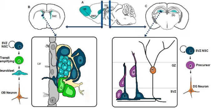
the hippocampal dentate gyrus (DG), the subventricular zone (SVZ) of the lateral ventricles and the posterior
periventricular area (Fig.1) [23,36,74,132]. Although these three are the most well established neurogenic
regions of adult mammalian brain, some studies indicated the presence of minute amounts of NSCs in other
brain areas, as striatum, spinal cord [133] and neocortex [76,97,132].
Figure 1 - Neurogenesis persists in adult mammals throughout life
a: the parasagittal section of a mouse brain demonstrates the brain regions involved in rodent adult neurogenesis (red). b,c: coronal sections at the approximate regions of the dashed lines. b: the SVZ, where neural stem cells persist throughout life. Slowly dividing type B cells (expressing glia fibrillary acidic protein – GFAP) give rise to rapidly dividing type C cells (that serve to increase the number of progeny), which in turn give rise to migratory neuroblasts (type A cells). These reach the olfactory bulb (OB) via the rostral migratory stream (RMS), and there preferentially differentiate into granule interneurons (that functionally integrate into local circuitry) and into dopamine periglomerular neurons. c: the dentate gyrus, where new neurons are also born throughout life. Type B cells in the subgranular zone (SGZ) give rise to precursor cells, which migrate up the radial projection of type B cells to become granule neurons in the granular zone (GZ). Adapted from Burns, et al, 2009[31]
NSCs can be segregated from the adult brain and then cultured in vitro, in the presence of basic fibroblast
growth factor (bFGF) and epidermal growth factor (EGF). In vitro, NSCs form cellular clusters named
9
vivo research revealed that NSCs have characteristics of astrocytes and that they are at the end of a
development continuum that starts with neuroepithelial cells in the neural tube, followed by radial glia that
function both as parent and radial migratory guide for newly born neurons in embryonic brain. Toward the end
of development, radial glia disappears in most parts of the brain, but some persist as multipotent astrocytes
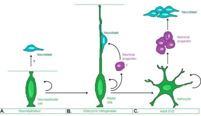
confined mainly to the adult brain regions mentioned above, where they are known thereafter as NSCs (Fig.2)
[23,132].
Figure 2 - Hypothesis for neural stem-cell development
A. Neural stem cells (green) in the early neuroepithelium expand from the ventricle to the pia. B. Radial glia (green) is thought to be a neural stem cell, possibly an elongated form of the neuroepithelial cell. These cells are known to divide symmetrically or asymmetrically to produce neurons (blue) that migrate into the cortex along the fibre of their progenitor. Radial glia may produce neurons directly or indirectly through transit amplifying cell types (violet). C. Later in the development, radial glia transform into cortical astrocytes. Cells derived from radial glia may come to reside in the adult subventricular zone (SVZ) (blue), these astrocyte-like cells behave as stem cells, since they self-renew and produce neurons, probably through intermediate cell types (violet). Adapted from Alvarez-Buylla et al 2001[6].
In the SVZ of the adult brain, NSCs are found nearby to a layer of ependymal cells that line the lateral
ventricles. This area has four different types of cells: type A, B, C and E. Of these, the most numerous are the
glial fibrillary acidic protein (GFAP)-positive type B cells, which are considered as the primary adult neural stem
cells. Type B cells are the astrocytes of the SVZ and form chains of young migratory neuroblasts, designated as
A cells, that migrate to the olfactory bulb (OB) through the rostral migratory stream (RMS), where they
differentiate into olfactory interneurons. The highly proliferative type C cells are at the base of the migratory
chain of A cells and are believed to be immature precursors, the intermediaries between B and A cells. Type E
cells correspond to ependymal cells [74,132]. It is interesting to realize that the neuroblast migration occurs
even in the absence of olfactory bulb, suggesting that the neural cell migration is not target mediated, but
10 In the dentate gyrus and the posterior periventricular area NSCs have quite limited self-renewing capacity
when compared to the NSCs of the SVZ. Besides, while the SVZ have four different cell types, DG is thought to
contain only one type of progenitor cells [74]. In DG, neural progenitors are situated nearby the hilus in the
SGZ, where they continuously proliferate, and then migrate to the granule cell layer, differentiate into mature
neurons and extend axonal projections into the Ammon’s horn of the CA3 area [74,132].
The subependymal layer of posterior periventricular area surrounding the hippocampus is thought to contain
the true stem cells that replenish the hippocampal neurons. Indeed, it was demonstrated that the newly
proliferated progenitors migrate and repopulate the hippocampal CA1 after ischemic cell death [132].
Knowing that the adult brain homes stem cells with neurogenic potential brings up the thrilling possibility of
exploiting them as a way to restore neurons and glia both lost following stroke [23,99]. However, in a normal
adult brain, neurogenesis, defined as the generation of new neurons, occurs in a limited capacity. Around 9000
new neuronal cells (0.1% of the granule cell population) are generated in the DG of mice per day [74].
Even though increased proliferation of NSCs frequently leads to augmented neurogenesis, it is not a direct
measure of neurogenesis, as differentiation of the newly born cells to a neural lineage is also a requisite. In the
same way, increased neurogenesis does not necessarily means that there will be an augment in the net
number of functional neurons. For that to happen newly born neurons must first survive, which usually is
associated with a successful synaptic integration into brain circuitry [23].
It appears that the major function of neurogenesis in the adult brain is to replace the neurons lost regularly in
certain brain areas. But then again, the resident neural progenitors can also be the emergency supply that
could be induced to replace neurons lost due to acute insults. It is fascinating to notice that a lot of conditions
may be capable of affecting neurogenesis in the adult brain, upregulating it (stroke, positive life experiences,
for instance, exercise, learning, enriched environment, caloric restriction) or downregulating it (negative life
experiences, such as stress, drug abuse, irradiation, hight-fat diet, diabetes, inflammation…) [91,132].
Ischemia-induced neurogenesis
Models of brain ischemia
Quite a few rodent models of brain ischemia were created. To generate “global ischemia” it can be used
transient bilateral carotid artery stenosis, which induces selective cell types destruction, such as CA1 pyramidal
cells in the hippocampus [23]. Nevertheless, most often the ischemic models are focal, generally involving
occlusion of the middle cerebral artery (MCAO). Differently from global ischemia, during focal ischemia blood
perfusion is only blocked to specific regions of brain. The MCAO model results in damage mostly to striatum
and, in a minor part, to the overlying cortex [23,76,132]. In order to create selective cortical lesions it can be
used occlusion distal to the striatal branches [23].
When aiming a permanent occlusion it can be used ligation, cauterization or laser-induced photothrombosis. If
a transient occlusion is intended to model an ischemia-reperfusion injury, it can be used an intraluminal
filament through the carotid artery into the proximal middle cerebral artery (MCA) [23]. When this transient
occlusion is shorter than half an hour the damage preferentially affects the striatal tissue, but if the occlusion
11
Increased neurogenesis in ischemic neurogenic regions
In 1998, a pioneering study by Liu et al [73], reported, for the first time, increased neurogenesis in the DG after
global ischemia. In order to determine whether ischemia affects neurogenesis, newly divided cells in the DG
were examined after transient global ischemia in adult gerbils. A 12-fold increase in DG cell birth was observed
1-2 weeks after 10 minutes of global ischemia (with bilateral common carotid artery occlusions), using
5-bromo-29-deoxyuridine-59-monophosphate (BrdU) immunohistochemistry. It was also demonstrated that
newborn cells differentiate into a neural phenotype by 26 days after the onset of ischemia and are primarily
located in the granule cell layer at 7 months post-ischemia. As ischemic preconditioning (which protects CA1
neurons from subsequent ischemic damage) did not alter the level of increased neurogenesis, neurogenesis in
this case was not attributable to actual neural loss. Thus, the level of progenitor proliferation in SGZ, although
affected by the duration of global ischemia, is not influenced by the intensity of CA1 cell death [73].
Afterwards, several studies confirmed that global cerebral ischemia stimulates neurogenesis in the DG of adult
rodents [58], monkeys and other species [74,122]. Strikingly, stroke induced neurogenesis has recently been
observed in the adult human brain [57].
Increased neurogenesis has also been reported in the SVZ and posterior periventricular areas following global
ischemia [23,52,74,76,90,132]. The newly generated progenitors in the SVZ, after global ischemia, express
many immature neural markers including highly poly-sialated neural cell adhesion molecule (PSA-NCAM),
doublecortin (DCX), nestin and β-III tubulin, and migrate along the RMS to the olfactory bulb [52,122].
Using models of focal ischemia it was also reported a significantly enhanced progenitor proliferation in the SVZ
as well as in the DG [58,98,136]. In fact, different groups observed that, after transient focal unilateral ischemia
enhanced proliferation begins bilaterally in both SVZ and SGZ as early as 2 days, reaches the peak at 1-2 weeks
and declines to the basal levels by the 3-4 week of reperfusion [33,58,132,136].
The degree of post-ischemic progenitor proliferation was shown to be influenced by the duration of MCAO,
with a 120 minutes occlusion inducing more proliferation than a 30 min MCAO [10].
Neurogenesis in ischemic non-neurogenic brain regions
Differently from the DG where there is no known evidence of migration of NSCs from the subgranular zone out
of the DG to restore other brain areas [56], in 2002, for the first time, it was observed, in a rat model of
transient MACO [98], that neuroblasts from an expanded ipsilateral SVZ are activated after ischemia, deviate
from their normal route, toward the olfactory bulb (via RMS), and migrate in chains toward the ischemic
penumbra of striatum and cerebral cortex. It was also observed that some of these neuroblasts persisted in
damaged neostriatum and with time differentiate into cells with neuronal morphologies, that expressed
markers of matures striatal medium spiny neurons. However, the functional integration of newly generated
neurons in the neostriatum was not shown [98].
Some previous studies had reported post-ischemic neurogenesis in the cerebral cortex following focal ischemia
[39,54]. However, the origin of the neurons present in the cortex is being debated [132], with some stating that
they could correspond to apoptotic neurons, instead of new born neurons [23]. A subsequent study showed
12 cortical stream, into the penumbra of ischemic cortex. But survival and maturation of these cells at later time
points was not investigated [56].
Currently it is accepted that changes in the SVZ leading to striatal neurogenesis following ischemic injury persist
long term, and new neurons continue to be added to the striatum for at least several months [70,121] or even
for a year [63] after stroke.
Nakatomi et al. [90] showed that the newly proliferated progenitors migrate from the posterior periventricular
area and repopulate the hippocampal CA1 following ischemic cell death. And later it was demonstrated that
this phenomena leads to recovery of spatial learning and memory function in adult rats. In fact, so far this study
was the most convincingly showing functional integration of the new neurons in the adult brain following
transient global ischemia. The great level of neuroregeneration shown in this study permitted prospective
labeling of periventricular NSCs with a green fluorescent protein (GFP) transfered by lentivirus, allowing direct
demonstration of the functionality of the new neurons via electrophysiological studies [23,132].
Regulation and augmentation of ischemia-induced neurogenesis
Presently it is widely accepted that neurogenesis occurs in response to ischemic lesions. However, it also seems
clear, that this limited response needs to be potentiated in order to achieve a significant level of regeneration.
In fact, differently from what happens with amphibians and reptiles that present a robust regeneration of
central nervous system after injury, achieving a post-injury brain that is structurally and functionally
comparable to the pre-lesion state, it remains uncertain whether mammalian baseline levels of stroke-induced
neuroregeneration is of any functional value at all. Some claim that this scarce neuroregenerative capacity is
the price paid for increased complexity of mammalian brain. More and more it has been shown that some
physiological stimuli, endogenous molecules and exogenous agents are capable of regulate adult neurogenesis
[23]. The significance of some of these factors will be briefly indicated, since no exhaustive review on this topic
is intended.
Factors that modulate post-ischemic neurogenesis
As previously mentioned, increased neurogenesis is reported not only in the ipsilateral DG and SVZ, but also
contralaterally, what suggests that diffusible factors may play a role in promoting post-ischemic proliferation.
Several diffusible mitogens including growth factors, cytokines and cell division modulators are known to be
upregulated during brain ischemia [132]. Some biologic characteristics of these diffusible factors will be briefly
reviewed.
Epidermal growth factor (EGF) and fibroblast growth factor-2 (FGF-2) arefactors that expand the stem cell pool, and thus appear to be useful to provide an adequate cellular substrate for neuroregenerative response.
Both growth factors are known to play a significant role in neurogenesis in vivo and regarded as essential to
preserve the pluripotence of neural stem cells cultures in vitro [132]. After cerebral ischemia the FGF-2
expression suffers a notable increase and it was shown that the FGF-2 knockout mice present an attenuation of
the neurogenesis induced by focal ischemia [132]. Leker et al [70] showed that adenovirus programmed to
13
accompanied by continuous improvement in behavior, suggesting FGF-2’s role in long-term neurological repair.
EGF may have an analogous effect [132]. Infusion of a combination of EGF and FGF-2 was shown to increase
neuroproliferative potential in the posterior periventricular area following global ischemia [90] and in the DG
and SVZ after focal ischemia [124]. The amplified progenitor proliferation in rats infused with both FGF-2 and
EGF was also shown to be associated with improved postischemic memory formation and retention [90].
Brain-derived neurotrophic factor (BDNF), glial cell line-derived neurotrophic factor (GDNF) and the stem cell factor (SCF) are endogenous substances which synthesis is induced by transient focal ischemia [132]. Also, it was demonstrated that BDNF promotes neurogenesis, leading to increased numbers of neurons in the
olfactory bulb and promoting the appearance of new neurons in the striatum [23]. The infusion of GDNF into
lateral ventricles after MCAO in the adult rats revealed a considerably enhance in the neurogenesis both in SVZ
and DG [33]. SCF is known to be a major player in the embryonic neurogenesis and it was demonstrated to be
upregulated in hypoxia-treated cultures of embryonic cells from mouse cerebral cortex [132]. Jin et al. showed
that SCF significantly stimulated BrdU uptake into cultured stem cells and when cultures were exposed to
hypoxia conditioned medium containing SCF antibodies, BrdU labeling was reduced. Moreover, SCF infusion
enhanced the BrdU incorporation in SVZ and SGZ and cells in which SCF stimulates BrdU incorporation were of
neuronal lineage [55].
Erythropoietin (EPO) and vascular endothelial growth factor (VEGF) promote the differentiation of bone-marrow stem cells into circulating mature red cells [76]. In adition, EPO is an important mitogen that is
upregulated in the SVZ following ischemia and promotes post-ischemic neurogenesis [23,132]. And besides
increasing the number of proliferating cells in the SVZ, EPO was shown to augment the levels of other growth
factors, such as VEGF and BDNF. EPO is known to promote differentiation of type B stem cells into type C
transient amplifying cells, leading to a decrease in the true stem cell population and increasing the number of
lineage protected neuronal precursors [114]. VEGF is an angiogenetic growth factor also upregulated by
cerebral ischemia that showed to promote neurogenesis both in vitro and in vivo and to increase the survival
and proliferation of transplanted human NSCs [76,132].
Insulin-like growth factor-1 (IGF-1) has well-established angiogenic, anti-inflammatory and anti-apoptotic properties. This factor is also upregulated in response to ischemia, and infusion of antibodies to IGF-1
attenuates ischemia-induced proliferation in the DG [33] and SVZ [134]. In fact, IGF-1 treatment has
neuroprotective effects in mice with ischemic stroke associated with improved long-term clinical outcome [76].
Nitric oxide (NO) is known to be formed in excess after cerebral ischemia and it is acknowledge as a modulator of stroke-induced neurogenesis [132]. NO is formed by 3 different isoforms of NO synthase (NOS):
neuronal NOS, inducible NOS and endothelial NOS. In relation to modulation of post-ischemic neurogenesis,
the role of these NOS isoforms seems to be different. In fact, studies indicate that NO produced by neuronal
NOS is inhibitory, while NO formed by inducible NOS is beneficial for post-ischemic neurogenesis [74].
Granulocyte colony-stimulating factor (G-CSF) is in charge of bone-marrow-derived stem cell differentiation in circulating neutrophilic granulocytes. Yet, in experimental models of focal ischemia, the
14 mechanisms including activation of anti-apoptotic pathways, reduction of focal inflammatory response,
neurogenesis and angiogenesis potentiation, enhancement of cell proliferation of the SGZ of the DG and
promotion of stem cell mobilization and homing to brain [76]. Furthermore, the concomitant administration of
G-CSF and SCF in an animal model of brain ischemia was associated with more than fifty percent infarct volume
reduction and significantly increased angiogenesis [123].
The significance ofneurotransmittersin modulating post-ischemic neurogenesis stills needs to be better understood. Disturbed glutamatergic neurotransmission, especially modifications on receptor expression and
function, is recognized to play a significant role in post-ischemic neuronal death. Other neurotransmitters such
as serotonin or dopamine are also thought to modulate neurogenesis in the adult brain. Thus serotonin
reuptake inhibitors, which are commonly used as antidepressants, have been shown to potentiate
neurogenesis presenting an association between increased serotonin and neurogenesis. Dopamine, a
neurotransmitter related with mood and motivation, has been suggested to play a significant role in
neurogenesis in adult brain [132].
Are there neural stem cells in non-neurogenic regions?
Several were the articles that showed compelling evidence of neuroblast migration from the SVZ or rostral
migratory stream (RMS) to areas of injury. Yet, in a number of them, one cannot rule out the possibility that
some newly generated neuronal cells may be born locally. Despite scarce evidence of normal neurogenesis in
the majority of brain regions, in truth, cells isolated from different areas in the brain (such as cerebellum,
cortex, white matter and spinal cord) show neural stem cells’ properties in vitro, since they can differentiate
into neurons, astrocytes and oligodendrocytes. What exact features do these cells have? Their precise identity
remains uncertain. They could correspond to quiescent NSCs or even to glial cells that display neural potential
within certain circumstances. Actually, oligodendrocytes precursor cells have been shown to generate neurons
in vitro, and endogenous NSCs now seem to belong to the astroglial lineage [23]. Furthermore, studies have suggested that genetic manipulation of parenchymal progenitors, such as oligodendrocyte precursor cells
(OPCs), may perhaps represent a way to promote neuroregeneration after stroke [23,95].
Effect of age on neuroregeneration
Although it is well known that aging is a risk factor for stroke and that human stroke frequently occurs in the
aged brain environment, the majority of rodent studies of stroke has been performed on young animals, and
thus may not fully replicate the effects of ischemia on neural tissue in aged subjects [22-23]. Several lines of
evidence have indicated that neurogenesis decreases abruptly in intact aged brain, both in the SVZ and the DG.
So, one may conjecture that in old age it may be observed less neurogenic response to stroke. However, data
indicates that decreased neurogenesis with age may be a reflection of brain microenvironment changes, rather
than modification in the number or properties of NSCs themselves [14,23]. This interpretation is supported by
the possibility of enhancing neurogenesis in the aged brain by suitable stimulation. While neurogenic regions of
aged brain provide poor support for grafted neural stem cells, improving the environment enhances survival of
15 Furthermore, with age, basal levels of corticosteroids increase, decreasing the NSC proliferation [22-23].
Correction of this augment by adrenalectomy translates into a considerable neurogenesis increase in the DG. In
addition, the levels of several cytokines decline with age, namely fibroblast growth factor 2 (FGF2), IGF-1, and
VEGF, as well as the level of EGF signaling, and infusion of such cytokines in aged brains reverses age-related
declines [23]. Moreover, isolation and in vitro culture of NSCs from young and old rodent brains results in
similar numbers of neurospheres (i.e. proliferative clusters of cells) [22-23] and it was shown that in vivo
magnitude of striatal neurogenesis after stroke is similar in young and old rats [31].
Besides, it has been observed stroke-induced neurogenesis in the human brain of aged patients [57,78,85].
Thus, it seems that the aged brain may present a similar neurogenic potential for response to injury [23].
Is there a role for endogenous bone marrow in neuroregeneration after stoke?
The role of endogenous bone marrow in stroke has been a topic of some controversy [23].
Different bone marrow derived cells have interesting relationships with the brain. Microglia derive from the
hematopoietic lineage and assume roles of immune surveillance (comparable to macrophages in other organs),
rapidly responding to brain injury and demonstrating both pro and anti-inflammatory properties, which are of
paramount importance to minimize injury following stroke [23]. Perivascular cells, inside the basal lamina of
vessels, play an active role in vascular regulation and remodeling and therefore are thought to have an
important function in stroke [23,64]. The number of circulating endothelial progenitor cells was shown to
increase in acute ischemic stroke patients, and seems to be associated with a good prognosis, consistently with
the increasingly understood importance of early angiogenesis after stroke [23,116].
Recently, several reports have advocated that bone marrow-derived cells may contribute to endogenous
neurogenesis, claiming that the cerebral ischemia promotes recruitment and mobilization of stem cells from
the bone marrow to brain [23,74]. However, while some groups, relying specially on fluorescent in situ
hybridization (FISH), presented evidence suggesting bone marrow plasticity, others categorically report a
nonexistence of bone marrow-derived neurogenesis, using transgenic labels that showed absence, following
stroke, of derived neurons and glia in the cerebrum after bone marrow transplantation or by demonstrating
that bone marrow-derived cells fuse with various somatic cells. Thus, although the idea of “turning blood into
brain” is intriguing, further investigation is needed to clarify it [23].
Putting it briefly…
Numerous endogenous molecular mechanisms synergistically promote the postischemic progenitor cell
proliferation, survival, migration to the areas of injury and eventually functional integration. Several exogenous
agents including a variety of growth factors promote the post-ischemic neurogenesis [132]. However
definitively assessing the role of neurogenesis in observed functional recovery remains an ongoing challenge
[23]. There are quite a few obstacles needing to be defeated, such as inducing sufficient proliferation to repair
the devasting neuronal damage following ischemia, promoting the survival of the newly formed cells in the
hostile environment, and more importantly to induce proper connectivity of the newly formed cells with the
16
E
X O G E N O U S S T E M C E L L SIntroduction to cell transplantation
So far, the majority of studies have shown relatively limited cell replacement from endogenous NSCs. In
addition, the technology for endogenous NSCs mobilization is quite recent, whereas replacement of lost neural
cells by transplantation has been studied for decades. This approach could have the advantages of greater
control over cell fate, ability to deliver the desired amount of cells, and reduce the risks associated with
mitogen infusion [23].
A number of cell sources for stem cell transplantation have been considered with different aims that vary from
replacement of the lost circuitry to delivery of neuroprotective or immunomodulatory compounds, as it will be
discussed further below [23].
However stroke poses special conditions that impact the potential success of transplantation to enhance
neurological recovery [110].
Transplantation variables
In order to maximize the number of surviving functional cells present at the appropriate site of injury it is
important to account for diverse parameters that may differ according to the cell type used or to the
predominant goal: neuroprotection or cell replacement [23].
Anatomy
As it was previously mentioned, since stroke affects multiple cell types (including neurons, glia and endothelial
cells), cells used to transplantation, ideally, need to maintain initially an immature state and differentiate into
several specific cell types after engraftment [76,110]. Furthermore, stroke may also affect simultaneously white
and grey matter and disrupt various neuroanatomical pathways including motor, sensory, cerebellar and visual
tracts as wells as networks for attention, language, and praxis. Therefore, patients may present a large
spectrum of clinical features, differently from what happens in many neurodegenerative conditions. So at least
one question arises: which stroke lesions are amenable to cell transplantation? The majority of preclinical
ischemia studies are conducted using a middle cerebral artery stroke model which represents damage mainly
to striatum and also, in a smaller proportion, to cortex [76,110]. The striatum and the rest of the basal ganglia
are anatomically well defined and stereotactically accessible by following a trajectory under the Sylvian fissure.
Cortical lesions also may be accessible to transplantation [110], but only a few authors have investigated cell
therapy for cortex infarcts and still there are not any conclusive results on the possibility on restoring cortical
damage [76].
On other words, it is necessary to make available therapeutic strategies depending on the zone affected by the
stroke.
Another aspect to account for is the size and extent of the infarct. Ideally, a small number of cells would
reasonable cover the affected area. Yet, in extensive cerebral damage, the number of cells need to restore
17
Timing
The optimal time for implant following the cerebral infarct is not yet well defined [40,76,110]. Dynamic changes
of the ischemic lesion’s environment occur over time. In the acute phase, the main threat to the new tissue
introduced into the peri-infarct region is the release of exocitotoxic neurotransmitters, free radicals, as well as
proinflammatory mediators [41,76,110]. Besides cells may be undergoing apoptosis in the penumbra over
several weeks following stroke [40,110]. The activated inflammatory response leading to microglial activation
may inhibit the growth and survival of both transplanted and endogenous cells. But on the other hand, it may
be better to take advantage of local repair processes that happen during the stroke acute phase, such as the
release of cytokines and neurotrophic factors, which could potentially favor implant growth, survival,
differentiation, and/or integration [76,110]. Further, the ischemic environment also promotes the generation
of new neurons in periventricular regions and in the cerebral cortex [110]. And also postponing transplantation
for weeks, poses the disadvantage of allowing the formation of scar tissue, which may adversely affect
implanted cells [76,110]. The way transplantation will affect on-going neurogenesis is unknown. The timing
choice should also take into consideration the natural course of recovery from stroke. Stroke impairments,
depending on the type and severity, have wide variety courses of improvement. And in addition, individual
brains are very differently wired. For instance, while some patients who become aphasic after an infarct of the
left caudate region regain some language function by using the intact cerebral hemisphere, others don’t [110].
Thus, for a lot of neurologists it is rational to delay transplantation until neurologic deficits have plateaued
[76,110].
Vascular supply
Vascular supply is vital to support graft survival [76,110]. Even before the challenge of neuron differentiation
and survival of neurons, the ischemic environment defies functional restoration. A large infarct with massive
necrosis may yield an infarct core that is inhospitable to newly delivered cells due to lack of blood supply and
absence of an appropriate extracellular support. One approach has involved the transplantation of
NSC-embedded scaffolding into the infarct cavity to promote the formation of reciprocal connections between graft
and host [23].
But on the other hand, the angiogenesis in the ischemic border, result of the vascular sprouting from mature
endothelial cells of pre-existing blood vessels, creates a hospitable microenvironment for neural plasticity [76].
In fact, it has been described that endothelial cells release factors that stimulate the self-renewal of both
embryonic and adult neural stem cells, inhibit their differentiation, and promote their production of neurons.
Besides affecting neurogenesis, angiogenesis influences also the migration of SVZ neuroblasts toward the
ischemic boundary where it takes place [120].
Route and site of stem cell delivery
Another crucial point in stem cell transplantation is the route of cell administration [23,40-41,76]. They may be
delivered systemically into the vasculature or locally into the brain [23]. Several studies indicate that stem cells
can home to sites of injury in the CNS and induce functional recovery after stereotactic intraparenchymal [61],
18 A comparative study [59] revealed that all routes resulted in cells targeting the lesion and in similar functional
recovery. However more cells were found at the lesion site when using the intracerebral route, followed by
intracerebroventricular and intravenous delivery [59]. Nevertheless, researchers in this study assessed
exclusively the absolute number of cells nearby the lesion. And some argue that they took no account of
whether these cells were therapeutically distributed to all injured areas of brain parenchyma on a microscopic
level. As a matter of fact, many believe that intravascular delivery of stem cells may lead to a wider distribution
of cells around the lesioned area as compared with focal perilesional transplants, thereby leading to superior
stem cell–injured tissue interactions. Cells travel in the bloodstream and follow a chemoattractant gradient generated by inflammation in the injured brain [41]. Unfortunately, stem cells delivered intravenously pass
through the systemic and pulmonary circulation systems and home to other organs as well (kidneys, lungs,
spleen), which significantly reduces cell homing to the injured brain [15,17,41]. An alternative route to
intravascular delivery is intra-arterial, which would circumvent body circulation, since the first pass of stem
cells injected into the carotid artery would be the brain [41].
Nevertheless, interestingly, entry of intravenously injected cells into the central nervous system is not required
for therapeutic effects, indicating that peripheral mechanisms play a role as well [17,49]. In fact, the majority of
the studies involving intravenous cell delivery either failed to demonstrate, or at best showed only a very small
proportion of, the injected cells entering the injured brain. But, despite this poor transendothelial migration,
intravenous cell delivery still leads to enhanced functional recovery [17,41].
Stem cell direct delivery into the infarct core generally yields poor cell survival, while injection into the
penumbra routinely yields both surviving cells and neuroprotection [23]. On the other hand, transplantation
might be performed distant from the infracted area, for instance at the contralateral side, characterized by
healthy and well vascularized surroundings. Veizovic et al [125] have shown long-term recovery, from deficits
induced occlusion of the middle cerebral artery, after grafts of the transgenic murine stem cell line MHP36
were placed in the intact hemisphere contralateral to the stroke lesion to avoid exposure to the inflammatory
environment of the developing lesion. Transplanted cells did not stay at the site of implantation, but
extensively migrated through the striatum and somatosensory cortex in both the intact and lesioned
hemispheres, with approximately a third of cells crossing from the intact to the lesioned side. The presence of
cells on both sides of the brain suggested that functional recovery may involve both limited repair of pathways
on the damaged side and interactions with processes of reorganization in the intact side [88,125].
While in one study [131], Willling et al, found out intravenous delivery of human umbilical cord stem cells at 24
hours was more beneficial than intracerebral delivery, Borlongan et al [16], observed the opposite when using
bone marrow cells.
The route of stem cells delivery may also influence the type of recovery. For instance, Modo et al [88], showed
that intraparenchymal grafts of the transgenic murine stem cell line MHP36 only enhanced sensoriomotor
function, whereas intracerebroventricular grafts only affected learning and memory.
Therefore, the optimum route of delivery still needs to be established considering the specific cell type or the
![Table 4 - Exclusion criteria for the treatment of acute ischemic stroke with recombinant tissue plasminogen activator (alteplase) within 3 hours of stroke onset [2]](https://thumb-eu.123doks.com/thumbv2/123dok_br/16777349.748456/44.892.137.756.628.1134/exclusion-criteria-treatment-ischemic-recombinant-plasminogen-activator-alteplase.webp)