2018;31(4):e1398
DOI: /10.1590/0102-672020180001e1398
From the 1Laboratório de Pesquisa Experimental, Hospital e Maternidade Angelina Caron, Campina Grande do Sul, PR; 2Programa de Pós-Graduação em Clínica Cirúrgica, Universidade Federal do Paraná, Curitiba, PR, Brasil. (1Experimental Research Laboratory, Maternity Hospital Angelina Caron, Campina Grande do Sul, PR; 2Program of Post-Graduation in Surgical Clinic of the Federal University of Paraná, Curitiba, PR, Brazil)
HEADINGS - Nephrectomy. Uremia. Wound healing. Colon. Rats Wistar.
ABSTRACT - Background: Chronic kidney disease affects more than 500 million people worldwide.
In this context, the uremic toxins present are related to worsening in tissue healing. Aim: Evaluate on healing of colonic anastomosis in uremic rats, serum and anatomopathological indicators, which may be related to the change tissue repair process. Methods: Twenty Wistar
rats, were randomly separated into two groups. In the sham group they were submitted to 5/6 nephrectomy simulation in left kidney, simulation right nephrectomy, median laparotomy, colotomy and colorraphy. In the uremia group, they were submitted to 5/6 nephrectomy of the left kidney, total nephrectomy of the right kidney and median laparotomy, colotomy and colorraphy. Were collected for serum urea, creatinine and CRP dosages and the colonic segments were studied for evaluation of granulation tissue, collagen maturation, microvascular and myofibroblasts density, and cell viability. Through histochemical processing, microvascular density was evaluated by anti-CD34 monoclonal antibody marking, cell viability by cell proliferation nuclear antigen screening and myofibroblasts density with monoclonal anti-α-actin antibody. Computerized histometry was used for evaluations of collagens type I and III by the coloration of picrosirius. Results: The group submitted to nephrectomy 5/6, compared to the sham group, show urea increase (p<0.0000) and higher C reactive protein (p=0.0142). Decrease of granulation tissue formation (border reepithelialization p=0,0196, angiofibroblast proliferation p=0.0379), mean collagen I (p=0,0009) and collagen III (p=0,016), microvascular density (p=0,0074), cell proliferation nuclear antigen (p<0,0000) and myofibroblasts (p<0,0001).Conclusion: The uremia induced by nephrectomy 5/6 model establishes negative impact in the colonic wound healing.
RESUMO - Racional: A doença renal crônica atinge mais de 500 milhões de pessoas em todo o mundo. Neste contexto, as toxinas urêmicas estão relacionadas ao comprometimento da cicatrização tecidual. Objetivo: Avaliar, na cicatrização de anastomoses colônicas de ratos urêmicos indicadores séricos e anatomopatológicos que possam estar relacionados com alteração do processo de reparação tissular. Métodos: Utilizaram-se 20 ratos Wistar divididos aleatoriamente em dois grupos. No grupo simulação eles foram submetidos à simulação da nefrectomia 5/6 do rim esquerdo, simulação de nefrectomia total do rim direito, laparotomia mediana, colotomia e colorrafia. No grupo uremia, eles foram submetidos à nefrectomia 5/6 do rim esquerdo, nefrectomia total do rim direito, laparotomia mediana, colotomia e colorrafia. Coletaram-se amostras de sangue para dosagens séricas da ureia, creatinina e proteína C reativa, e do cólon para processamentos histológicos e histoquímicos na avaliação do tecido de granulação, maturação de colágeno, densidade microvascular e de miofibroblastos, viabilidade celular cicatricial. Empregou-se a histometria computadorizada para as avaliações de colágenos tipos I e III, densidade microvascular pela marcação com anticorpo monoclonal anti-CD34, viabilidade celular pela pesquisa do antígeno nuclear de proliferação celular e a densidade de miofibroblastos com anticorpo monoclonal anti-α-actina. Resultados: O grupo submetido à nefrectomia 5/6, em comparação ao grupo simulação, demonstraram aumentos da ureia sérica (p<0,0000) e proteína C reativa (p=0,0142), redução da formação de tecido de granulação (reepitelização de bordas p=0,0196, proliferação angiofibroblástica p=0,0379), porcentagens de colágeno I (p=0,0009) e colágeno III (p=0,016), densidade microvascular (p=0,0074) e miofibroblastos (p<0,0001) e antígeno nuclear de proliferação celular (p<0,0000). Conclusão: A uremia induzida pelo modelo de nefrectomia 5/6 determina impacto negativo no processo de cicatrização colônico.
Correspondence: Carlos Eduardo da Silva
E-mail: [email protected]
Financial source: The first author was a Fellow of the Foundation for the Improvement of Higher Education Personnel (Capes) during the execution of this work.
Conflict of interest: none
Received for publication: 19/06/2018 Accepted for publication: 05/09/2018
DESCRITORES - Nefrectomia. Uremia. Cicatrização. Cólon. Ratos Wistar.
INTRODUCTION
H
ealing is a complex process, which has begun to be understood to a greater extent in recent years. However, the knowledge thereof must still be extended in view of the innovative preventive and curativemeasures available to surgeons, thus reducing the possibility of complications in
the handling of patients who need surgical aggression to cure their ills24,28.
Since cicatrization is developed by a harmonic set of local cellular and biochemical events, common to several sectors of the organism, it can be said
How to cite this article: Silva CE, Repka JCD, Souza CJF, Matias JEF. Effects of renal dysfunction on healing of colonic anastomosis: experimental study in wistar rats. ABCD Arq Bras Cir Dig. 2018;31(4):e1398. DOI: /10.1590/0102-672020180001e1398
Original Article
EFFECTS OF RENAL DYSFUNCTION ON HEALING OF
COLONIC ANASTOMOSIS: EXPERIMENTAL STUDY IN
WISTAR RATS
Efeitos da disfunção renal na cicatrização de anastomoses colônicas: estudo experimental em ratos wistar
that these influence its basic intermediary mechanisms such as hemostasis, inflammation, cell proliferation and
wound remodeling.
In this context, uremic toxins, generated in renal dysfunction, are responsible for the progression of chronic renal disease (CKD) by inducing loss of residual renal function, triggering systemic and vascular inflammatory responses and thus, increasing renal endothelial dysfunction. Uremic toxins are responsible for the progression of CKD and loss
of residual renal function; however, no specific time points to the onset of uremia in patients with progressive loss
of renal function19. Other adverse effects of CKD include
decreased phagocytic activity of polymorphonuclear cells, impaired tissue healing, delayed cicatricial inflammatory
process, low proliferation of fibroblasts and endothelial cells, low tissue levels of hydroxyproline and collagen,
subcutaneous connective tissue and granulation tissue30.
Among surgical procedures, gastrointestinal operations
are among the most frequently performed. In patients with CKD when they need some intestinal surgical approach, even under uremic conditions, this will be the only decision to
be made by the surgeon in the search for a solution for the patient, as it occurs in emergency situations or in cases of renopancreatic transplantation. Among the gastrointestinal surgical complications, the most described are the failures in anastomotic healing represented by 3.4% to 12% of
dehiscence, with the main cause being the metabolic
disorders of uremia secondary to CKD16.
Although a large number of scientific information on the surgical induction of renal dysfunction in rats is
available, there are still few studies on the effects of uremia
on intestinal healing. Animal models of renal dysfunction approach the human condition and are important for the understanding of the disease and for the development of
new therapeutic strategies3,4.
Therefore, the objective of the present study was to
evaluate, in an experimental model of uremia in rats, specific serum and anatomopathological aspects in the healing of colonic anastomosis.
METHOD
Twenty rats (Rattus norvegicus albinus, Rodentia mammalia)
of the Wistar lineage, with ages between 143-152 days and weights of 249.2±13.80 g were used. They were separated into two groups.
Surgical procedure
Anesthesia was given in two stages. Firstly, they were
submitted to sedation by inhalation and after intramuscular inoculation in both posterior calves of the anesthetics ketamine
hydrochloride, associated with xilasin hydrochloride.
In order to induce uremia in the animals of the uremia group, the surgical procedure called nephrectomy 5/6, described by Viana et al.33 was used, which consists of the
following steps: partial nephrectomy, when both renal poles
are resected, and seven days after complete nephrectomy
of the contralateral kidney. In D0, partial nephrectomy was
performed by means of a left lumbar incision of about 3 cm
in extension and the peritoneal cavity was accessed for the exposure of the left kidney that was drawn out of the cavity,
decapsulated preserving the gland adrenal axis and ablation
of the renal poles with Argon plasma electrocoagulator (Argon
4 - WEN ®) corresponding to approximately 2/3 renal mass.
The thread was preserved, as well as the vascularization and
the ureters.
In the simulation group, only a 4 cm extension incision
was made and the peritoneal cavity was accessed with exposure
of the kidney, which was drawn out. In both groups, wall closure was done by continuous suturing with monofilament nylon 3.0 wire in musculoaponeurotic and cutaneous planes.
Sample collection
On the 7th day of post-colotomy and colorraphy (D21)
evolution, the rats were again weighed on analytical balance and underwent closed-loop halothane inhalation sedation
and anesthetized by intramuscular injection of ketamine hydrochloride.
Cardiac puncture was then performed with the collection of 8-10 ml of blood, which corresponded to an exanguinative
puncture and induction of cardiorespiratory arrest. Blood
samples were immediately sent to the laboratory for serum
levels of urea, creatinine, and C-reactive protein.
Also under anesthesia and with evidence of death, the abdominal cavity was extensively opened, an inventory of the cavity was made, the colonic segment was located, which was incised and rayed, resected, extended on filter paper, washed with phosphate buffered saline solution (PBS) pH 7,4 and fixed in formalin.
Evaluations
To evaluate the evolution, initial and periodic weighing were used on days D0, D2, D4, D7, D9, D12, D14, D17 and D19, as well as the observation of the animals evaluating the search for food, water and ambulation as indicators of normality in comparison between groups. Evidence of uremic state induction was made by serum urea and creatinine at
the end of the study period, using an automated method
with specific reagents for urea, creatinine and C-reactive
protein6,25.
Microscopic evaluations of the healing process of
the colonic anastomosis were performed through tissue
granulation analysis, formation of collagens types I and III,
microvascular density, cell proliferation and myofibroblasts
density.
Sections were made perpendicular to the largest
axis of the suture, in triplicates for each of the histological
determinations, with a microtome 4 µm thick and fixed in slides to be stained according to the evaluation to be made.
The formation of granulation tissue by H&E staining was
evaluated by microscopy31. Was used computerized histometry
for the analysis of collagen types I and III by Picrosirius18,
microvascular density by anti-CD3415,23 monoclonal antibody
labeling, cell viability by nuclear proliferation cell antigen5,15
and myofibroblasts density with anti-α-actin monoclonal
antibody15,21.
Statistical analysis
The results were expressed as mean±standard deviation and the ANOVA and Student T tests were used with p<0.05 for comparisons between groups using the GraphPad InStat software.
RESULTS
Weight evaluation
The rats of the uremia group presented greater weight loss during the experiment, but without statistical differences in the weighing moments.
Biochemical evaluations
Dosages of urea
The model employed was able to induce uremia in
the experimental group in relation to the simulation group
(p<0.0000), although the higher creatinine levels in the uremia group were not significantly different (p=0.0904) simulation
FIGURE 1 - Graph showing the arithmetic means of the urea (mg/ ml) and creatinine (mg/ml) dosages
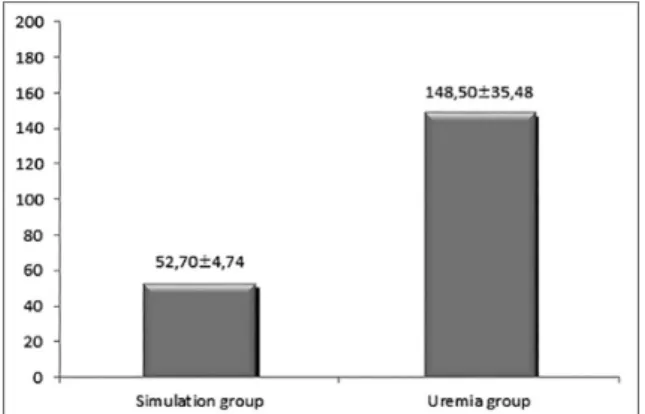
C-reactive protein dosages
As shown in Figure 2, the rats in the uremia group had significantly higher C-reactive protein values than the simulation group (p=0.0142)
FIGURE 2 - Graph showing the arithmetic means of the dosages of ultra-sensitive C reactive protein (mg/ml)
Microscopic evaluations
Histopathological evaluation of granulation tissue
In the evaluation of the colonic cicatrization process, the
superiority of the simulation group over the uremia group was clearly observed, with a statistically significant impact on reepithelialization of borders (p=0.0196) and angiofibroblast proliferation (p=0.0379).
Histometric evaluation of the percentage of collagen types I and III
As shown in Figure 3, the rats in the uremia group presented significantly worse results than the simulation group in the mean percentages of collagen I (p=0.0009) and in relation to collagen III (p=0.016).
FIGURE 3 - Graph showing the arithmetic means of the percentages of collagens type I and III in the intestinal scar tissue
Microvascular density evaluation
Animals from the uremia group had significantly lower microvascular density (p=0.0074) than those from the simulation
group (Figure 4).
FIGURE 4 - Graph showing the arithmetic means of the microvascular
density quantification in the intestinal cicatricial tissue
Tissue evaluation of nuclear proliferation nuclear antigen (pcna)
Uremia-inducing animals demonstrated a significant
(p<0.0000) negative impact on cell proliferation in the intestinal cicatricial tissue when analyzed by nuclear proliferation cell
antigen expression (Figure 5).
FIGURE 5 - Graph showing the arithmetic averages of cellular
proliferation of nuclear antigen quantification in
intestinal cicatricial tissue
Myofibroblast density evaluation
The quantified density of myofibroblasts in the intestinal cicatricial tissue had a sharp and significant reduction (<0.0001)
in animals that had uremia induced in relation to those that
did not present with uremia (Figure 6).
FIGURE 6 - Graph showing the arithmetic means of quantifications
of myofibroblasts in the intestinal cicatricial tissue
DISCUSSION
Was adopted the experimental model proposed by Vianna et al.14,17,33 with modification. The differential of the present
study of this model is the non-accomplishment of ablation
use of argon plasma electrocoagulator for the ablation of 2/3 of renal mass. Experimental studies have demonstrated the
superiority of this mode of electrocauterization due to efficiency
and limited tissue trauma13.
The reproduction of this technique in rats similar to that
performed in the present study was performed by Fleck et
al10, demonstrating a decrease in the glomerular filtration rate
after 10 weeks of the procedure, with the particularity of the analysis between genders. The study verified that there was a significant difference between male and female rats, assuming
greater fragility and susceptibility in females10.
Although other experimental models are also effective
in demonstrating renal injury, nephrectomy 5/6 has some advantages such as reproducing renal dysfunction due to loss
of nephrons as well as in humans, providing more complete
data on proteinuria and hypertension secondary to decreased
glomerular filtration, and compare renal damage to the amount of renal mass withdrawn. When performed nephrectomy 1/2,
2/3 and 5/6 analysis of the biochemical and morphological
effects of the remaining kidney after progressive renal mass
ablation, demonstrates that compensatory renal hypertrophy and glomerular injury are closely related to the volume of the
remaining kidney, and therefore, being more evident when a
greater fraction of renal tissue is extracted20,29.
Urea is formed in the liver as the main end product of the metabolism of nitrogenous substances and is subsequently excreted by the kidneys2. Experimental and clinical studies have
demonstrated the negative effect of uremia on wound healing.
Colin et al.7 concluded that rats with uremia were delayed in
the healing of small intestine anastomoses and aponeurosis.
Therefore, the presence of uremic toxins has a negative effect
by decreasing cell proliferation, the amount of hydroxyproline
in the operative wound, as well as causing alteration in the formation of fibroblasts7,22.
Uremia can cause intestinal mucosal injury, edema,
inflammation, ulceration with loss of mucosal barrier and as a consequence, bacterial translocation may occur, a factor known
to be detrimental to the healing process9.
Studies have shown that granulation tissue was reduced in uremic mice by performing histological analysis after five days of the procedure for induction of renal dysfunction, as well as inhibition of cell proliferation in fibroblasts and granulation
tissue endothelial cells22.
To date, 19 isoforms of collagen have been identified,
and type I collagen predominates and constitutes 80-90% and the remaining 10-20% are type III12. Type I collagen is the most
frequent and predominant in bones and tendons is considered mature collagen. Type III is most commonly found in soft tissues such as blood vessels, dermis and fascia. The granulation tissue expresses 30-40% of type III collagen, being considered immature
collagen. The most important is type I, which is responsible
not only for the maintenance of the integrity of most tissues in function of their mechanical properties26, but also for their
active participation in the functionality of the tissues as a
function of their interaction with the cells present in the matrix
extracellular. It is present in all vertebrates, and included in the list of the largest and most complex macromolecules found in
the animal kingdom, along with other macromolecules form
the extracellular matrix.
Russell et al.27, after 10 weeks of nephrectomy and
contralateral segmental renal ischemia in rats, demonstrated that there is an evident alteration in the formation of the bone mineral matrix and in the maturation of collagen in the group
with uremia.
In a study to evaluate the tensile strength and collagen
formation in rats after five, 10 and 15 days of uremia, there was a considerable and significant reduction in collagen accumulation, verified by the quantification of hydroxyproline
on the 10th and 15th days27.
The production of new vessels is essential in wound repair,
and is observed in healing tissues and characterized by a complex process involving multiple stages: vasodilatation, endothelial permeability, rupture of the endothelial cell connection, proliferation and migration of these cells, and subsequent remodeling forming conduits for the passage of nutrients11.
Nuclear cell proliferation antigen plays an important role in nucleic acid metabolism. It is a protein synthesized mainly during the S-phase of the cell cycle and is essential for chromosomal chromatin replication, transcription and assembly8.
In a study that evaluated the chondrocyte proliferation activity by the expression of cellular proliferation nuclear
antigen in the zone of cartilage growth after 30 days of subtotal 5/6 nephrectomy in rats, it was possible to conclude that the number of proliferating cells in the growth plateau was significantly lower in uremic rats when compared to control
rats1. Another experimental study in rats that investigated
the density of myofibroblasts in wound healing by the same
method employed in this study concluded that metronidazole,
applied topically to wound healing with second intention, does not interfere with the contraction of the wound and delays the appearance of myofibroblasts32.
CONCLUSION
The experimentally induced uremic state is capable of
negatively influencing the healing process of the large intestine.
Uremia compromises the formation of granulation tissue, delays maturation of type I collagen, induces microvascular density reduction in the anastomosis, reduces cell viability in
the area of healing, and density of myofibroblasts present in
colonic healing.
REFERENCES
1. Barbosa APF, Silva JDP, Fonseca EC, Lopez PM, Fernandes MBC, Balduino A et al. Response of the growth plate of uremic rats to human growth hormone and corticosteroids. Braz J Med Biol Res 2007;40(8):1101-1109. 2. Baum N, Dichoso CC, Carlton JR CE. Bood urea nitrogen and serum
creatinina. Urology 1975;5: 583-588.
3. Becker GJ, Hewitson TD. Animal models of chronic kidney disease: useful but not perfect. Nephrol Dial Transplant 2013;28(10):2432-2438. 4. Boudet J, Man NK, Pils P, Sausse A, Brentano JLF. Experimental chronic
renal failure in the rat by eletrocoagulation of the renal córtex. Kidney Int 1978;14(1):82-86.
5. Bravo R. Synthesis of the nuclear protein cyclin (PCNA) and its relationship with DNA
6. Brusilow SW. “Inborn errors of urea synthesis”. In: Glew RH, Ninomiya Y. Clinical studies in medical biochemistry. 2nd ed. [New York]:Oxford University Press, 1997;260-267.
7. Colin JF, Elliot P, Ellis H. The effect of uraemia upon wound healing: an experimental study. Br J Surg 1979;66:793-797.
8. Dou L, Bertrand E, Cerini C, Faure V, Sampol J, Vanholder R, Berland Y, Brunet P. The uremic solutes p-cresol and indoxyl sulfate inhibit endothelial proliferation and wound.
9. Duarte JBA, Nascimento JEA, Nascimento M, Nochi Jr RJ. Bacterial translocation in experimental uremia. Urol Res 2004;32(4):266-270. 10. Fleck C, Appenroth D, Jonas P, Koch M, Kundt G, Nizze H et al. Suitability
of 5/6 nephrectomy (5/6nx) for the induction of interstitial renal fibrosis in rats - influence of sex, strain, and surgical procedure. Exp Toxicol Pathol 2006;57(3):195-205.
11. Folkman J. Tumor angiogenesis: therapeutic implications. N Engl J Med 1971;285(21):1182-1126.
12. Giaquinto MGC, Mota DSC. Cicatrização de feridas. In: MARQUES RM. Técnica operatória e cirurgia experimental. 1.ed. [Rio de Janeiro]:Guanabara Koogan, 55-70; 2005.
13. Grund KE, Straub T, Farin G. New haemostatic techniques: argon plasma coagulation. Baillieres Best Pract Res Clin Gastroenterol 1999;13(1):67-84. 14. Hermann JB, Woodward SC, Pulaski EJ. Healing of colonic anastomosis
in the rat. Surg Gynecol Obstet 1964;119:269-275.
16. Iwanaga TC, Aguiar JL, Martins-Filho ED, Kreimer F, Silva-Filho FL, Albuquerque AV. Analysis of biomechanical parameters in colonic anastomosis. ABCD Arq Bras Cir Dig 2016;29(2):90-92.
17. Jiborn H, Ahonen J, Zederfeldt B. Healing of experimental colonic anastomoses. III. Collagen metabolism in the colon after left colon resection. Amer J of Surg 1980;139(3):398-405.
18. Junqueira LCU, Montes GS, Sanchez EM. The influence of tissue thickness on the study of collagen by the picrosirius-polarization method. Histochemistry 1982;74(1):153-156.
19. /DOQI clinical practice guidelines for chronic kidney disease: evaluation, classification, and stratification. Am J Kidney Dis. 2002;Suppl 1:S1-266. 20. Kaufman JM, DiMeola HJ, Siegel NJ, Lytton B, Kashgarian M, Hayslett JP.
Compensatory adaptation of structure and function following progressive renal ablation. Kidney Int 1974;6(1):10-17.
21. Lorena D, Uchio K, Costa AM, Desmoliere A. Normal scarring: importance of myofibroblasts. Wound Repair Regen 2002;10(2):86-92.
22. McDermott FT, Nayman CM, De Boer WGRM. The effect of acute renal failure upon wound healing: histological and autoradiographic studies in the mouse. Annals of Sur, 1968;168(1):142-146.
23. Molgaard HV, Spurr NK, Greaves MF. The hemopoietic stem cell antigen CD34 is encoded by a gene located on chromosome 1. Leukemia 1989;3(11):773-776.
24. Nery RA, Kahlow BS, Skare T, Tabushi FI, Castro A. Uric Acid and Tissue Uric Acid and Tissue Repair. ABCD Arq Bras Cir Dig 2015;28(4):290-292. 25. Pincus MR, Henry JB. Química clínica. In: Henry JB. Diagnósticos Clínicos
e tratamento por métodos laboratoriais. 20 ed. 289-293;2008.
26. Ramachandran GN, Mitra AK. An explanation for the rare occurrence of cis peptide units in proteins and polypeptides. J Mol Biol 1976;107(1):85-92. 27. Russell JE, Avioli LV. Effect of Experimental Chronic Renal Insufficiency on
Bone Mineral and Collagen Maturation. J Clin Invest 1972;51(12):3072-3079. 28. Salgado FL, Artigiani-Neto R, Lopes-Filho GJ. Growth factors and COX2
in wound healing: an experimental study with Ehrlich tumors. ABCD Arq Bras Cir Dig 2016;29(4):223-226.
29. Santos LS, Chin EWK, Ioshii SO, Filho RT. Surgical reduction of the renal mass in rats. Morphologic and functional analysis on the remnant kidney. Acta Cir Bras 2006;21(4): 253-256.
30. Sesso RC, Lopes AA, Thomé FS, Lugon JR, Watanabe Y, Santos DR. Relatório do Censo Brasileiro de Diálise Crônica. J Bras Nefrol 2014;36(1):48-53.. 31. Stevens A, Lowe J. Respostas teciduais ao dano. Patologia. 2. ed. [São
Paulo]: Manole, 2000;35-50
32. Trindade LCT, Biondo-Simões MLP, Sampaio CPP, Farias RE, Pierin RJ, Netto MC. Avaliação do uso tópico do metronidazol no processo de cicatrização de feridas: um estudo experimental. Rev Col Bras Cir 2010;37(5):358-363.