Mem Inst Oswaldo Cruz, Rio de Janeiro, Vol. 113(10): e180160, 2018 1|11
online | memorias.ioc.fiocruz.br ORIGINAL ARTICLE
Protein mass spectrometry extends temporal blood meal detection
over polymerase chain reaction in mouse-fed Chagas disease vectors
Judith I Keller1, Justin O Schmidt2, Anna M Schmoker1, Bryan A Ballif1, Lori Stevens1/+
1University of Vermont, Department of Biology, Burlington, VT, United States of America 2Southwestern Biological Institute, Tucson, AZ, United States of America
BACKGROUND Chagas disease is highly prevalent in Latin America, and vector control is the most effective control strategy to date. We have previously shown that liquid chromatography tandem mass spectrometry (LC-MS/MS) is a valuable tool for identifying triatomine vector blood meals.
OBJECTIVES The purpose of this study was to determine blood meal detection ability as a function of method [polymerase chain reaction (PCR) vs. LC-MS/MS], time since feeding, and the effect of molting in mouse-fed triatomine insect vectors targeting hemoglobin and albumin proteins with LC-MS/MS and short interspersed nuclear elements (SINE)-based PCR.
METHODS We experimentally fed Triatoma protracta on mice and used LC-MS/MS to detect hemoglobin and albumin peptides
over time post-feeding and post-molting (≤ 12 weeks). We compared LC-MS/MS results with those of a standard PCR method
based on SINEs.
FINDINGS Hemoglobin-based LC-MS/MS detected blood meals most robustly at all time points post-feeding. Post-molting, no
blood meals were detected with PCR, whereas LC-MS/MS detected mouse hemoglobin and albumin up to 12 weeks.
MAIN CONCLUSIONS In our study, the hemoglobin signature in the insect abdomen lasted longer than that of albumin and DNA. LC-MS/MS using hemoglobin shows promise for identifying triatomine blood meals over long temporal scales and even post-molting. Clarifying the frequency of blood-feeding on different hosts can foster our understanding of vector behavior and may help devise sounder disease-control strategies, including Ecohealth (community based ecosystem management) approaches.
Key words: LC-MS/MS - Chagas disease - blood meals - hemoglobin - albumin - SINE-PCR
Chagas disease is a major neglected tropical disease, with high endemic prevalence in Latin America where most transmission is by insect vectors in the subfamily Triatominae, also known as kissing bugs. Although au -tochthonous cases of Chagas disease in the United States are thought to be rare, Chagas incidence is likely, for a variety of reasons, to be underreported.(1-3) Chagas dis-ease is caused by the protozoan parasite Trypanosoma cruzi, with the most common mode of transmission via a triatomine insect defecating while taking a blood meal or through the oral transmission route. The para-site is subsequently introduced into the new host’s blood stream through a break in the skin or mucous membrane.
Identifying the blood meal sources and feeding pat-terns of native insect vectors provides valuable data for understanding the ecology and behavior of the vector, presenting the need for research aimed at developing better blood meal identification methods. In addition,
doi: 10.1590/0074-02760180160
Financial support: NIH [grant 8P20GM103449 (BAB) from the INBRE program of the NIGMS, NIH grant R03AI26268/1-2 (LS)] from the NIAID and National Science Foundation grant BCS-1216193 (LS) as part of the joint NSF-NIH-USDA (United States Department of Agriculture) Ecology and Evolution of Infectious Diseases Program.
+ Corresponding author: [email protected] Received 28 March 2018
Accepted 3 August 2018
blood meal source prevalence can help elucidate local transmission cycles and can provide data for evidence-based vector control strategies.(4,5) Insecticide spraying is often effective in the short term, but reinfestation of insect vectors and the financial burden of frequent large-scale spraying, as well as pyrethroid resistance make this not the most strategic option for battling Chagas disease.(6-8) While massive campaigns of indoor residual insecticide spraying have effectively reduced introduced vectors in many regions, such as Rhodnius prolixus
in Central America and Triatoma infestans in parts of South America,(7,8) for native vectors such as Triatoma dimidiata in Central America, a recent study suggests a control strategy for Chagas disease includes a holistic Ecohealth approach that focuses on community partici-pation, education, and vector control.(5,9-12) As such, with-in the Ecohealth framework, vector control policies need to consider domestic vectors and perhaps underestimate sylvatic vector influence.(13) To develop effective native vector control strategies, a knowledge of the vector ecol -ogy, including blood meal sources of vectors collected in various ecotopes, such as sylvatic, peridomestic, or domestic environments is important.(5)
Judith I Keller et al. 2|11
and transportation of insect samples to maintain high quality of DNA or antigens commonly used for blood meal identification. Thus, time since feeding and stor-age of material could be two of the major reasons why in many studies upwards of 50% of samples do not have a blood meal detected.(19-23)
Blood meal sources have traditionally been detected by a number of methods including immunological approach-es such as precipitin tapproach-ests and enzyme-linked immuno -sorbent assay (ELISA)(24) which are dependent on protein antibodies of blood meal sources present in an area, and DNA-based methods often focusing on polymerase chain reaction (PCR) amplification of mitochondrial or nuclear DNA. In addition, PCR amplification of a species-spe-cific repetitive sequences of nuclear DNA, such as short interspersed nuclear elements (SINEs) using standard PCR,(17,18) PCR amplification followed by sequencing of vertebrate mitochondrial DNA(25) or ribosomal subunits such as 12 S,(26) genomics,(27) and other next-generation
sequencing tools(28) are emerging. Recently, we have shown the usefulness of a protein-based method based on liquid chromatography tandem mass spectrometry (LC-MS/MS) for blood meal source identification.(29) A few studies have compared different methodologies directly. For example, Lucero et al. compared 12 S sequencing and qPCR,(30) while Stevens et al. compared 12 S sequencing and cytochrome b.(25) We compared LC-MS/MS with 12 S-based DNA sequencing and found LC-MS/MS identi-fied blood meals from insects collected alive and dead; however, blood meals were not detected with DNA se-quencing methods for three of the four samples.(29) Blood meal detection ability can also vary depending on the spe-cies of host blood. A study examining the effect of both blood meal host species and time elapsed since a recent feeding, reported detection ability can drop off as early as 1-2 weeks for some species.(18)
Blood is a complex fluid with components that decay at different rates. Because iron stabilises molecules, blood proteins such as hemoglobin can be remarkably stable and have been detected in a 46-million-year-old fossil-ised mosquito.(16,31,32) Peptides from hemoglobin, the most abundant protein in red blood cells, have been detected 309 days post-molt in ticks; and peptides from albumin, the most abundant blood serum protein, have been detect-ed 85 days post-molt in ticks.(16) Other components such as
transferrin and immunoglobulins are known to degrade rapidly, while keratin, actin, histones, and tubulins were too conserved across the animal kingdom to be informa-tive when evaluated for mass-spectrometry-based detec-tion in ticks.(16) Abundance, stability, and species-specific
sequence variation make hemoglobin and albumin mol -ecules great targets for LC-MS/MS-based techniques.
(16,29,32) LC-MS/MS has led to promising results when
com-pared with DNA-based methods.(29) A major benefit of us-ing protein-based techniques is the quantity and quality of data gained from a single LC-MS/MS run.(16)
Information about blood meal perseverance in the in-sect gut is limited. Nevertheless, the longevity of blood meal detection ability in Chagas vectors is critical infor-mation to aid in the knowledge of the parasite transmis-sion and overall feeding habits of the insect. For
exam-ple, the average time for a blood meal digestion has been estimated to be approximately 14 days in adult female
T. infestans,(33) but different components of blood may vary and blood meals have been detected up to 10 weeks post-feeding in adult male and female T. pallidipennis, T. barberi, T. dimidiata, T. phyllosoma, and T. longipennis.
(15) Molting behavior of insects can also affect blood meal
detection. Kissing bugs are hemimetabolous, emerging wingless from an egg, and successively molt through five nymphal instars into winged adults.(34) Experimen-tally evaluating blood meal detection post-molt would indicate whether or not we can detect feeding across molts and if the decrease in albumin and hemoglobin peptides over time can provide temporal information about the last blood meal.
In this study we determine how the ability to detect and identify a blood meal declines over time and is affect-ed by molting. We assess Triatominae insect vector blood meals for the first time comparing two identification methods, protein LC-MS/MS and PCR of SINE-DNA in two experiments: (1) recently molted adult Triatoma pro-tracta (Hemiptera: Reduviidae) fed once on mouse and assayed 0-4 weeks post-feeding; and (2) recently molted (fed approximately 1 week prior) T. protracta not fed after eclosion and assayed 0-12 weeks after molting. In addi-tion to comparing LC-MS/MS with PCR, within LC-MS/ MS we compare the ability of hemoglobin and albumin peptides to identify the source of a blood meal.
MATERIALS AND METHODS
Ethics - White inbred ICR (CD-1) Mus musculus
(house mouse) (Harlan Laboratories, Madison, WI) were used for feeding experiments. All procedures using mice were first approved by the Southwestern Biological Institute, Tucson, Arizona, USA Animal Care and Use Committee and follow international standards.(35) Mice were immobilised in small mesh cages and placed in the enclosure containing the insect vectors. Insects were al-lowed to feed until satiated, or approximately for 30-60 minutes, at which point mice were removed.
Mem Inst Oswaldo Cruz, Rio de Janeiro, Vol. 113(10), 2018 3|11
representing the two experiments: (1) post-feeding or (2) post-molting. After the experimental mouse feeding, the two experimental groups were kept separately in equiva -lent aforementioned containers. No non-experimental in-sects were housed in the experimental groups.
The post-feeding (F) insects were collected after eclos-ing as adults and allowed a seclos-ingle blood meal on M. mus-culus within 19-37 days. Within an hour of feeding, the
adult individuals (hereafter referred to as F0wk) were pre -served in 95% ethanol and 5% glycerol and stored at 4ºC. Additional individuals were collected at each of the fol-lowing time points: one week (F1wk), two weeks (F2wk), and four weeks (F4wk) post-feeding without access to an additional blood meal. Insects that died between sampling times were not analysed (see Supplementary data I, Table II for details of sample sizes and longevity). None of the insects in the F group survived past four weeks, except one specimen that was not analysed due to a mishap in the preparation. We analysed four insects from each time period, except F0wk where we analysed three.
Adult post-molting (M) insects were collected from the colony after feeding on M. musculus as 5th instar nymphs, and most molted to adults within one week of feeding. After molting, insects in this group were not fed, but were collected and preserved at the same time intervals (M0wk, etc) as the fed insects. These post-molt insects survived longer than the fed insects and sampling was extended to eight and 12 weeks (Table I). The longev-ity of post-molt bugs was a little longer, of the nine re-maining at eight weeks, eight died before week 12 and we were able to analyse the one specimen alive at 12 weeks (Supplementary data I, Table II). Although most adult Tri-atoma species live much longer than 4-8 weeks, starva -tion has been shown to reduce adult longevity in Rhod-nius prolixus,(36) and the difference between the post-molt and post-fed specimen may be an artifact of small sample size. We sampled two insects from each time period, 0, 1, 2, 4, 8 and one surviving individual at 12 weeks. Samples were stored at 4ºC and within 1-4 weeks of collection, were shipped to the University of Vermont by priority mail in insulated containers, where they were stored at -20ºC until dissection in August 2016 and May 2017.
The 15 post-feeding and 11 post-molting T. protracta
were evaluated using methods similar to Keller et al.(29) Results were visualised with graphs made using JMP®, Version 13 (SAS Institute Inc., Cary, NC, 1989-2016). Below we highlight important aspects of the methods and indicate changes from our previous study.(29)
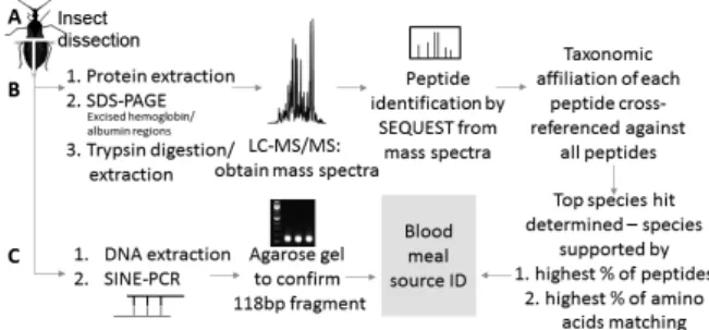
Dissection of insect vectors - For each insect, the abdomen was cut into left and right halves. Abdomen halves were randomly assigned to LC-MS/MS protein analysis or mouse-specific SINE-DNA PCR.
Hemoglobin and albumin protein extraction, sodi-um dodecyl sulfate polyacrylamide gel electrophoresis (SDS-PAGE), and mass spectrometry - We extracted protein from T. protracta insect abdomen halves as previously described(29) except that 200 µL of denatur-ing sampldenatur-ing buffer was added per 0.1 g of insect tis -sue. For samples weighing 0.05 g and below, 100 µL of 95ºC denaturing sampling buffer was added. Denaturing
SDS-PAGE using gel regions surrounding the molecular weight of hemoglobin (~16 kDa) and albumin (~65 kDa) were excised and prepared for mass spectrometry analy-sis as previously described.(29) In brief, because LC-MS/ MS works on peptides smaller than the hemoglobin and albumin proteins, following in-gel digestion with tryp-sin and peptide extraction, LC-MS/MS was performed using a linear ion trap-orbitrap (LTQ-Orbitrap; Thermo Electron, Waltham, Massachusetts, USA) where spec-tra all were collected in the orbispec-trap.(29,37) Samples were subjected to 15 min of isocratic loading in 2.5% MeCN, 0.15% FA (Solvent A), and peptides were subsequently eluded with a 0-50% gradient of 99% MeCN, 0.15% FA (Solvent B) over 45 min (400 mL/min flow rate average across a flow splitter), followed by 10 min 100% Solvent B, and a 15 min equilibration with Solvent A.
LC-MS/MS does not directly sequence peptides, but rather infers amino acid sequences of short peptides based on theoretical peptide masses present in an underlying database, in our case, GenBank(38) hemoglobin and
al-bumin entries. We searched these mass spectra using the SEQUEST algorithm (Thermo Electron V26.12) against a custom forward and reverse concatenated database containing vertebrate hemoglobin sequences (20 January 2016, 17,000+ entries) extracted from GenBank as pre -viously described(29) and “serum albumin” (26 October 2016, 1600+ entries) in any curated field. Peptide identifi -cation and stringent filtering of peptides was as described in Keller et al.,(29) where no reverse database matches re-sulted and false discovery of peptides was below 0.01%.
Blood meal sources were identified as previously described with a pipeline to infer the most likely blood source(29) (Fig. 1). The pipeline considered the potential taxa represented by the peptide inferred from the mass spectra and cross-referenced the most likely blood meal source based on individual peptides in a sample. We sub-sequently quantified the identified protein coverage at the peptide and amino acid level. This provided the per-centage support for a particular blood source (see Keller et al.(29) for details).
DNA extraction and SINE-based PCR - DNA extrac-tion used the DNeasy Blood and Tissue Kit (Qiagen, Valencia, CA) as previously described.(17,30) Briefly, the
Fig. 1: workflow describing liquid chromatography tandem mass
Judith I Keller et al. 4|11
manufacturer’s instructions for extracting tissue were followed using insect abdomens chopped finely with scissors. DNA was eluted with two separate sequential elutions of 100 µL each. DNA concentration was mea -sured using a Nanodrop ND-1000 instrument (Thermo Scientific, Waltham, MA, USA), and the instrument was calibrated using the elution buffer (Qiagen, Valencia, CA) DNA was stored in.
DNA extracts were subjected to PCR amplification after optimising previously published methods for our equipment and reagents.(18,39,40) The 12 µL PCR reaction contained 1 µL of DNA template, 0.2 µM each of for -ward (5’AGATGGCTCAGTGGGTAAAGG3’) and
re-verse (5’GTGGAGGTCAGAGGACAAACTT3’) prim
-ers, and 6 µL 1X EconoTaq PLUS GREEN (Lucigen, Middleton, WI, USA). PCR conditions were as follows: initial denaturisation for 5 min at 95ºC, 30 cycles of 95ºC for 30 s, annealing for 30 s at 55ºC, and extension for 30 s at 72ºC, followed by a final extension of 5 min at 72ºC. Nancy-520 stained 1.5 % agarose gels (Sigma-Aldrich, Milwaukee, WI, USA) were used to verify the 118 bp PCR fragments. Positive (DNA extracted from mouse tissue) and negative (PCR-grade water) controls were in-cluded in each set of PCR amplifications.
RESULTS
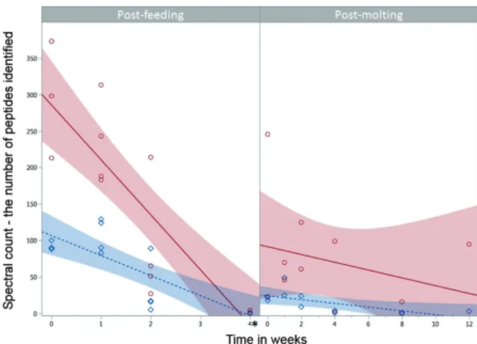
The purpose of this study was to determine blood meal detection ability as a function of method (PCR vs. LC-MS/MS), time since feeding, and the effect of molt-ing in Triatominae insect vectors targetmolt-ing hemoglobin and albumin proteins with LC-MS/MS and SINE-based PCR. This is the first study to compare protein-based and DNA-based detection methods of experimentally-fed ar-thropod vectors. Briefly, hemoglobin-based LC-MS/MS yielded the most robust mouse blood meal detection and identification over time. In our experiments none of the post-feeding specimens lived longer than four weeks; however, we were able to identify the M. musculus blood meals based on hemoglobin and albumin peptides four and two weeks post-feeding, respectively (Figs 2-3, Sup-plementary data I, Fig. 2, Table III). With SINE-based PCR we were able to detect M. musculus blood meals up to one week post-feeding. The post-molting triatomines lived longer, and we were able to identify M. musculus
hemoglobin and albumin peptides both up to 12 weeks post-molting, while SINE-based PCR detected no M. musculus blood meals at any time post-molting (Fig. 3,
Supplementary data I, Fig. 2).
Post feeding
LC-MS/MS - hemoglobin - Using LC-MS/MS we were able to identify hemoglobin peptides throughout the entire sampling time of four weeks post-feeding, except for one F4wk sample; however the number of hemoglobin peptides decreased over time from an average of over 300 at F0wk to less than 10 at F4wk (Fig. 2, Table I, Supple-mentary data II, Table IV). For all four of the biological replicates at the 0, 1, and 2 wk time points and one of the F4wk samples the combination of peptides unambigu-ously identified the blood meal to the species level, M. musculus. With the other three F4wk samples, for two
we were able to identify 2-6 species as the most probable
blood source (which in both cases included the correct M. musculus blood meal - Sample ID 49 equally supported six species: M. musculus, Otospermophilus beecheyi,
Mus spretus, Mus minutoides, Jaculus jaculus, Callo-spermophilus lateralis; sample ID 51 equally supported M. musculus and M. spretus), while as stated above, one replicate did not contain hemoglobin peptides (Table I).
LC-MS/MS - albumin - We were less successful de-tecting albumin peptides over time. We were only able to identify albumin peptides up to two weeks post-feeding and like hemoglobin, the number decreased over time from an average of over 100 at F0wk to 0 at F4wk (Fig. 2, Table II, Supplementary data III, Table V). For all the rep-licates at F0wk and F1wk, and three of the four reprep-licates at F2wk, we identified the blood meal to the species level (Fig. 3). For the other F2wk sample, there was equally strong support for Rattus norvegicus and M. musculus
as the blood meal source (Table II). Overall, hemoglo-bin and albumin peptide abundance varied significantly between each time point post-feeding (Least Squares Re-gression, p < 0.001), and albumin was significantly lower in abundance than hemoglobin (Least Squares Regres-sion, p < 0.001) (Table III).
SINE-DNA - The M. musculus-specific-SINE based PCR was the least successful in detecting the M. mus-culus blood meal, we were only able to detect the blood meal in the F0wk and F1wk samples (Fig. 3).
Post molting
LC-MS/MS - hemoglobin - Using LC-MS/MS, we were able to identify hemoglobin peptides throughout the entire sampling time of 12 weeks post-molting, ex-cept for one M8wk samples. As expected, for the M0, 1,
Fig. 2: detection of hemoglobin and albumin peptides over time from a known Mus musculus blood meal source. The number of hemoglo-bin (red open circle/solid line) and albumin (blue diamond/dotted line) spectral counts decreased over time post-feeding (left) and
post-molt-ing (right). There were more peptides at 0 and 2 wk in the post-feedpost-molt-ing specimens, but more at 4 wk for the post-molt. Linear regression lines
were fit and 95% confidence intervals are shown (shading) with an alpha level of 0.05. Hemoglobin and albumin peptide abundance var-ied significantly between each time point post-feeding (Least Squares
Regression, p < 0.001), and albumin was significantly lower in abun
-dance than hemoglobin (Least Squares Regression, p < 0.001). Note
the difference in time scales on the x-axis (*), none of the analysed
Mem Inst Oswaldo Cruz, Rio de Janeiro, Vol. 113(10), 2018 5|11
2wk specimens, the number of hemoglobin peptides was lower than for the post-feeding experiment and gener-ally decreased over time from an average of over 100 at M0wk to less than 10 at M8wk (Fig. 2, Table I). Spectral counts were more variable in post-molting individuals. For example, M8wk averaged around eight hemoglobin peptides while the single replicate M12wk contained 95 hemoglobin peptides. For all replicates except one M8wk sample we identified the blood meal to the species level. One M8wk sample contained a single hemoglobin pep-tide matching 261 species (Table I).
LC-MS/MS - albumin - As with the post-feeding speci-mens, we were less successful detecting albumin peptides post-molting. We were able to detect albumin peptides over all time points, but the number decreased from a range of 18-49 at the early times to 0-3 at the later (Fig. 2, Table II). For all the replicates at 0, 1, and 2 wk and one of the two M4wk, we identified the blood meal to the species level. For the other samples, we narrowed the likely blood meal sources to 2-7 species, again, all including known blood meal source M. musculus (sample ID 54 equally supported M. musculus and R. norvegicus; sample ID 23 and 30 equally supported M. musculus, R. norvegi-cus, Sorex araneus, Octodon degus, Ochotona princeps,
Dipodomys ordii,and Cricetulus griseus). Albumin pep-tides appeared to decrease significantly after the 2 week timepoint (Table II). Overall, hemoglobin and albumin peptide abundance did not vary significantly post-molting (Least Squares Regression, p > 0.05) (Table III).
SINE-DNA - Our M. musculus-specific SINE-based PCR did not detect M. musculus blood meals at any time points post-molting (Fig. 3).
DISCUSSION
Ideally, a combination of assays gives researchers a diverse toolbox for identification of blood meal sources. For blood-feeding arthropod disease vectors, including the triatomine vectors of Chagas disease, increasing our understanding of vector blood meal sources facilitates the design of evidence-based control strategies. Few papers have compared different blood meal detection techniques directly, e.g.,(30,31) and this is the first paper to compare a LC-MS/MS protein-based approach to a well-established DNA-based approach. The assay based on protein identified the blood meal source for a longer time post-feeding as well as post-molting.
This study shows variation in detection of blood meals by LC-MS/MS and mouse-specific SINE-based PCR as a function of time since last feeding and time since molting. Overall, LC-MS/MS based on hemoglo-bin gave precise blood meal identification for the longest amount of time post-feeding and post-molting. Albumin peptides were also present for the majority of time points in both experiments, however, detection of albumin pep-tides dropped off sharply two weeks post-feeding. DNA-based detection using mouse-specific SINE-DNA was only successful in detecting fresh blood meals up to one week post-feeding and did not detect a blood meal at any time points for the post-molt experiment. To our knowl-edge, this study is the first to explore post-molt blood meal detection in triatomines, as only incidental find-ings have been reported previously.(19)
Using hemoglobin peptides opens the door to ex-ploring blood sources of early stage nymphs that have molted into later stages and possibly adults. Many stud-ies of blood meal sources, e.g.,(41) focus on later stage tri-atomines, which are more likely to be encountered and are more mobile. Because we were able to detect peptides from a previous life stage up to 12 weeks post-molting with LC-MS/MS but not DNA, we suggest further studies that explore blood sources of early stage nymphs. Such future studies could include examining the nymphs them-selves, or through experimental feeding to determine if blood sources of early stage nymphs could be detected in later nymphs or even adults. The SINE-based PCR did not detect any blood meals post-molt. Albumin did have a lingering signature post-molt, but recovered peptides were significantly lower in number. While protein analy-ses do not have the luxury of amplification technologies that exist for DNA, LC-MS/MS is becoming more sensi-tive. Because of the low fmol (femtomole = 10-15) amount
of material needed for LC-MS/MS, future studies should examine the ability to detect blood proteins over the life cycle of the insect. The development and standardisation of methods for LC-MS/MS detection will increase our ability to detect blood sources in field-collected samples, and can boost the design of control strategies for the dis-tinct transmission cycles maintained by different vector species in different localities.
An increasing number of studies are trying mass spectrometry-based techniques for blood meal identifi-cation in insect vectors.(16,42,43) Our mass spectrometry-based technique differs from DNA sequencing in that the molecule is not sequenced per se, but rather highly
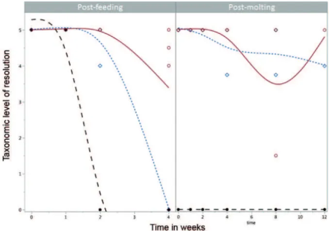
Fig. 3: taxonomic level of resolution for Mus musculus blood meals over time. Taxonomic level of resolution by liquid chromatography tandem mass spectrometry (LC-MS/MS) varied between hemoglobin (red open circle/solid line) and albumin (blue diamond/dotted line) proteins and from short interspersed nuclear elements-based
poly-merase chain reaction (SINE-based PCR) (black closed circle/lined
line) post-feeding (left). DNA, albumin, and hemoglobin provided species-specific blood meal identification up to one, two, and four
weeks post-feeding, respectively. Post-molting (right), the taxonomic
level of resolution for M. musculus blood meals by LC-MS/MS was stronger for hemoglobin and albumin than SINE DNA, which never detected a blood meal at any post-molting time point. A best-fit line
(cubic smoothing spline, lambda of 0.0855, standardised X values)
Ju
d
ith I K
e
lle
r e
t a
l.
6|
11
TABLE I
Summary statistics for hemoglobin peptides identified in adult Triatoma protracta abdomen halves in post-feeding and post-molting specimens
Mus musculus match
Sample ID
Unique peptides match/ total uniquea
(nº)
Peptides matching
M. musculus
(%)
Total AA identified based on no. of unique peptidesb
AA mis- matches
(nº)
Amino acid matching to M. musculus
(%)
Spectral countc
Average spectral count
1. Post-feeding F0wk 33 16/20 80.00 299 8 97.3 298 294.7
F0wk 34 15/18 83.33 292 9 96.9 373
F0wk 35 18/24 75.00 399 16 96.0 213
F1wk 38 16/20 85.00 311 12 96.1 188 231.8
F1wk 40 20/26 76.92 390 12 96.9 183
F1wk 41 15/19 78.95 300 8 97.3 243
F1wk 42 18/21 85.71 340 9 97.4 313
F2wk 43 14/15 93.33 213 1 99.5 65 89.3
F2wk 44 13/14 92.86 200 1 99.5 51
F2wk 45 17/22 77.27 314 9 97.1 214
F2wk 46 4/4 100.00 56 0 100.0 27
F4wk 49 1/1d 100.00 13 0 100.0 1 2.3
F4wk 50 4/4 100.00 56 0 100.0 4
F4wk 51 4/4d 100.00 53 0 100.0 4
F4wk 52 no peptides identified 0
2. Post-molting M0wk 15 23/28 82.14 422 15 96.4 246 134.0
M0wk 4 10/12 83.33 186 6 96.8 22
M1wk 11 19/22 86.36 320 9 97.2 70 58.0
M1wk 10 19/21 90.48 326 9 97.2 46
M2wk 17 20/22 90.91 293 6 98.0 126 93.5
M2wk 19 10/12 83.33 197 5 97.5 61
M4wk 24 17/21 80.95 295 14 95.3 99 50.5
M4wk 23 2/2 100.00 31 0 100.0 2
M8wk 27 1/1d 100.00 0 0 100.0 1 8.5
M8wk 30 8/10 80.00 151 6 96.0 16
M12wk 54 18/21 85.71 317 9 97.2 95 95.0
AA: amino acid; a: the number of unique peptides identified in a sample that match the known blood meal source, Mus musculus, of the approximately 23 detectable peptides from
trypsin digestion of 142 aa alpha and 147 aa beta hemoglobin (depending on the amino acid variation; based on GenBank entries NP_032244.2, BAG16710.1); b: number of amino acids
of the unique peptides identified that match mouse; c: spectral count, or number of hemoglobin peptides identified in each liquid chromatography tandem mass spectrometry (LC-MS/ MS) run; d: M. musculus was not uniquely identified as the most likely blood meal source. Sample ID 49 equally supported six species: M. musculus, Otospermophilus beecheyi, Mus spretus, Mus minutoides, Jaculus jaculus, Callospermophilus lateralis; Sample ID 51 equally supported M. musculus and M. spretus; The single peptide from sample ID 27 was a
Mem Inst Oswaldo Cruz
, Rio de Janeiro, V
ol.
113
(10), 2018
7
|
11
TABLE II
Summary statistics for albumin peptides identified in adult Triatoma protracta abdomen halves in post-feeding and post-molting specimen
Mus musculus match
Sample ID
Unique peptides match/ total uniquea
(nº)
Peptides matching
M. musculus
(%)
Total AA identified based on no. of unique peptidesb
AA mis- matches
(nº)
Amino acid matching to M. musculus
(%)
Spectral countc
Average spectral count
1. Post-feeding F0wk 33 14/14 100.00 194 0 100.0 90 95
F0wk 34 14/14 100.00 176 0 100.0 102
F0wk 35 14/14 100.00 166 0 100.0 93
F1wk 38 8/8 100.00 100 0 100.0 90 108.25
F1wk 40 5/5 100.00 66 0 100.0 83
F1wk 41 15/15 100.00 182 0 100.0 129
F1wk 42 15/15 100.00 189 0 100.0 131
F2wk 43 9/9 100.00 116 0 100.0 17 30.25
F2wk 44 9/9 100.00 116 0 100.0 17
F2wk 45 12/12 100.00 181 0 100.0 81
F2wk 46 3/3d 100.00 40 0 100.0 6
F4wk 49 no peptides identified 0
F4wk 50 no peptides identified
F4wk 51 no peptides identified
F4wk 52 no peptides identified
2. Post-molting M0wk 15 14/14 100.00 172 0 100.0 23 20.5
M0wk 4 11/11 100.00 138 0 100.0 18
M1wk 11 19/19 100.00 248 0 100.0 49 37
M1wk 10 13/13 100.00 160 0 100.0 25
M2wk 17 10/10 100.00 134 0 100.0 25 17
M2wk 19 7/7 100.00 83 0 100.0 9
M4wk 24 2/2 100.00 22 0 100.0 4 2.5
M4wk 23 1/1d 100.00 13 0 100.0 1
M8wk 27 no peptides identified 0.5
M8wk 30 1/1d 100.00 13 0 100.0 1
M12wk 54 3/3d 100.00 40 0 100.0 3 3
AA: amino acid; a: of the approximately 58 detectable peptides from trypsin digestion of the 609 AA albumin protein (depending on AA variation, based on GenBank sequence
CAD29888.1), shown are the number of unique peptides identified in a sample that match the known blood meal source, Mus musculus; b: number of amino acids of the unique
peptides identified that match mouse; c: spectral count, or number of albumin peptides identified in each liquid chromatography tandem mass spectrometry (LC-MS/MS) run;
d: M. musculus was not uniquely identified as the most likely blood meal source. Sample ID 46 and 54 equally supported M. musculus and Rattus norvegicus; Sample ID 23 and 30
Judith I Keller et al. 8|11
accurate mass measurements are used to match observed mass spectra with theoretical ones based on protein se-quences in GenBank.(29,32) An alternative approach,
matching spectral libraries using measurements made by Matrix Assisted Laser Desorption/Ionisation time of flight mass spectrometry (MALDI-TOF) has also been used(42,43) but requires blood controls from animal spe-cies likely encountered in the field to make the matching spectral libraries. However, obtaining such material can be problematic and precludes identifying unanticipated taxa. LC-MS/MS matching theoretical to observed spec-tra provides a practical approach using sequence search-ing with data readily available in GenBank,(29) which we
only expect to grow in sequence information over time. In addition, as management decisions are often based at higher taxonomic levels (e.g. rodents in general, birds in general), identifying closely related species through conservation of hemoglobin sequences of related species allows for comprehensive vector management using LC-MS/MS data. In our study, the hemoglobin signature in the blood lasted longer than that of albumin, which has also been previously shown.(16) We show that hemoglo-bin is detected in T. protracta even several weeks after
the insect had fed, and was detected even longer, notably from fewer peptides, in insects that had molted but not subsequently fed (four and 12 weeks, respectively).
Given hemoglobin is more abundant in blood and is highly stable, it is not necessarily surprising that it is detectable longer than albumin. However, this might not have been the case, particularly as albumin is also stable and as a larger molecule compared to hemoglobin (~608 vs. ~289 amino acids) offers more total tryptic peptides for identification by LC-MS/MS.(38) Hemoglobin has a second advantage regarding peptide identification in that the size of the underlying database, in this case all hemoglobin entries currently in GenBank, is larger than that for al-bumin. Albumin has an order of magnitude fewer entries (> 17,000 for hemoglobin (20 January 2016) vs. < 1,700 for albumin (26 October 2016)), which could be a problem when examining samples from sylvatic vectors collected in regions with little molecular data on vertebrate biodi-versity. Even if less useful for species identification, the presence or absence of albumin peptides could provide an estimate of the time window in which the insect vector fed, although this would require more controlled experi-ments to develop a range for time window estimates.
Blood meals have only occasionally been detected post-molting in triatomine insect vectors,(19) and there is a gap in literature on this subject. Although we used positive controls (DNA extracted from mouse tissue) and checked for PCR inhibition with our samples (internal, same-tube controls), we did not detect a blood meal post-molt. Theo-retically PCR can amplify from a single molecule, but it is likely that in the insect vector, DNA from mouse blood was not of sufficient quality or too low in abundance, and PCR requires an intact DNA strand for the sequence between the primers. With DNA we targeted a 118 bp fragment with PCR, however, this SINE transposable ele-ment has an estimated 2000 copies in the mouse genome and a detection limit of 0.01 ng 10-5 g using qPCR.(39) In
contrast, LC-MS/MS easily analyses small peptides and surveys all peptides extracted from the insect digestive system for a match to hemoglobin peptides reported in GenBank. Experimental feeding studies looking at blood meal detection at times post-feeding by Pinto et al.(18) had similar results. They were able to detect a mouse blood meal in T. infestans 14, but not 21, days after feeding and
reported a detection limit of 10 ng with ethidium bromide stained agarose gels, using the same mouse-specific PCR assay used in this study. Hemoglobin-based LC-MS/MS can potentially fill the gap in knowledge of an insect vec-tor’s previous blood meal after the insect has molted or not fed for long periods of time. Triatomine nymphs need at least one blood meal to molt to the subsequent life stage and female vectors generally need a blood meal before egg laying, although autogeny has been recorded in some species of kissing bugs.(44)
Triatomine vector life spans vary, but have been re-corded to last from several months to over a year(45,46) and feeding patterns can change from various nymphal stages to adult stages, as well as over the life span of an adult.(47) In addition, triatomine species can exhibit opportunistic feeding behaviors related to the relative abundance and proximity of animal blood sources.(48) Therefore, detection of blood meals at various times post-feeding as well as post-molting, and elucidating blood meal sources from previous life stages, is an im-portant aspect of making Ecohealth-based management decisions especially for native vectors.
In this study we show that LC-MS/MS allows correct identification of blood meal sources to the species level but the taxonomic level of resolution decreases with time TABLE III
Least square regression of albumin and hemoglobin peptide abundance in post-feeding and post-molting experiment of Triatoma protracta
Term Estimate t Ratio Prob > |t|
1. Post-feeding Time -51.33298 -7.97 < 0.0001*
Molecule [Albumin, Hemoglobin] -44.86667 -4.79 < 0.0001*
Time x Molecule interaction 23.879202 3.71 0.0010*
2. Post-molting Time -3.960947 -1.35 0.1938
Molecule [Albumin, Hemoglobin] -28.45455 -2.59 0.0183*
Mem Inst Oswaldo Cruz, Rio de Janeiro, Vol. 113(10), 2018 9|11
and as a function of molting and the molecule examined. Although > 95% of amino acids identified were previous-ly reported in GenBank in mouse, not all peptides identi-fied matched the known blood source. Indeed, even with sequencing, a 100% match with DNA is also not always possible because of previously unidentified DNA poly-morphisms. We chose to use mouse for our controlled blood meal as mice are easily available and have been used in previous feeding studies.(15,18) However, they also come with challenges such as heterozygosity and various chromosome locations of hemoglobin genes.(49)
As we are critically examining the strengths and weaknesses of this LC-MS/MS technique, we spent con-siderable energy investigating the few instances when the peptide identified was not known to match to the known blood meal source. Two likely explanations for these mis-matches are: previously unknown polymor-phisms, and, misidentification by the SEQUEST pro-gram. When our sample pool is corrected for likely cases of misidentification by SEQUEST, our lowest identify of 95.3% amino acids matching mouse increases to 99.7% (294/295 amino acids). If the same approach is applied to all samples, all blood source identification confidence based on amino acids increase to greater than 99.5%, and it may be a fair assumption that the 0.5% represent addi-tional unknown polymorphisms. We detail in the supple-mentary material specific examples of misidentification by SEQUEST (See Supplementary data I, Table I, Fig. 1). Furthermore, for a field study if several species match as the most likely blood meal source (e.g., Samples 26, 30, Table II), one could easily rule out those whose biogeog-raphy does not overlap with the species of Chagas vector examined, e.g., the Eurasian shrew, Sorex araneus. For others, e.g., American pika, Ochotona princeps, there is equal support from a single specimen for both pika and mouse. One would be able to comment on the likeli-hood of each based on other specimens examined in the same study. In addition, published host records, e.g.,(26) would indicate if the host had been previously reported for Chagas vectors, although novel blood meal sources are regularly detected and need to be considered.
The use of a blood meal detection technique known to accurately detect blood meals across long temporal scales such as LC-MS/MS can lead to a better under-standing of vector biology and for developing informed strategies for vector control. Chagas and other arthro-pod disease vectors have often not fed recently, but may contain remnants of a blood meal from some time ago. Indeed, in some studies field-collected triatomines were mostly found unfed (Rhodnius prolixus)(50) and while 5th instar nymphs feed most frequently (Meccus pallidipen-nis),(51) nymphs especially are capable of surviving long period of starvation.(50) In addition to time since feeding, the quantity of a blood meal is likely to affect detection. Although in our experiment vectors were allowed to feed until satiated, this is not always the case in the wild.
In addition to enhancing our ability to detect blood meals for longer times after feeding, hemoglobin-based LC-MS/MS might also be able to detect multiple blood meals. Wild vectors often have multiple blood meal sources, a topic we have yet to address with our
detec-tion technique. As suggested previously(29) further stud-ies could examine the possibility of using synthetic pep-tides such as AQUA (Absolute QUAntification) peptides for quantification of a blood meal. Spiking a synthetic AQUA peptide into a blood meal sample could aid in quantification of a blood meal and allow more detailed detection of multiple blood meals. Our previous study showed the feasibility(29) of this approach.
Freshly fed triatomine specimens are ideal for blood meal analysis. Storage conditions may affect detection ability and fieldwork conditions are not perfect for the storage and transportation of insect samples to main-tain high quality of DNA or antigens commonly used for blood meal identification,(52) however, we showed previously(29) and in this study that LC-MS/MS seem less sensitive to the storage condition of the vectors. In addition, detection of non-recent blood meals is im-portant to developing Ecohealth management decisions and developing vector control strategies. Therefore, us-ing a technique such as hemoglobin-based LC-MS/MS has strong advantages in some situations for identify-ing blood meal sources, such as the ability of a sidentify-ingle LC-MS/MS run to identify all blood meals at the same time and is also reasonably priced (see Keller et al.,(29) Supplementary data I, Table III, and Önder et al.,(42) for review on cost analysis) and proteomics resources are available in many areas with endemic triatomine popu-lations.(53) LC-MS/MS assays based on hemoglobin and potentially other proteins are a powerful tool for evalu-ating blood meals in Chagas disease vectors, and could be applied to other vector disease systems. The ability to detect blood proteins over long temporal scales and in molted individuals opens the door to using LC-MS/ MS hemoglobin-sequence-based techniques in field-collected specimens and is a valuable part of the diverse toolbox for identification of blood meal sources.
ACKNOWLEDGEMENTS
To Bethany Ahlers for helpful guidance with mass spec-trometry analysis and the reviewers for helpful comments.
AUTHORS’ CONTRIBUTION
JK conceived and designed study, performed experimental
lab work, prepared drafts of the manuscript and wrote final
draft; JS performed experimental feeding study, experimental design and edited manuscript draft; AS ran samples on mass spectrometer; BB conceived and designed study, edited drafts of the manuscript; LS conceived and designed study, edited drafts of the manuscript. All authors have read and approve the manuscript.
REFERENCES
1. Cantey PT, Stramer SL, Townsend RL, Kamel H, Ofafa K, Todd
CW, et al. The United States Trypanosoma cruzi infection dtudy: evidence for vector-borne transmission of the parasite that causes Chagas disease among United States blood donors. Transfusion.
2012; 52(9): 1922-30.
2. Stimpert KK, Montgomery SP. Physician awareness of Chagas dis
-ease, USA. Emerg Infect Dis. 2010; 16(5): 871.
3. Edwards MS, Stimpert KK, Montgomery SP. Addressing the chal -lenges of Chagas disease: an emerging health concern in the
Judith I Keller et al. 10|11
4. Lucero DE, Morrissey LA, Rizzo DM, Rodas A, Garnica R, Ste-vens L, et al. Ecohealth interventions limit Triatomine reinfesta-tion following insecticide spraying in La Brea, Guatemala. Am J
Trop Med Hyg. 2013; 88(4): 630-7.
5. Pellecer MJ, Dorn PL, Bustamante DM, Rodas A, Monroy MC. Vector blood meals are an early indicator of the effectiveness of the Ecohealth approach in halting Chagas transmission in
Guate-mala. Am J Trop Med Hyg. 2013; 88(4): 638-44.
6. Yoshioka K, Nakamura J, Pérez B, Tercero D, Pérez L, Tabaru Y.
Effectiveness of large-scale Chagas disease vector control pro-gram in Nicaragua by residual insecticide spraying against Tri-atoma dimidiata. Am J Trop Med Hyg. 2015; 93(6): 1231-9.
7. Nakagawa J, Cordón-Rosales C, Juárez J, Itzep C, Nonami T.
Impact of residual spraying on Rhodnius prolixus and Triatoma dimidiata in the department of Zacapa in Guatemala. Mem Inst
Oswaldo Cruz. 2003; 98(2): 277-82.
8. Hashimoto K, Cordón-Rosales C, Trampe R, Kawabata M. Impact
of single and multiple residual sprayings of pyrethroid insecticides against Triatoma dimidiata (Reduviiade; Triatominae), the prin-cipal vector of Chagas disease in Jutiapa, Guatemala. Am J Trop
Med Hyg. 2006; 75(2): 226.
9. Gürtler RE, Yadon ZE. Eco-bio-social research on community-based approaches for Chagas disease vector control in Latin
America. Trans R Soc Trop Med Hyg. 2015; 109(2): 91-8. 10. Waleckx E, Camara-Mejia J, Ramirez-Sierra MJ, Cruz-Chan V,
Rosado-Vallado M, Vazquez-Narvaez S, et al. An innovative eco-health intervention for Chagas disease vector control in Yucatan,
Mexico. Trans R Soc Trop Med Hyg. 2015; 109(2): 143-9. 11. Vazquez-Prokopec GM, Spillmann C, Zaidenberg M, Kitron U,
Gürtler RE. Cost-effectiveness of Chagas disease vector control
strategies in northwestern Argentina. PLoS Negl Trop Dis. 2009; 3(1): e363.
12. Gurevitz JM, Gaspe MS, Enriquez GF, Provecho YM, Kitron U,
Gürtler RE. Intensified surveillance and insecticide-based control of the Chagas disease vector Triatoma infestans in the
Argentin-ean Chaco. PLoS Negl Trop Dis. 2013; 7(4): e2158.
13. Henao-Martínez AF, Colborn K, Parra-Henao G. Overcoming re -search barriers in Chagas disease - designing effective
implemen-tation science. Parasitol Res. 2017; 116(1): 35-44.
14. Mesquita RD, Vionette-Amaral RJ, Lowenberger C, Rivera-Po -mar R, Monteiro FA, Minx P, et al. Genome of Rhodnius prolixus, an insect vector of Chagas disease, reveals unique adaptations to hematophagy and parasite infection. Proc Natl Acad Sci USA.
2015; 112(48): 14936-41.
15. Mota J, Chacon JC, Gutiérrez-Cabrera AE, Sanchez-Cordero V,
Wirtz RA, Ordonez R, et al. Identification of blood meal source and infection with Trypanosoma cruzi of Chagas disease vectors using a multiplex cytochrome b polymerase chain reaction assay.
Vector Borne Zoonotic Dis. 2007; 7(4): 617-27.
16. Laskay ÜA, Breci L, Vilcins I-ME, Dietrich G, Barbour AG, Pies -man J, et al. Survival of host blood proteins in Ixodes scapularis
(Acari: Ixodidae) ticks: a time course study. J Med Ent. 2013; 50(6): 1282-90.
17. Pizarro JC, Lucero D, Stevens L. A method for the identification
of guinea pig blood meal in the Chagas disease vector, Triatoma infestans. Kinetoplastid Biol Dis. 2007; 6: 1.
18. Pinto J, Roellig DM, Gilman RH, Calderón M, Bartra C, Sala -zar R, et al. Temporal differences in blood meal detection from the midguts of Triatoma infestans. Rev Inst Med Trop São Paulo.
2012; 54: 83-8.
19. Kjos SA, Marcet PL, Yabsley MJ, Kitron U, Snowden KF, Logan
KS, et al. Identification of bloodmeal sources and Trypanosoma
cruzi infection in triatomine bugs (Hemiptera: Reduviidae) from residential settings in Texas, the United States. J Med Entomol.
2013; 50(5): 1126-39.
20. Breniere SF, Pietrokovsky S, Gastelum EM, Bosseno MF, Soto
MM, Ouaissi A, et al. Feeding patterns of Triatoma longipennis Usinger (Hemiptera, Reduviidae) in peridomestic habitats of a
rural community in Jalisco state, Mexico. J Med Entomol. 2004; 41(6): 1015-20.
21. Buitrago NLR, Bosseno MF, Waleckx E, Brémond P, Vidaurre P, Zoveda F, et al. Risk of transmission of Trypanosoma cruzi by wild Triatoma infestans (Hemiptera: Reduviidae) in Bolivia sup-ported by the detection of human blood meals. Infect Genet Evol.
2013; 19: 141-4.
22. Peña VH, Fernández GJ, Gómez-Palacio AM, Mejía-Jaramillo AM, Cantillo O, Triana-Chávez O. High-resolution melting
(HRM) of the cytochrome B gene: a powerful approach to identify blood-meal sources in Chagas disease vectors. PLoS Negl Trop
Dis. 2012; 6(2): e1530.
23. Gonçalves TCM, Rocha DS, Cunha RA. Feeding patterns of Tri-atoma vitticeps in the state of Rio de Janeiro, Brazil. Rev de Saúde
Pública. 2000; 34(4): 348-52.
24. Rabinovich JE, Kitron UD, Obed Y, Yoshioka M, Gottdenker N,
Chaves LF. Ecological patterns of blood-feeding by kissing-bugs (Hemiptera: Reduviidae: Triatominae). Mem Inst Oswaldo Cruz.
2011; 106(4): 479-94.
25. Stevens L, Dorn PL, Hobson J, de la Rua NM, Lucero DE, Klotz
JH, et al. Vector blood meals and Chagas disease transmission
po-tential, United States. Emerg Infect Dis. 2012; 18(4): 646-9. 26. Georgieva AY, Gordon ERL, Weirauch C. Sylvatic host associa
-tions of Triatominae and implica-tions for Chagas disease reser-voirs: a review and new host records based on archival specimens.
Peer J. 2017; 5: e3826.
27. Kieran TJ, Gottdenker NL, Varian CP, Saldaña A, Means N, Ow -ens D, et al. Blood meal source characterization using Illumina Sequencing in the Chagas disease vector Rhodnius pallescens
(Hemiptera: Reduviidae) in Panamá. J Med Entomol. 2017; 54(6): 1786-9.
28. Dumonteil E, Ramirez-Sierra M-J, Pérez-Carrillo S, Teh-Poot C,
Herrera C, Gourbière S, et al. Detailed ecological associations of triatomines revealed by metabarcoding and next-generation se-quencing: implications for triatomine behavior and Trypanosoma cruzi transmission cycles. Sci Rep. 2018; 8(1): 4140.
29. Keller JI, Ballif BA, Clair RMS, Vincent JJ, Monroy MC, Stevens
L. Chagas disease vector blood meal sources identified by protein mass spectrometry. PLoS ONE. 2017; 12(12): e0189647.
30. Lucero DE, Ribera W, Pizarro JC, Plaza C, Gordon LW, Pena Jr R,
et al. Sources of blood meals of sylvatic Triatoma guasayana near
Zurima, Bolivia, assayed with qPCR and 12S cloning. PLoS Negl Trop Dis. 2014; 8(12): e3365.
31. Greenwalt DE, Goreva YS, Siljestrom SM, Rose T, Harbach
RE. Hemoglobin-derived porphyrins preserved in a Middle
Eo-cene blood-engorged mosquito. Proc Natl Acad Sci USA. 2013; 110(46): 18496-500.
32. Laskay ÜA, Burg J, Kaleta EJ, Vilcins IM, Telford Iii SR, Barbour
AG, et al. Development of a host blood meal database: de novo sequencing of hemoglobin from nine small mammals using mass
spectrometry. Biol Chem. 2012; 393(3): 195-201.
33. Lehane MJ. The biology of blood-sucking in insects. New York: Cambridge University Press; 2005.
34. Belles X. Origin and evolution of insect metamorphosis. Ency -clopedia of Life Sciences (ELS). Chichester: John Wiley & Sons,
Mem Inst Oswaldo Cruz, Rio de Janeiro, Vol. 113(10), 2018 11|11
35. National Research Council. Guide for the care and use of labora
-tory animals: Washington: National Academies Press; 2010. 36. Feliciangeli MD, Rabinovich J, Fernandez E. Resistencia al ayuno
en Triatominos (Hemiptera: Reduviidae) venezolanos. Rev Inst
Med Trop São Paulo. 1980; 22(2): 53-61.
37. Ballif BA, Carey GR, Sunyaev SR, Gygi SP. Large-scale identi -fication and evolution indexing of tyrosine phosphorylation sites
from murine brain. J Proteome Res. 2008; 7(1): 311-8.
38. Benson DA, Karsch-Mizrachi I, Lipman DJ, Ostell J, Sayers EW. GenBank. Nucleic Acids Res. 2009; 37(Database issue): D26-31. 39. Walker JA, Hughes DA, Hedges DJ, Anders BA, Laborde ME,
Shewale J, et al. Quantitative PCR for DNA identification based on genome-specific interspersed repetitive elements. Genomics.
2004; 83(3): 518-27.
40. Pizarro JC, Stevens L. A new method for forensic DNA analysis of the blood meal in Chagas disease vectors demonstrated using Triatoma infestans from Chuquisaca, Bolivia. PLoS One. 2008;
3(10): e3585.
41. Gürtler R, Cecere M, Lauricella M, Cardinal M, Kitron U, Cohen J.
Domestic dogs and cats as sources of Trypanosoma cruzi infection in
rural northwestern Argentina. Parasitology. 2007; 134(Pt 1): 69-82. 42. Önder Ö, Shao W, Kemps BD, Lam H, Brisson D. Identifying
sources of tick blood meals using unidentified tandem mass spec
-tral libraries. Nat Commun. 2013; 4: 1746.
43. Niare S, Berenger J-M, Dieme C, Doumbo O, Raoult D, Parola P,
et al. Identification of blood meal sources in the main African
ma-laria mosquito vector by MALDI-TOF MS. Malar J. 2016; 15(1): 1.
44. Noriega FG. Autogeny in three species of Triatominae: Rhodnius prolixus, Triatoma rubrovaria, and Triatoma infestans
(Hemip-tera: Reduviidae). J Med Entomol. 1992; 29(2): 273-7.
45. Nattero J, Leonhard G, Rodríguez CS, Crocco L. Influence of the
quality and quantity of blood ingested on reproductive parame-ters and life-span in Triatoma infestans (Klug). Acta Trop. 2011;
119(2): 183-7.
46. Zeledón R, Guardia VM, Zuñiga A, Swartzwelder JC. Biology
and ethology of Triatoma dimidiata (Latreille, 1811) II. Life span of adults and fecundity and fertility of females. J Med Entomol.
1970; 7(4): 462-9.
47. Gürtler RE, Cecere MC, Vazquez DP, Chuit R, Cohen JE. Host-feeding patterns of domiciliary Triatoma infestans (Hemiptera: Reduviidae) in Northwest Argentina: seasonal and instar
varia-tion. J Med Entomol. 1996; 33(1): 15-26.
48. Gürtler RE, Cardinal M. Reservoir host competence and the role of domestic and commensal hosts in the transmission of Trypano-soma cruzi. Acta Trop. 2015; 151: 32-50.
49. Hardison RC. Evolution of hemoglobin and its genes. Cold Spring
Harb Perspect Med. 2012; 2(12): a011627.
50. Luz C, Fargues J, Romaña C. Influence of starvation and blood
meal-induced moult on the susceptibility of nymphs of Rhodnius prolixus Stål (Hem., Triatominae) to Beauveria bassiana (Bals.)
Vuill. infection. J Appl Entomol. 2003; 127(3): 153-6.
51. Martínez-Ibarra JA, Grant-Guillén Y, Nogueda-Torres B,
Trujillo-Contreras F. Influence of the blood meal source on the biology of Meccus longipennis (Hemiptera: Reduviidae) under laboratory
conditions. J Am Mosq Control Assoc. 2004; 20(3): 328-30. 52. Post RJ, Flook PK, Millest AL. Methods for the preservation of
insects for DNA studies. Biochem Syst Ecol. 1993; 21(1): 85-92. 53. Padrón G, Domont GB. Two decades of proteomics in Latin
America: a personal view. J. Proteomics. 2014; 107: 83-92. 54. Steen H, Mann M. The ABC’s (and XYZ’s) of peptide sequencing.