Vanessa Clemente Almeida
Licenciada em Química Aplicada
Dry powder formulations containing
bioactive compounds from marine
Actinobacteria
Dissertação para obtenção do Grau de Mestre em Biotecnologia
Orientador(es): Prof. Doutora Ana Aguiar-Ricardo, FCT-UNL
Doutora Susana P. Gaudêncio, FCT-UNL
Júri:
Presidente: Prof. Doutor Pedro Miguel Ribeiro Viana Baptista
Arguente: Doutora Ana Alexandra Figueiredo Matias
Vogais: Prof. Doutora Ana Isabel Nobre Martins Aguiar de Oliveira Ricardo
Doutora Susana Maria Pereira Gaudêncio
Dry powder formulations containing bioactive compounds from marine
A
ctinobacteria
Copyright © Vanessa Clemente Almeida, FCT-UNL, FCT
I
Acknowledgments
First of all I would offer my sincerest gratitude to my supervisor Prof. Doctor Ana Aguiar-Ricardo, for giving me the opportunity to work in the Polymer Synthesis and Processing laboratory and for all the guidance and support given throughout this year. Likewise, I want to thank to my supervisor Doctor Susana Gaudêncio, whom positive and enthusiastic supervision was very important to keep me on working without giving up.
Funding from Fundação para a Ciência e Tecnologia (FC&T-Lisbon) and FEDER through contracts PTDC/EQU-EQU/116097/2009, Pest-C/EQB/LA0006/2013, PTDC/QUI-QUI/119116/2010, UID/QUI/50006/2013 and UID/Multi/04378/2013), is acknowledged.
In relation to NMR spectroscopy, I would like to thank the NMR Service being the spectrometers NMR part of "The National NMR Facility", with support from the Fundação para a Ciência e a Tecnologia (RECI/BBB-BQB/0230/2012).
Dra Teresa Casimiro who always helped me by sharing ideas and by the interesting discussions with some relevant points contributing with a input in my work
Also, would like to thank Doctor Florbela Pereira for the guidance with such sympathy and enthusiasm, whose help and time I appreciate very much.
Dra Rosário Mato who provided the vancomicin resistant Enterococcus faeciu strain. To my dearest friend, Márcia Tavares, who taught me so many useful things to my work, who supported me and who was always available to help with my SASD problems. Also, for the support, friendship and mostly patience to deal with me on a daily basis. I love you girl!
A special gratitude to Rita Pires, a person with a great heart and patience. She was inspiring, gave me a different motivation and energy by her presence. Thank you a lot dear Rituxa!
I would also like to thank Patrícia Morgado, Pedro Lisboa, Sara Correia and Vanessa Correia for all the incentive, advice and friendship, you are the best work buddies.
To all the 510/508 lab team: Sofia Silva, Dr. Vasco Bonifácio, Raquel Viveiros, Carmen Montoya, Gosia Zakrzewska, Fabiana Gonçalves and Marta Silva and to Tiago Dias from lab 333.
To Mrs. Maria José Carapinha, Mrs. Conceição and Mrs. Idalina for all the assistance. To all my friends, in and outside the university campus, for the support and for the good moments that I had with all of you this last year.
III
Abstract
Lower respiratory infections were the leading cause of sickness and mortality in 2013.1 The
treatment of such infections relies on antibiotic therapy. However, antibiotic resistance to human pathogens and the prevalence of new cancer types continues to increase, so it is imperative not only to discover new lead-like drugs agents, but also develop new drug delivery systems for pulmonary diseases.2,3 In order to achieve such goal, it was isolated and elucidated antibacterial compounds
from a marine-sediment-derived Actinobacteria, collected along Madeira archipelago. These bioactive compounds were obtained from Streptomyces aculeolatus, PTM-029, belonging to the MAR4 group. The structures of these compounds were elucidated by 1D and 2D NMR, HR-MS and other spectroscopic data. The antibacterial activity against methicillin-resistant Staphylococcus aureus (MRSA) and vancomycin-resistant Enterococcus faecium EF82 (VRE) were determined for the most promising compound isolated from PTM-029, with a minimum inhibitory concentration (MIC) of 1,95 µg/mL for both bacterial pathogens. Subsequently, chitosan and cholesterol-based dry powder formulations were manufactured, containing encapsulated POxylated polymer, efficiently synthesized using a supercritical-assisted polymerization in carbon dioxide (CO2),
end-capped to a model drug ibuprofen (IBP) and a marine bioactive compound, PTM-029, F4, F39. The dry powder formulations (DPF) were then synthesized through the Supercritical Assisted Spray-Drying (SASD) technique. All the produced DPFs were characterized in detail in relation to their morphology, physical-chemical properties and aerodynamic performance. The resulting particles showed good aerodynamic diameters between the 1 and 7 μm, yields up to 45% and FPF percentages rounding the 71%, as well as the required morphology to make them suitable for pulmonary delivery.
V
Resumo
As infeções respiratórias constituíram uma das principais causas de doença e mortalidade em 2013.1 O tratamento para este tipo de infeções realiza-se através do recurso a antibióticos. No
entanto, devido ao aumento da resistência dos patogénicos humanos a antibióticos, bem como ao aparecimento de novos tipos de cancro, torna-se imperativo não só descobrir novos fármacos mas também desenvolver novos sistemas de administração de fármacos para doenças pulmonares.2,3 De
modo a alcançar este objetivo, isolaram-se compostos antibacterianos a partir de sedimentos marinhos derivados de Actinobacteria, recolhidos ao largo do arquipélago da Madeira. Os compostos bioactivos foram obtidos a partir da estirpe Streptomyces aculeolatus, PTM-029, que pertence ao grupo MAR4. Elucidou-se a estrutura de um dos compostos por RMN a 1D e 2D, por HR-MS e outros dados espectroscópicos. A atividade antibacteriana contra Staphylococcus aureus
resistente à meticilina (MRSA, de “Methicillin Resistant Staphylococcus aureus”) e Enterococcus
faecium EF82 resistente à vancomicina (VRE, de “Vancomycin Resistant Enterococci”) foram determinados para o composto mais promissor isolado da PTM-029, com uma concentração inibitória mínima (MIC) de 1,95 µg/mL, para ambos os agentes patogénicos humanos. Consequentemente, foram produzidas formulações de pó seco à base de quitosano e colesterol, contendo um polímero POxilado encapsulado. Este polímero foi sintetizado de forma eficiente através de uma polimerização supercrítico-assistida em dióxido de carbono (CO2), ligado a uma
droga modelo, ibuprofeno (IBP) e a um composto bioactivo marinho PTM-029, F4, F39. As formulações de pó seco (FPS) foram sintetizadas através da técnica de secagem assistida por fluídos supercríticos (SASD). Todas as FPS produzidas foram caracterizadas em detalhe em relação à sua morfologia e às suas propriedades físico-químicas, bem como o seu desempenho aerodinâmico. As partículas obtidas apresentaram bons diâmetros aerodinâmicos entre 1 e 7 μm, rendimentos até os 45% e valores de FPF a rondar os 71%. Em geral, as micropartículas produzidas apresentam os requerimentos necessários para administração via pulmonar.
VII
Contents
Acknowledgments ... I
Abstract ... III
Resumo ... V
Index of Figures ... XI
Index of Tables...XV
List of Abbreviations ... XVII
Overview ... 1
Part I - Isolation of bioactive compounds from marine Actinobacteria ... 3
1. Introduction ... 3
1.1 Natural products ... 3
1.2 Actinomycetess from marine sources ... 4
1.3 Actinomycetes terpenoids ... 5
1.4 Hybrid isoprenoids classes obtained from marine environmental bacteria sources ... 6
1.5 Phylogenetic and chemical diversity of a hybrid-isoprenoid-producing streptomycete lineage ... 7
1.6 Biosynthesis of hybrid isoprenoids from actinomycetes sources... 11
1.7 Napyradiomycins... 16
2. Experimental Section ... 19
2.1 Materials ... 20
2.2 General Experimental Procedures ... 20
2.3 Collection, Identification, Cultivation, and Extraction of Strain PTM-029 ... 20
2.4 Isolation of Napyradiomycins ... 21
2.5 Antibiotic Assay ... 21
3. Results ... 22
3.1. PTM-029 crude extract, fractionation, and antibacterial activity assessment ... 22
3.2. Isolation of PTM-029 compounds by HPLC and its antibacterial activity ... 23
3.3. PTM-029 Structure elucidation ... 25
4. Discussion ... 26
VIII
6. References ... 32
Annex 1 ... 38
Annex 2 ... 46
Annex 3 ... 54
Annex 4... 59
Annex 5 ... 73
Part II - Dry powder formulations containing encapsulated bioactive-agent by supercritical assisted spray-drying (SASD) ... 79
1. Introduction ... 79
1.1 Dry Powder Inhalers ... 80
1.2 Characterization of inhaled particles... 82
1.3 Cholesterol and Chitosan excipients ... 84
1.4 PLGA ... 86
1.5 Living polymer end-capped with different compounds ... 87
1.6 Particle Production ... 89
1.7 Supercritical Fluid Technology ... 90
1.8 Supercritical assisted spray-drying (SASD)Supercritical Fluid Technology ... 91
2. Experimental Section ... 94
2.1 Materials ... 94
2.2 Polymer synthesis in scCO2 ... 94
2.2.1 Synthesis of the living polymer... 94
2.2.2 Synthesis of the living polymer end-capped with water ... 95
2.2.3 Synthesis of the living polymer with (S)-(+)-ibuprofen salt ... 95
2.2.4 Synthesis of the living polymer with PTM-029, F4, F39... 96
2.3 Microparticles preparation ... 96
2.3.1 CLT Microparticles ... 96
2.3.2 CHT Microparticles... 98
2.3.3 SASD Apparatus ... 98
2.4 Microparticles Characterization ... 100
2.4.1 Particle Size Distribution ... 100
IX
2.4.3 Fourier Transform Infra Red (FT-IR) ... 100
2.4.4 Water Cotent Determination ... 101
2.4.5 Aerodynamic Properties ... 101
2.4.5.1 Emitted Fraction ... 101
2.4.5.2 Anderson Cascade Impactor ... 102
2.4.6 Pharmocokinetic Studies ... 103
2.4.6.1 CLT and CHT microparticles... 103
3. Results and Discussion ... 104
3.1 CLT Microparticles ... 104
3.1.1 Morphology ... 106
3.1.2 Aerodynamic Properties ... 112
3.1.3 Physical-Chemical Properties ... 114
3.1.4 Pharmocokinetic Studies ... 115
3.2 CHT Microparticles ... 117
3.2.1 Morphology ... 118
3.2.2 Aerodynamic Properties ... 122
3.2.3 Physical-Chemical Properties ... 123
3.2.4 Pharmocokinetic Studies ... 125
3.2.4 Biologial Activity ... 126
4. Discussion ... 127
5. Conclusion ... 130
6. References ... 131
XI
Index of Figures
Part I
Figure I. 1 - Radial tree depicting the phylogenetic relationships of 13 groups of actinomycetes within six different families.7 ... 5
Figure I. 2 - Structures of hybrid isoprenoids produced by actinomycetes. Isoprenoid portions of compounds are indicated in red11 ... 7
Figure I. 3 - 16S rRNA gene phylogeny of the MAR4 clade. * and ** indicate that the clades represented by the S. aculeolatus and S. synnematoformans type strains, respectively. 8 ... 9
Figure I. 4 - Hybrid isoprenoids structure classes detected from MAR4 strains. The terpene-derived portion of each molecule is highlighted in red. 8... 10
Figure I. 5 - (A) Mevalonate pathway diagram showing the conversion of acetyl-CoA into isopentenyl pyrophosphate, the essential building block of all isoprenoids. The eukaryotic variant is shown in black. Archaeal variants are shown in red and blue. (B) Non-Mevalonate pathway. 10 ... 13
Figure I. 6 - Examples of isoprenoids and their biosynthesis. 14 ... 15
Figure I. 7 - Example of structures from napyradiomycins family A series (A), B series (B) and C series (C).15 ... 16
Figure I. 8 - UV profile of napyradiomycins family. ... 17 Figure I. 9 -Structures and proposed biosynthetic pathway of the chlorinated dihydroquinones 1–3.16 ... 18 Figure I. 10 - Diagram of experimental section: previously performed work is marked in blue and
the one achieved in this project is presented in orange. ... 19 Figure I. 11 - (A) PTM-029 Streptomyces aculeolatus strain, (B) PTM-029 culture in 15 L medium
and (C) PTM-029 crude extraction with EtOAc.. ... 21 Figure I. 12 - Proposed structure of the pure compound PTM-029, F4, F39 based on NMR spectra. ... 26 Chromatogram 1 - (A) Chromatographic profile at λ= 260nm and (B) DAD of fraction 2 (F2) from
PTM-029 performed by HPLC. Fractions in highlight are pure compounds belonging to the napyradiomycins family. ... 54 Chromatogram 2 - (A) Chromatographic profile at λ= 260nm and (B) DAD of fraction 3 (F3) from
PTM-029 performed by HPLC. Fractions in highlight are pure compounds belonging to the napyradiomycins family. ... 54 Chromatogram 3 - (A) Chromatographic profile at λ= 260nm and (B) DAD of fraction 4 (F4) from
PTM-029 performed by HPLC. Fractions in highlight are pure compounds belonging to the napyradiomycins family. ... 55 Chromatogram 4 - (A) Chromatographic profile at λ= 260nm and (B) DAD of fraction 5 (F5) from
PTM-029 performed by HPLC. Fractions in highlight are pure compounds belonging to the napyradiomycins family. ... 55 Chromatogram 5 - (A) Chromatographic profile at λ= 260nm and (B) DAD of fraction 6 (F6) from
PTM-029 performed by HPLC. Fractions in highlight are pure compounds belonging to the napyradiomycin family. ... 56 Chromatogram 6 - (A) Chromatographic profile at λ= 260nm and (B) DAD of fraction 7 (F7) from
PTM-029 performed by HPLC. Fractions in highlight are pure compounds belonging to the napyradiomycins family. ... 57 Chromatogram 7 - (A) Chromatographic profile at λ= 260nm and (B) DAD of fraction 8+9 (F8+9,
XII Chromatogram 8 - (A) Chromatographic profile at λ= 260nm and (B) DAD of fraction 8+9 (F8+9,
F2) from PTM-029 performed by HPLC... 58
Annex 5. 1 - 1H-NMR spectrum of PTM-029, F4, F39 in CDCl 3. ... 73
Annex 5. 2 - 13C-NMR spectrum of PTM-029, F4, F39 in CDCl 3. ... 73
Annex 5. 3 - 13C-NMR DEPT 135 spectrum of PTM-029, F4, F39 in CDCl 3. ... 74
Annex 5. 4 - COSY spectrum of PTM-029, F4, F39. ... 74
Annex 5. 5 - TOCSY spectrum of PTM-029, F4, F39. ... 75
Annex 5. 6 - HSQC spectrum of PTM-029, F4, F39. ... 75
Annex 5. 7 - HMBC spectrum of PTM-029, F4, F39. ... 76
Annex 5. 8 - NOESY spectrum of PTM-029, F4, F39. ... 76
Annex 5. 9 - ROESY spectrum of PTM-029, F4, F39. ... 77
Annex 5. 10 - HR-Mass spectrum of PTM-029 ……….……... 77
Part II
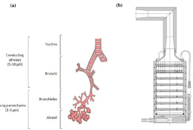
Figure II. 1 - Example of different inhalers (a) is a nebulizer, (b) a pDMI and (c) a dry powder inhaler.41………..………81Figure II. 2 - (a) Representation of the different stages of the respiratory tract and the particles’ deposition according to size. (b) Schematic representation of the Andersen Cascade Impactor, adapted from the Europeanpharmacopoeia. ……….……….83
Figure II. 3 - Cholesterol structure. ... 84
Figure II. 4 - Chitosan structure, where n is related to the DD % and m to (100 – DD %). Ideal chitosan would have m=0 and chitin would have n=0. Adapted from M. Dash et al. 53 and J. Kumirska et al. 52... 85
Figure II. 5 - PLGA structure, with the lactic acid between the left-side brackets and the glycolic acid between the right-side brackets. x and y represent the number of times the unit it repeated. ... 87
Figure II. 6 - Mechanism of the living cationic ring-opening polymerization of 2-ethyl-2-oxazoline. 67 ... 88
Figure II. 7 - Phase diagram of CO2, adapted from W. Leitner et al. 83... 90
Figure II. 8 - Representation of the atomization mechanism, adapted from E. Reverchon et al. 77 . 92 Figure II. 9 - VLE of water-CO2-ethanol system, adapted from C. Duarte et al.79 ... 92
Figure II. 10 - Schematic representation of the SASD apparatus: (CB) cryogenic bath; (LP) liquid pump; (HB) heating bath; (TC) temperatute controller; (M) manometer; (S) saturator; (P) precipitator; (c) cyclone. ... 99
Figure II. 11 - Schematic representation of the DUSA, adapted from Copley Scientific. ...101
Figure II. 12 - Experimental set-up to perform ACI (adapted from European Pharmacopeia). ...103
Figure II. 13 - 0.25% (w/v) CLT-IBP (QCO2/Qsol=5) microparticles. (A) SEM images of with a magnification of (a) 1,500x (b) 5,000x and (c) 10,000x and (B) Morphologi G3 images with magnification of (a) 20,000 and (b) 50,000 respectively. ...107
Figure II. 14 - 0.25% (w/v) CLT-IBP (QCO2/Qsol=8.3) microparticles. (A) SEM images of with a magnification of (a) 1,500x (b) 5,000x and (c) 10,000x and (B) Morphologi G3 images with magnification of (a) 20,000 and (b) 50,000 respectively. ...108
Figure II. 15 - 0.5% (w/v) CLT-IBP (QCO2/Qsol=5) microparticles. (A) SEM images of with a magnification of (a) 1,500x, (b) 5,000x and (c) 10,000x and (B) Morphologi G3 images with magnification of (a) 20,000 and (b) 50,000 respectively. ...108
XIII Figure II. 17 - 0.5% (w/v) CLT-PLGA (40:60) microparticles. (A) SEM images of with a
magnification of (a) 1,500x, (b) 5,000x and (c) 10,000x. ...109
Figure II. 18 - 0.5% (w/v) CLT-PLGA (50:50) microparticles. (A) SEM images of with a magnification of (a) 1,500x, (b) 5,000x and (c) 10,000x. ...109
Figure II. 19 - Figure 3.7 - 1% (w/v) CLT microparticles. (A) SEM images of with a magnification of (a) 1,500x, (b) 5,000x and (c) 10,000x and (B) Morphologi G3 images with magnification of (a) 20,000 and (b) 50,000 respectively. ...110
Figure II. 20 - 1% (w/v) CLT-PEtOx-OH microparticles. (A) SEM images of with a magnification of (a) 1,500x, (b) 5,000x and (c) 10,000x and (B) Morphologi G3 images with magnification of (a) 20,000 and (b) 50,000 respectively. ...110
Figure II. 21 - Figure 3.9 - 1% (w/v) CLT-PEtOx-IBP microparticles. (A) SEM images of with a magnification of (a) 1,500x, (b) 5,000x and (c) 10,000x and (B) Morphologi G3 images with magnification of (a) 20,000 and (b) 50,000 respectively. ...111
Figure II. 22 - Graphical representation of the powder distribution in the ACI stages for the CLT microparticles. (I.P. represents the induction port). ...112
Figure II. 23 - FTIR. (a) CLT microparticles, (b) PEtOx-OH microparticles and (c) CLT-PEtOx-IBP microparticles. ...114
Figure II. 24 - (A) In vitro drug release studies of PEtOx-IBP loaded in CLT microparticles at pH 7.4 and 35 ºC and (B) release profile fitted through Korsmeyer-Peppas mathematical model for the first 60% of release. ...116
Figure II. 25 - Korsmeyer-Peppas Model for mechanism of PEtOx-IBP release from the CLT microparticles at pH 7.4 and 35º C. ...117
Figure II. 26 - 1% (w/v) CHT microparticles. (A) SEM images of with a magnification of (a) 1,500x, (b) 5,000x and (c) 10,000x and (B) Morphologi G3 images with magnification of (a) 20,000 and (b) 50,000 respectively. ...120
Figure II. 27 - 1% (w/v) CHT-PEtOx-OH microparticles. (A) SEM images of with a magnification of (a) 1,500x, (b) 5,000x and (c) 10,000x and (B) Morphologi G3 images with magnification of (a) 20,000 and (b) 50,000 respectively. ...120
Figure II. 28 - 1% (w/v) CHT-PEtOx-IBP microparticles. (A) SEM images of with a magnification of (a) 1,500x, (b) 5,000x and (c) 10,000x and (B) Morphologi G3 images with magnification of (a) 20,000 and (b) 50,000 respectively. ...121
Figure II. 29 - 1% (w/v) CHT-PEtOx-PTM-029, F4, F39 microparticles. (A) Morphologi G3 images with magnification of (a) 20,000 and (b) 50,000 respectively. ...121
Figure II. 30 - Graphical representation of the powder distribution in the ACI apparatus for the CHT microparticles...122
Figure II. 31 - FTIR. (a) CHT microparticles, (b) PEtOx-OH microparticles and (c) CHT-PEtOx-IBP microparticles. ...124
Figure II. 32 - (A) In vitro drug release studies of PEtOx-IBP loaded in CLT microparticles at pH 7.4 and 35 ºC and (B) release profile fitted through Korsmeyer Peppas mathematical model for the first 60% of release. ...125
Figure II. 33 - Korsmeyer-Peppas Model for mechanism of PEtOx-IBP release from the CLT microparticles at pH 8 and 35º C. ...126
Annex 1. Figure 1 - 1H-NMR spectrum of PEtOx-OH in DMSO-d6………..137
Annex 1. Figure 2 - 13C-NMR spectrum of PEtOx-OH in DMSO-d6………..137
Annex 1. Figure 3 - 1H-NMR spectrum of PEtOx-IBP in DMSO-d6. ...138
Annex 1. Figure 4 - 13C-NMR spectrum of PEtOx-IBP in DMSO-d6. ...138
Annex 1. Figure 5 - 1H-NMR spectrum of PEtOx-PTM-029, F4, F39 in DMSO-d6. ...139
XV
Index of Tables
Part I
Table I. 1 - Antibacterial activities and mass of the PTM-029 fractions obtained from the crude extract. ... 22 Table I. 2 - The eight fractions from PTM-029 with their total of pure compounds. ... 23 Table I. 3 - Antibacterial activity and respectively mass of pure compounds of F4 from PTM-029…. ... …24 Table I. 4 - NMR spectroscopic data for a napyradiomycin derivative PTM-029, F4, F39 a ...27
Part II
Table II. 1 - Comparison between the different inhalers relatively to different
characteristics………80
Table II. 2 - Composition of casting solutions with various concentrations of 0.25%, 0.5% and 1% (w/v) of total solids used for the preparation of CLT microparticles……...………..97 Table II. 3 - Composition of casting solutions with 1% (w/v) of total solids used for the preparation
of CHT microparticles ……… 98
Table II. 4 - Operating parameters of the CLT assays and the respective yields. ...105 Table II. 5 - Properties of CLT microparticles. ...106 Table II. 6 - Representation of the aerodynamic properties determined by ACI and DUSA for the
CLT microparticles. ...113 Table II. 7 - Water content values for 1% (w/v) CLT, 1% (w/v) CLT-PEtOx-OH and 1% (w/v)
CLT-PEtOx-IBP microparticles. ...115 Table II. 8 - Kinetic values obtained from the Korsmeyer equation for the 1% (w/v) CLT-PEtOx-IBP microparticles. ...116 Table II. 9 - Operating parameters of the CHT assays and the respective yields. ...118 Table II. 10 - Properties of CHT microparticles. ...118 Table II. 11 - Representation of the aerodynamic properties determined by ACI and DUSA for the
CHT microparticles. ...123 Table II. 12 - Water content values for the CHT, CHT-PEtOx-OH and CHT-PEtOx-IBP
XVII
List of Abbreviations
ACI: Andersen cascade impactor API: Active pharmaceutical ingredient ACN: Acetonitrile
BSA: Bovine serum albumin CHT: Chitosan
CLT: Cholesterol EtOx: 2-ethyl-oxazoline
13C-NMR: Carbon-13 nuclear magnetic resonance
CROP: cationic ring-opening polymerization COPD: Chronic obstructive pulmonary disease COSY: Correlation Spectroscopy
Da: Aerodynamic diameter DAD: Diode array detector
DMAPP: Dimethylallyl diphosphate DMSO: Dimethyl Sulfoxide
Dn,50: Particle mean numeric diameter
DPI: Dry powder inhaler
DUSA: Dosage unit sampling apparatus Dv,50: Particle mean volumetric diameter
EF: Emitted fraction
FDA: Food and Drug Administration FPF: Fine particle fraction
FPP: Farnesyl diphosphate FTIR: Fourier transform infrared GGPP: Geranylgeranyl diphosphate GPP: Diphosphate
GSD: Geometric standard deviation HI: Hybrid isoprenoids
HMBC: Heteronuclear Multiple-Bond Correlation HPLC: High-performance liquid chromatography
1H-RMN: Proton nuclear magnetic resonance
XVIII IBP: Ibuprofen
IPP: Isopentenyl diphosphate
LC-MS: Liquid Chromatography–Mass Spectrometry MEP: Methylerythritol phosphate pathway
MMAD: Mass median aerodynamic diameter MIC: Minimum Inhibitory Concentration
MRSA: Methicilin-resistant Staphylococcus aureus NGI: Next Generation Impactor
pDMI: Pressurized metered dose inhalers PEG: Polyethylene glycol
PEtOx-IBP: 2-ethyl-oxazoline end-capp with Ibuprofen salt
PEtOx-PTM-029, F4, F39: 2-ethyl-oxazoline end-capp with marine bioactive compound PEtOx-OH: 2-ethyl-oxazoline end-capp with water
PLGA: Poly (lactic-co-glycolic acid) PTases: Prenyltransferases enzymes
PTM- # - the strain code and /or extract obtained from ocean sediments of Archipelago Madeira PS: Particle size
PSD: Particle size distribution
RESS: Rapid expansion of a supercritical solution SAS: Supercritical antisolvent
SASD: Supercritical assisted spray-drying scCO2: Supercritical carbon dioxide SEM: Scanning electron microscope
THN: Pentaketide 1,3,6,8-tetrahydroxynaphthalene Tg: Glass transition temperature
TOCSY: Total Correlation Spectroscopy
V-CIPOs: Vanadium-dependent haloperoxidases VLE: High-pressure vapor liquid equilibria
1
Overview
This thesis theme emerges from the need to fight lower respiratory infections which affects the lower airways (trachea, bronchi, bronchioles and lungs) caused by pathogenic bacteria, virus or fungi.4 In 2013 infections such as pneumonia, pulmonary tuberculosis, bronchitis, among others were the leading cause of sickness and mortality, both in children and adults, causing 2.7 million deaths worldwide, 4.8% of the overall deaths.1 Therefore consists in a health, societal and economic
problem. The treatment of such infections relies on antibiotic therapy. However, antibiotic resistance to human pathogens continues to increase, as a consequence of the misuse of antibiotics and pathogen evolution.2,5 It is, more than ever, imperative not only to discover new lead-like drug
3
Part I - Isolation of bioactive compounds from marine
Actinobacteria
1. Introduction
1.1 Natural products
Natural products are chemical compounds produced by living organisms that are found in nature, from prokaryotic bacteria and eukaryotic microorganisms to fungi, plants and animals.6
These compounds are produced by the pathways of primary or secondary metabolism from living organisms. The primary metabolites (amino acids, nucleotides, and vitamins) are common in all biological systems. The secondary metabolites are, however, low molecular weight (MW<3000), chemically diverse compounds, usually exhibiting a wide range of biological activities.6
Since the discovery of the antibiotic properties of penicillin by Alexander Fleming in 1928, through the “golden age” of antibiotics and into to our days, the result of this frenzied research lead to the discover of ten thousands of natural products derived from microbial sources. Even though, drug discovery strategies changed in the 1990s, techniques such as combinatorial chemistry, high-throughput screening, and computer-assisted design of small-molecule ligands created alternatives to traditional drug discovery paradigms. Yet like many trends, the interest towards these fields showed some cyclic features with successes and failures associated with the evolution around the clinical needs and new enabling technology. The renewed interest in drug discovery comes at a time when it is widely perceived that the pipeline for new antibiotics discovery is running dangerously low. It is also a response to the realization that bacterial diversity and major environmental habitats have not been efficiently explored.6,7
Given the vastness of the world’s oceans (70% of the Earth’s surface) harboring most of the planet’s biodiversity, the oceans are a highly complex microbiological environment with typical microbial abundances of 106 and 109 per ml in seawater and ocean-bottom sediments, respectively.
As DNA sequencing based methods are applied to the field of marine microbial ecology, it is known how complex, exquisitely unique and highly adapted these organisms are.
4
1.2 Actinomycetes from marine sources
Bacteria are an exceptional source of chemical diversity. Among them, species of the Order Actinomycetales (commonly called actinomycetes), a group of Gram-positive bacteria, are the single most productive source of microbial derived natural products, accounting for ca. 75% of all antibiotics discovered from the genus Streptomyces, as well as a broad range of anticancer agents.6,7,8 Therefore, marine bacteria are an important resource for drug discovery.
Although actinomycetes are best known as soil bacteria, a growing interest in its distribution and ecological role in the marine environment has been observed. Thus, in the last decades several research efforts have been done to tackle the marine actinomycetes biodiversity, which allowed to discover a significant number of new marine-derived actinomycetess belonging to the genus Micromonospora, Streptomyces, Nocardia, Rhodococcues, Dietzia, Prauserella, Serinicoccus, Marinophilus, Solwaraspora, Lamerjespora, Aeromicrobium,
5 Figure I. 1 -Radial tree depicting the phylogenetic relationships of 13 groups of actinomycetes within six
different families.7
1.3 Actinomycetes terpenoids
Terpenoids play important roles in all living organisms; they function as steroid hormones in mammals, carotenoids in plants, and ubiquinone or menaquinone in bacteria. They are the most ubiquitous class of metabolites observed in nature that provide diverse functions in both primary and secondary metabolism. Also, they are assembled from five-carbon isoprene subunits into a wide assortment of structures that generally range from monoterpenes (two isoprene units) to triterpenes (six isoprene units), and in certain cases can also be larger [monoterpenes (C10), sesquiterpenes (C15), and diterpenes (C20)].10,11,12
Although actinomycetes bacteria are one of the primary sources of microbial derived secondary metabolites, they rarely produce compounds with this biosynthetic class.10 Terpenoid
moieties can be attached to molecules produced via non-terpenoid biosynthetic routes forming products known as hybrid isoprenoids (HIs). As secondary metabolites, they are common in plants, insects, fungi, and some marine invertebrates such as soft corals.13 They are known to play diverse
6 odors and flavors associated with plants and fruits. HIs include significant structural diversity and biological activity and thus are important targets for drug discovery.11
1.4 Hybrid isoprenoids classes obtained from marine
environmental bacteria sources
7 Figure I. 2 - Structures of hybrid isoprenoids produced by actinomycetes. Isoprenoid portions of compounds
are indicated in red11
1.5 Phylogenetic and chemical diversity of a
hybrid-isoprenoid-producing streptomycete lineage
8 A phylogenetic analysis of HI producers reveals how they are sporadically distributed throughout the Streptomyces phylogenetic tree. From all the thirteen MAR groups, a robust clade enriched in strains is able to produce this class of compounds. From phylogenetic studies this clade is identified as a marine lineage within the genus Streptomyces, and it was given the designation MAR4. 8,11
The MAR4 clade currently includes 57 strains, 47 of which were isolated from marine sediments. To date, are recognized within this clade, the type strains of S. aculeolatus and S. synnematoformans, which were isolated from a terrestrial soil sample and a sand dune sample, respectively, being the only formally described species within the clade (Figure I. 3). The 57 members of the MAR4 clade share as little as 96.2% 16S rRNA gene sequence identity and thus contain as much phylogenetic diversity as many bacterial genera. 8
9 Figure I. 3 - 16S rRNA gene phylogeny of the MAR4 clade. * and ** indicate that the clades represented by
the S. aculeolatus and S. synnematoformans type strains, respectively. 8
10 Figure I. 4 - Hybrid isoprenoids structure classes detected from MAR4 strains. The terpene-derived portion of
each molecule is highlighted in red. 8
11
1.6 Biosynthesis of hybrid isoprenoids from actinomycetes
sources
Terpenoids are synthesized by consecutive condensations of an isoprene unit, isopentenyl diphosphate (IPP), to its isomer, dimethylallyl diphosphate (DMAPP). 10
13 Figure I. 5 - (A) Mevalonate pathway diagram showing the conversion of acetyl-CoA into isopentenyl pyrophosphate, the essential building block of all isoprenoids. The eukaryotic variant is shown in black.
Archaeal variants are shown in red and blue. (B) Non-Mevalonate pathway.10
In eukaryotes such as plants and fungi, which produce the vast majority of isoprenoids, the polyprenyl diphosphate is generally cyclized by an organism-specific isoprenoid cyclase to form a basic skeleton. This is followed by successive reactions, such as hydroxylation, methylation, and glycosylation that give rise to many thousands of compounds. In contrast to eukaryotes, prokaryotes are known to produce a limited number of isoprenoids. Moreover, it was recently revealed that among prokaryotes, actinomycetes produce many types of isoprenoids in relatively large numbers, and the structures of these compounds are unique and different from those of eukaryotic origin. In particular, isoprenoid moieties of the compounds produced by actinomycetes are generally attached to other moieties, such as an aromatic ring, an amino acid, and a phenazine moiety.12,14
16
1.7 Napyradiomycins
Within MAR4 clade, a group of novel antibacterial agents, napyradiomycins, was first isolated from the soil-derived bacterium Chainia rubra G802-AF, from Japan in 1986. As time passed, a series of congeners were isolated from different actinomycetes, such as Streptomyces antimycoticus, S. aculeolatus, and other Streptomyces species, and also from a marine source derived strain. The napyradiomycins are meroterpenoids composed of polyketide and terpenoid components which are formed by a process that involves halogen-induced cyclization. In annex 1 are listed all the napyradiomycins discovered since 1986 until now, in the MAR4 clade. 2
Structurally, napyradiomycins consist of a semi-napthoquinone core, a prenyl unit attached at C-4a that is cyclized to form a tetrahydropyran ring in most cases, and a monoterpenoid substituent attached at C-10a. Structure variations of napyradiomycins arise mainly from the 10-carbon monoterpenoid subunit, which is linear (napyradiomycin A series), or cyclized to a 6-membered ring (napyradiomycin B series), or cyclized to a 14-6-membered ring (napyradiomycin C series) (Figure I. 7). The different halogenation patterns also contribute largely to structure variations of napyradiomycins. 15
17 Napyradiomycins family has a characteristic UV profile at maximum of absorbance of 260 nm which allows their easy identification (Figure I. 8).
Figure I. 8 -UV profile of napyradiomycins family.
Biosynthetically, napyradiomycins originate from a HI / polyketide pathway which is rare in nature. Bacterial meroterpenoid antibiotics belonging to the napyradiomycin family of chlorinated dihydroquinones suggests that the biosynthetic cyclization of their terpenoid subunits is initiated via a chloronium ion. The vanadium-dependent haloperoxidases that catalyze such reactions are distributed in fungi and marine algae. 16
Winter et al. in 2007 studied the napyradiomycin biosynthetic cluster (nap) and, not only demonstrated that the enzymes were able to initiate cyclization of a terpene by a bromonium ion, but also proved that the halogenation reaction occurred with stereochemical control.16
Napyradiomycins family of chlorodihydroquinones are biosynthesized from the symmetrical pentaketide 1,3,6,8-tetrahydroxynaphthalene (THN) and isoprenoid units. In most cases, the isoprenoid building blocks are derived from the mevalonic acid biosynthetic pathway. A biosynthetic scheme for the production of this compound was proposed on the basis of the molecular logic of the nap cluster (Figure I. 9). The dihydroquinone core is catalyzed by the type III polyketide synthase homologous THN synthase NapB1, which condenses five malonyl-CoA molecules to THN. The monooxygenase NapB2 putatively oxidizes THN to flaviolin, which is then methylated by the methyltransferase NapB5. Attachment of the first isoprene unit, using dimethylallyl pyrophosphate as the substrate, occurs through a nucleophilic attack involving one of the two prenyltransferases (NapT8/T9). Hydrogenation or chlorination at C-2 by the
FADH2-Wavelength (nm)
A
bs
orba
nc
18 dependent halogenase NapH2 may facilitate the second prenylation reaction at C-3 with geranyl pyrophosphate by the second nap prenyltransferase to yield the diprenylated intermediate SF2415B1. Cyclization of the hemiterpene subunit via a chloronium ion is putatively catalyzed by one of the three nap V-ClPOs (NapH1, H3, H4) giving rise to the 7-methyl derivative of napyradiomycin A1. The monoterpene subunit of 7-methylnapyradiomycin A1 putatively undergoes a related V-ClPO-facilitated cyclization to form 1 and 3, in which a molecule of water is incorporated into the cyclohexanol moiety. Further dechlorination of 1 likely gives rise to 2 (Figure I. 9). 16
Figure I. 9 - Structures and proposed biosynthetic pathway of the chlorinated dihydroquinones 1–3.16
Napyradiomycins exhibit a diverse range of biological activities, including antimicrobial activity against methicillin-resistant Staphylococcus aureus (MRSA) or vancomycin-resistant Enterococcus (VRE) , cytotoxicity against HCT-116 human colon adenocarcinoma cells (Annex 2), gastric (H+-K+) ATPases inhibiting activities, as estrogen receptor antagonists, as well as
19
2. Experimental Section
This work was conducted under the project: "Tesouros Oceânicos- Sedimentos oceânicos do arquipélago da Madeira: nova fonte de compostos inovativos e bioactivos" ref. PTDC / QUI-QUI / 119116/2010. The global procedure steps are outlined in the following diagram:
Sample collection of ocean sediments
Inoculation of samples from ocean sediments Isolation and purification of actinobacteria
Achievement of the chemical extracts
Taxonomic description. DNA extraction and sequencing of
16S rRNA Determination of
the biological activity Purification of the
active chemical extracts Determination of the biological activity Purification and isolation of compounds from bioactive fractions
Determination of the biological
activity
Structural elucidation of bioactive compounds
20
2.1 Materials
All chemicals and solvents were used without further purification. Ethyl acetate (99.8% purity), isooctane (99.5% purity), methanol (99.9% purity), water and acetonitrile (99.9% purity, HPLC grade) were purchased from ProLabo. Silica gel 60 was purchased from Merck, sea and sand washed from Panreac, chloroform-D (≥ 99.8% purity) and dimethyl Sulfoxide (99.9% purity) from Cambridge Isotope Lab. and trifluoroacetic acid (≥ 99.9% purity) from Sigma-Aldrich.
2.2 General Experimental Procedures
1D and 2D NMR spectroscopic data were performed with a Bruker Advance 400MHz with samples in CDCl3. Low-resolution LC/MS data were measured using a Finnigan Surveyor by
Thermo ScientificTM equipped with a reversed-phase C
18 column (Phenomenex Luna, 4.6 x 100 mm,
5 µm) at a flow rate of 0.9 mL/min. High-resolution mass spectra were obtained using an GC-TOF method. HPLC purifications were performed using a Dionex Ultimate 3000 system controller and pumps with a model 300 spectrophotometer, using Phenomenex Luna semipreparative C18 (250 ×
10 mm, 5 μm) column at flow rates of 1.5 mL/ min with various ratios of ACN/H2O gradient.
2.3 Collection, Identification, Cultivation, and Extraction of
Strain PTM-029
21
2.4 Isolation of Napyradiomycins
The extract was fractionated by silica flash chromatography using various ratios of isooctane/EtOAc (100:0, 80:20, 60:40, 40:60, 20:80 and 0:100) and methanol/EtOAc (10:90, 50:50 and 100:0) to produce 9 fractions of increasing polarity. These fractions were then analyzed by LC-MS. Pure compounds were obtained by HPLC (Table I. 2).
2.5 Antibiotic Assay
The antibiotic assays were performed by the laboratory technician Tiago Dias.
Methicillin-resistant Staphylococcus aureus (MRSA)17 and vancomycin-resistant
Enterococcus faecium EF82 (VRE) strains were grown overnight at 37 °C in BHI media (17.5g brain heart infusion, 10g enzymatic digest of gelatin, 2g dextrose, 5g sodium chloride and 5g disodium phosphate). The culture was diluted to 0.04-0.06 OD, further diluted 1:10, and then added to the top row of a 96-well microtiter plate. Test materials were added to the top row, serially diluted (1:2) downward, and incubated for 18-24 h at 37 °C. DMSO was used to monitor the viability of the culture without the crude extract, vancomycin solution was used to monitor the cell growth inhibition of MRSA strain and if does not inhibit the culture of VRE EF82 strain. Optical density (OD) was measured at 600 nm using a Ultrospec™ 3100 pro UV/Visible reader.
(A) (B) (C)
Figure I. 11 - (A) PTM-029 Streptomyces aculeolatus strain, (B) PTM-029 culture in 15 L medium and (C)
22
3. Results
3.1. PTM-029 crude extract, fractionation, and antibacterial
activity assessment
After PTM-029 fractionation, biological tests were performed to determine the minimum inhibitory concentration (MIC) as described in chapter 2.5, against pathogenic bacteria MRSA and VRE EF82. Table I.1 describes the biological activity obtained for the PTM-029 fractions.
Table I. 1 - Antibacterial activities and mass of the PTM-029 fractions obtained from the crude extract.
PTM-029 fraction Mass of the fraction (g)
MIC (µg/mL)
MRSA VRE EF82
F1 0.0492 - -
F2 0.1717 62.50 62.50
F3 0.1689 15.60 31.30
F4 0.1016 7.81 3.91
F5 0.0532 15.60 3.91
F6 0.0664 31.30 3.91
F7 0.1809 0 31.30
F8 0.1126 0 250
23
3.2. Isolation of PTM-029 compounds by HPLC and its
antibacterial activity
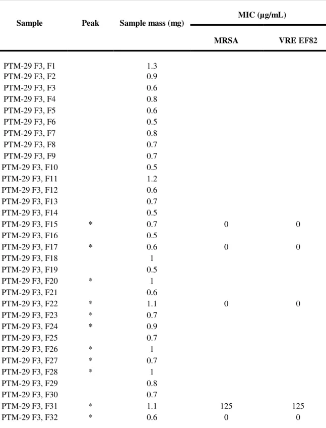
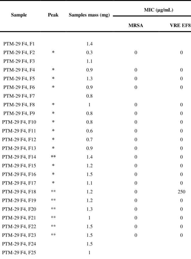
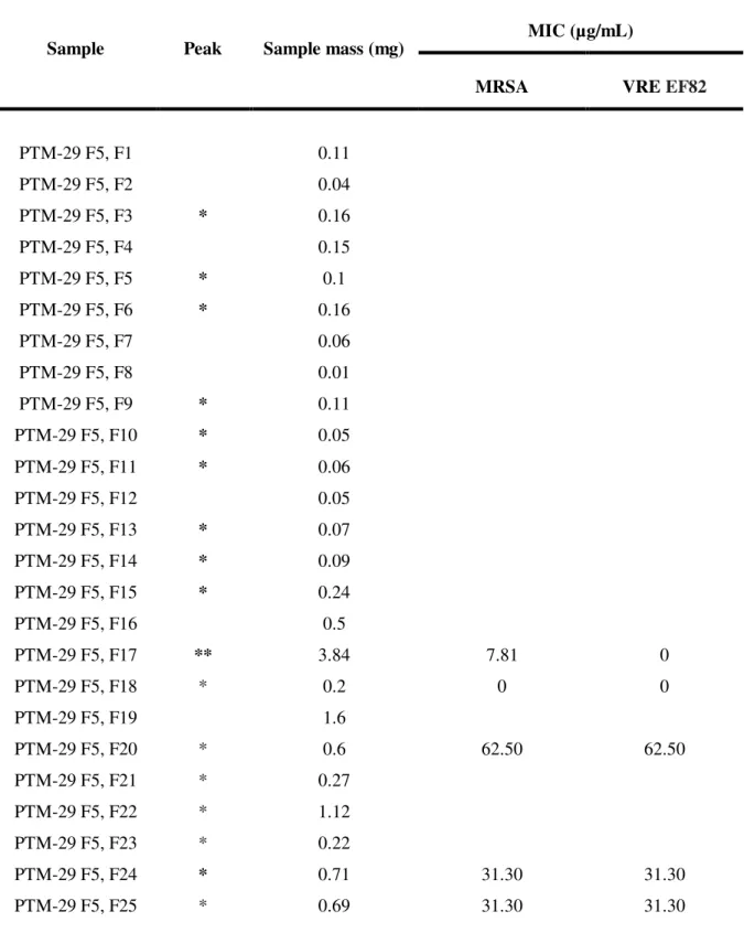
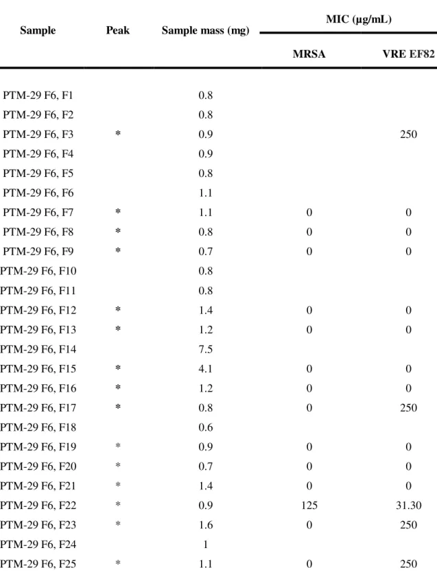
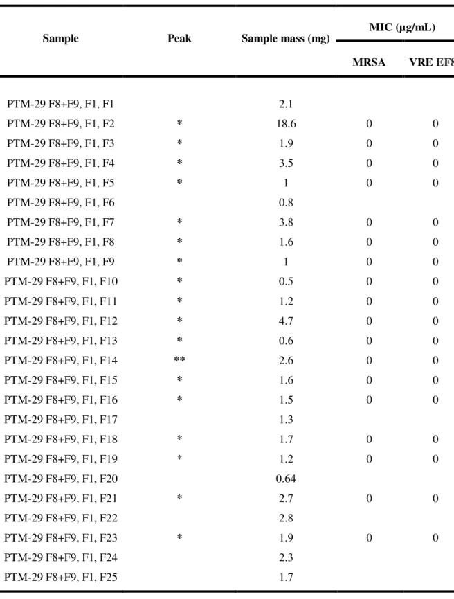
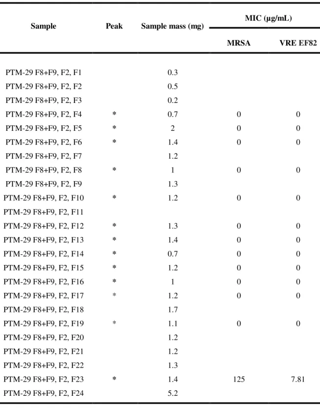
The fractions from PTM-029 that showed biological activity, proceeded to separation of the existing compounds by HPLC, as described in chapter 2.2 and 2.4. The HPLC chromatograms of PTM-029 fractions can be seen in Annex 3, as well as the pure compounds that are highlighted in each one.
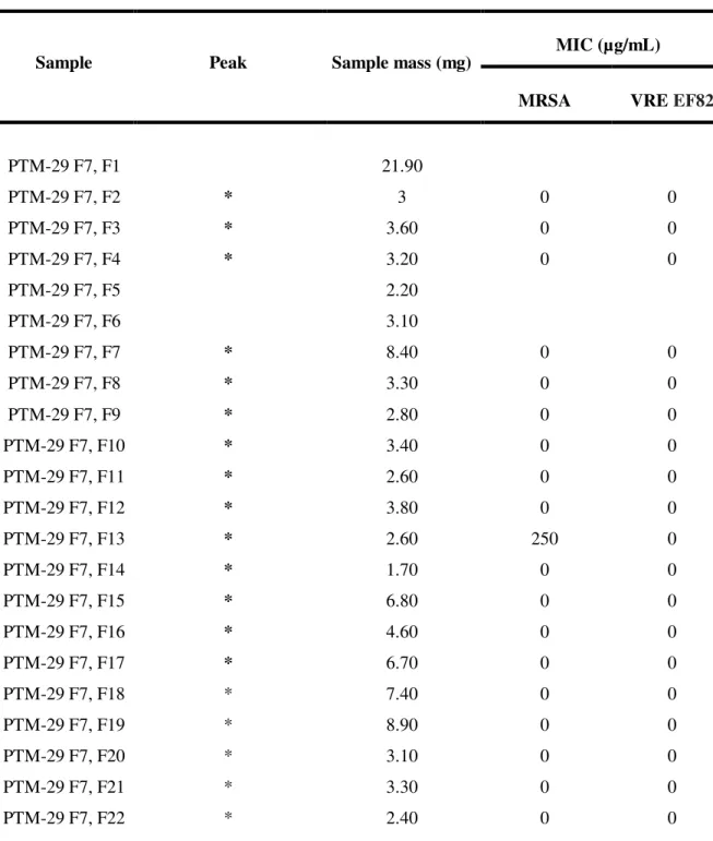
The pure compounds were also subjected to biological tests against pathogenic bacteria MRSA and VRE EF82, as described in section 2.5 and the values of the biological activity can be seen in Annex 4. In Table I. 2 are presented the total pure compounds obtained from each fraction of PTM-029 and in Table I. 3 the antibacterial activity of fraction 4, and the most promising compound PTM-029,F4,F39 proceeded for structure elucidation.
Table I. 2 - The eight fractions from PTM-029 with their total of pure compounds.
Fraction Total pure compounds
Number of
bioactive compounds
Napyradiomycin
UV profile
F2 34 8 5
F3 63 24 8
F4 49 21 18
F5 46 23 6
F6 60 35 4
F7 46 20 9
24 Table I. 3 - Antibacterial activity and respectively mass of pure compounds of F4 from PTM-029.
Sample Samples mass (mg)
MIC (µg/mL)
MRSA VRE EF82
PTM-29 F4, F27 2 62.50 125
PTM-29 F4, F28 2.80 31.30 0
PTM-29 F4, F29 2.60 62.50 62.50
PTM-29 F4, F30 2.50 62.50 62.50
PTM-29 F4, F31 2 31.30 15.60
PTM-29 F4, F32 3.10 250 0
PTM-29 F4, F33 2.60 31.25 250
PTM-29 F4, F34 2.40 62.5 125
PTM-29 F4, F35 1.20 31.30 15.60
PTM-29 F4, F36 3.10 62.50 250
PTM-29 F4, F37 1.40 62.50 15.60
PTM-29 F4, F38 1.80 3.91 0.24
PTM-29 F4, F39 13.20 1.95 1.95
PTM-29 F4, F40 3.40 3.91 0.98
PTM-29 F4, F42 5.30 3.91 0.98
PTM-29 F4, F44 6.10 3.91 0.98
PTM-29 F4, F45 3.40 3.91 1.95
PTM-29 F4, F46 1.60 15.60 7.81
25
3.3 PTM-029,F4,F39 Structure elucidation
It was performed the structural elucidation in a selected pure compound PTM-029 F4, F39 by NMR spectroscopy ( 1H, 13C, DEPT 135, COSY 1H-1H, TOCSY 1H- 1H, HSQC 1H-13C, HMBC 1H-13C and ROESY 1H-1H), the spectra are in Annex 5.
PTM-029, F4, F39: Yellow oil (13.2 mg); UVmax: 260 nm; 1H NMR (400 MHz, CDCl3):δ 12.28
(s, 3H), 7.93 (s, 3H), 6.55 (s, 3H), 4.57 (dd, J = 11.9, 3.7 Hz, 3H), 3.43 (dd, J = 12.1, 3.5 Hz, 3H), 2.73 – 2.35 (m, 10H), 2.22 (s, 9H), 2.20 – 1.66 (m, 16H), 1.66 – 1.55 (m, 11H), 1.38 (dd, J = 30.7, 12.0 Hz, 27H), 1.29 – 0.75 (m, 16H), 0.98 – 0.84 (m, 3H), 1.08 – 0.75 (m, 11H), 0.73 (d, J = 18.6 Hz, 10H), 0.47 (s, 1H), 0.42 (s, 9H). 13
C NMR (101 MHz, CDCl3): δ 192.62, 190.98, 164.37,
26
4. Discussion
PTM-029, F4 was the fraction with the best antibacterial activity, since it shows the lowest MIC against the two pathogenic bacteria, MRSA (7.8 μg/mL) and VRE EF82 (3.9 μg/mL). PTM-029, F8 was the fraction with the lower antibacterial activity with a MIC of 250 μg/mL against VRE
EF82.
The focus of this work turned to isolate pure bioactive compounds belonging to the napyradiomycins family by HPLC. PTM-029, F4 was also the one with the higher amount of pure compounds possible belonging to the napyradiomycins family. From all the 49 isolated compounds, obtained from PTM-029,F4, the compound with the higher antibacterial activity against MRSA was PTM-029, F4, F39 with a MIC of 1.9 μg/mL and against VRE EF82 was fractions PTM-029, F4, F40; PTM-029, F4, F42 and PTM-029, F4, F44 with the same MIC of 0.98 μg/mL.
PTM-029, F4, F39 was the most promising compound not only due to the presence of napyradiomycins UV profile (Annex 3, Chromatogram 3), but also because it was the compound obtained in higher amount (13.2 mg) and the most active, i.e, with the lower MIC against MRSA (1.95 µg/mL) and VRE EF82 (1.95 µg/mL). Exhibiting or meeting, the requirements to procedure the structure elucidation.
27 Table I. 4 - NMR spectroscopic data for a napyradiomycin derivative PTM-029, F4, F39 a
Carbon
no.
δc, type
δH, mult.
(J in Hz) COSY TOCSY HMBC ROESY
1 72.28, C -
2 40.79, CH2
1.78, m
1.94, m H3 H3,H4,H6 C1
3 29.79, CH2
1.44, m
1.94, m H2, H4 H2, H4 C1, C5
4 71.03, CH 3.43 dd
(12.2, 3.5) H3 H2, H3, H22
C2, C22,
C23 H21, H22
4 6.55, s (OH) C5 H6, H24,
H25
5 40.51, C -
6 52.21, CH 1.44, m H7 H7, H22 C1, C7,
C22, C23
H4, OH4, H22
7 38.22, CH2 1.59, m
2.51, m
H6 H6, H22, H23 C1, C6, C8, C20
8 85.58, C -
9 81.52, C -
10 57.89, CH 4.57 dd
(11.9, 3.5)
H11 H11, H24,
28 Carbon
no.
δc, type
δH, mult.
(J in Hz) COSY TOCSY HMBC ROESY
11 42.06, CH2
2.51, m 2.64, dd (14.2, 3.8)
H10 H10, H11 C12, C10
12 80.33, C -
13 192.63, C -
14 107.05, C -
15 163.34, C 12.28,s (OH) C14, C15,
C16
16 120.40, C -
17 164.37, C
18 108.69, CH 7.93, s H26 C10, C14,
C16,
19 131.81, C -
20 190.98, C -
21 24.38, CH3 1.33, s H3, H4
C1, C5,
C6 OH4, H4
22 28.51, CH3 0.42, s
H4, H6, H7, H23
C4, C6, C5, C23
H4, H6, H23
23 15.68, CH3 0.76, s H6, H7, H22
C4, C6,
29 Carbon
no.
δc, type
δH, mult.
(J in Hz) COSY TOCSY HMBC ROESY
25 22.95, CH3 1.37, s H24 C9,C10,
C24
26 8.44 CH3 2.22, s H18 C15, C16,
C17, C18
a Spectra were recorded in CDCl
3 at 400 MHz (1H) and 101 MHz (13C). Assignments were made on
the basis of DEPT, HSQC and HMBC sequence experiments.
The structure of this pure bioactive compound, PTM-029, F4, F39 was established by interpretation of spectroscopic data, especially 2D NMR spectroscopic data. The complex molecular ion cluster in the ESI mass spectrum of PTM-029, F4, F39 clearly showed the presence of three chlorine atoms in the molecule. HR-Mass analysis suggested the molecular formula C26H33Cl3O6 ([M - H]- m/z (obsd) 545.127), indicating 9° of unsaturation. PTM-029, F4, F39
showed strong UV absorptions at 210, 260, 320 and 360 nm, consistent with
characteristic UV
profile of a napyradiomycin derivative (
Figure I. 8). Analysis of 1H and 13C NMR and 2Dspectral data (Table I. 4) and comparison with the same data from compound A-80915C (Annex 1, compound (25)) allowed the structure of PTM-029, F4, F39 to be assigned. 18 The 1H NMR
spectrum of PTM-029, F4, F39 illustrated a single aromatic proton (C-18, δ 1H 7.93 s), one
aromatic methyl group (C-26, δ 1H 2.22 s), five quaternary methyl groups (C-21-C-25), one methine
proton adjacent to chlorine (C-10 δ 1H 4.57 dd, J = 11.9 Hz) and another one methine proton
adjacent to a hydroxyl group (C-4 δ 1H 3.43 dd, J = 12.1 Hz). Analysis of HMBC and HSQC NMR
data confirmed these assignments and allowed other proton assignments to be made. Two low field aromatic carbons, C-15 and C-17 (δ 13C 163.34 and 164.37, respectively), suggested the presence of
two phenolic hydroxyl groups. Besides, 1H NMR spectrum showed the lability of OH-15 proton (δ 1H 12.28 s) since it formed a hydrogen bridge with quinone’s ketone C- 13, changing the chemical
shift to lower field compared with the other OH-4 (δ 1H 6.55, s). These observations suggested that
C-30 21, -22, -32) in PTM029, F4, F39 showed that it formed a typical chair cyclohexane ring. Analysis of NMR data showed this ring is not identical to the one observed in the structure of A80915C18,19
(Annex 1, compound (25)) suggesting that in this ring the chlorine and the hydroxyl group can switch positions confirmed by the appearance of a new signal 6.55 ppm by 1H NMR spectrum,
corresponding to the hydroxyl group attached to C-4 and it correlation with C-5 in HMBC spectrum. The relative stereochemistry was assigned by interpretation of 2D NMR ROESY data. ROESY correlations between the C-22 methyl protons and the methine protons at C-4 and C-6 showed these protons were on the same face of the cyclohexane ring. Correlations between the C-21 and C-23 methyl group protons showed that the orientation of the C-21 and C-23 methyl goups were α (axial) and on the bottom face of the ring.
Relatively to the antibacterial activity between A80915C19 (Annex 2, compound (25)) and
31
5. Conclusion
In this work 323 compounds were isolated and purified from the Streptomyces aculeolatus, PTM-029 obtained from ocean sediments and 148 were bioactive compounds being 51 from the napyradiomycins family. Napyradiomycin-derived secondary metabolites were the most predominantly produced by this strain. A novel napyradiomycin derivative, PTM-029, F4, 39, exhibiting antimicrobial activity against pathogenic bacteria MRSA (1.95 µg/mL) and VRE
EF82 (1.95 µg/mL) was structurally elucidated. This compound is structurally related with the previously reported compound A80915C 18, 19 (Annex 1, compound (25)). In future work, it
would be interesting to study the biosynthetic differences between compound A80915C and PTM-029, F4, F39, performing full genome sequencing.
32
6. References
1. Draft, C. et al. Global , regional and national levels of age-specific mortality and 240 causes of death , 1990-2013 : A systematic analysis for the Global Burden of Disease Study 2013. The Lancet 385, (Elsevier Ltd, 2015).
2. Cheng, Y.-B., Jensen, P. R. & Fenical, W. Cytotoxic and Antimicrobial Napyradiomycins from Two Marine-Derived Streptomyces Strains. European J. Org. Chem. 2013, 3751–3757 (2013).
3. Ungaro, F. & Vanbever, R. Improving the efficacy of inhaled drugs for severe lung diseases: Emerging pulmonary delivery strategies. Adv. Drug Deliv. Rev. 75, 1–2 (2014).
4. Dasaraju PV, L. C. in Medical Microbiology (ed. S, B.) 93 (1996).
5. Haste, N., Farnaes, L., Perera, V. & Fenical, W. Bactericidal kinetics of marine-derived napyradiomycins against contemporary methicillin-resistant Staphylococcus aureus. Mar. Drugs 9, 680–689 (2011).
6. Bérdy, J. Bioactive microbial metabolites. J. Antibiot. (Tokyo). 58, 1–26 (2005).
7. Fenical, W. & Jensen, P. R. Developing a new resource for drug discovery: marine actinomycete bacteria. Nat. Chem. Biol. 2, 666–673 (2006).
8. Gallagher, K. a, Rauscher, K., Pavan Ioca, L. & Jensen, P. R. Phylogenetic and chemical diversity of a hybrid-isoprenoid-producing streptomycete lineage. Appl. Environ. Microbiol. 79, 6894–902 (2013).
9. Subramani, R. & Aalbersberg, W. Marine actinomycetes: An ongoing source of novel bioactive metabolites. Microbiol. Res. 167, 571–580 (2012).
10. Kuzuyama, T. & Seto, H. Diversity of the biosynthesis of the isoprene units. Nat. Prod. Rep. 20, 171–183 (2003).
11. Gallagher, K. a, Fenical, W. & Jensen, P. R. Hybrid isoprenoid secondary metabolite production in terrestrial and marine actinomycetes. Curr. Opin. Biotechnol. 21, 794–800 (2010).
12. Kawasaki, T. & Hayashi, Y. Biosynthesis of a natural polyketide-isoprenoid hybrid compound, furaquinocin A: identification and heterologous expression of the gene cluster. J. Bacteriol. 188, 1236–1244 (2006).
13. Pawlik, J. R. Marine chemical ecology. Mar. Ecol. Prog. Ser. 207, 225–226 (2000).
33 15. Wu, Zhengchao; Li, Sumei, et al. Antibacterial and Cytotoxic New Napyradiomycins from
the. Mar. Drugs 11, 2113–2125 (2013).
16. Winter, Jaclyn M., Moffitt, Michelle C., et al. Molecular Basis for Chloronium-mediated Meroterpene Cyclization CLONING, SEQUENCING,ANDHETEROLOGOUS EXPRESSION OF THE NAPYRADIOMYCIN BIOSYNTHETIC GENE CLUSTER. J. Biol. Chem. 282, 16362–16368 (2007).
17. Gill SR, et al. Insights on evolution of virulence and resistance from the complete genome analysis of an early methicillin-resistant Staphylococcus aureus strain and a biofilm-producing methicillin-resistant Staphylococcus epidermidis strain. J. Bacteriol. 187, 2426– 2438 (2005).
18. Soria-Mercado, I. Antibiotic terpenoid chloro-dihydroquinones from a new marine actinomycete. J. Nat. Prod. 68, 904–910 (2005).
19. Soria-Mercado, I. E., Jensen, P. R., Fenical, W., Kassel, S. & Golen, J. 3,4a-Dichloro-10a- (3-chloro-6-hydroxy-2,2,6-trimethylcyclohexylmethyl)-6,8-dihydroxy-2,2,7-trimethyl-3,4,4a,10a-tetrahydro-2 H -benzo[ g ]chromene-5,10-dione. Acta Crystallogr. Sect. E Struct. Reports Online 60, o1627–o1629 (2004).
20. Shiomi, K. et al. STRUCTURES OF NEW ATIBIOTICS NAPYRADIOMYCINS. J. Antibiot. (Tokyo). XXXIX, 494–501 (1986).
21. Shiomi, K., Nakamura, H. & Iinuma, H. NEW ANTIBIOTIC NAPYRADIOMYCINS A2 AND B4 AND STEREOCHEMISTRY OF NAPYRADIOMYCINS. J. Antibiot. (Tokyo). XL, (1987).
22. Shiomi, K. I. H. Biosynthesis of Napyradiomycins. J. Antibiot. (Tokyo). XL, 1740–1745 (1987).
23. Mynderse, J. S. Antibiotic A80915 and process for its production. (1990). at <http://scholar.google.com/scholar?hl=en&btnG=Search&q=intitle:No+Title#0>
24. Hori, Yasuhiro, Abe, Yukiko, et al. NAPYRADIOMYCINS A AND B1: NON-STEROIDAL ESTROGEN-RECEPTOR ANTAGONISTS PRODUCED BY A Streptomyces. J. Antibiot. (Tokyo). 46, 1890–1893 (1993).
25. Motohashi, K., Sue, M. & Furihata, K. Terpenoids produced by actinomycetes: Napyradiomycins from Streptomyces antimycoticus NT17. J. Nat. Prod. 71, 595–601 (2008).
26. Farnaes, L. & Coufal, N. Napyradiomycin Derivatives, Produced by a Marine-Derived Actinomycete, Illustrate Cytotoxicity by Induction of Apoptosis. J. Nat. Prod. 77, 15–21 (2013).
34 28. Takemura, S., Hirayama, A. & Tokunaga, J. A concise total synthesis of (±)-A80915G, a member of the napyradiomycin family of antibiotics. Tetrahedron Lett. 40, 7501–7505 (1999).
29. Shirai, M. et al. Terpenoids produced by actinomycetes: isolation, structural elucidation and biosynthesis of new diterpenes, gifhornenolones A and B from Verrucosispora gifhornensis YM28-088. J. Antibiot. (Tokyo). 63, 245–250 (2010).
30. Griffith, L. G. Polymeric biomaterials. Acta Mater. 48, 263–277 (2000).
31. Buttini, F., Colombo, P., Rossi, A., Sonvico, F. & Colombo, G. Particles and powders: Tools of innovation for non-invasive drug administration. J. Control. Release 161, 693–702 (2012).
32. Takami, T. & Murakami, Y. Development of PEG-PLA/PLGA microparticles for pulmonary drug delivery prepared by a novel emulsification technique assisted with amphiphilic block copolymers. Colloids Surfaces B Biointerfaces 87, 433–438 (2011). 33. Sung, J. C., Pulliam, B. L. & Edwards, D. a. Nanoparticles for drug delivery to the lungs.
Trends Biotechnol. 25, 563–570 (2007).
34. Pilcer, G. & Amighi, K. Formulation strategy and use of excipients in pulmonary drug delivery. Int. J. Pharm. 392, 1–19 (2010).
35. Beck-Broichsitter, M. et al. Characterization of novel spray-dried polymeric particles for controlled pulmonary drug delivery. J. Control. Release 158, 329–335 (2012).
36. Chow, a. H. L., Tong, H. H. Y., Chattopadhyay, P. & Shekunov, B. Y. Particle engineering for pulmonary drug delivery. Pharm. Res. 24, 411–437 (2007).
37. Geiser, M. et al. Cellular uptake and localization of inhaled gold nanoparticles in lungs of mice with chronic obstructive pulmonary disease. Part. Fibre Toxicol. 10, 19 (2013).
38. Newman, S. P. & Busse, W. W. Evolution of dry powder inhaler design, formulation, and performance. Respir. Med. 96, 293–304 (2002).
39. Shoyele, S. a. & Slowey, A. Prospects of formulating proteins/peptides as aerosols for pulmonary drug delivery. Int. J. Pharm. 314, 1–8 (2006).
40. Healy, A. M., Amaro, M. I., Paluch, K. J. & Tajber, L. Dry powders for oral inhalation free of lactose carrier particles. Adv. Drug Deliv. Rev. 75C, 32–52 (2014).
41. Malcolmson, R. J. & Embleton, J. K. Dry powder formulations for pulmonary delivery. Pharm. Sci. Technol. Today 1, 394–398 (1998).
35 43. Choi, H. S. et al. Rapid translocation of nanoparticles from the lung airspaces to the body.
Nat. Biotechnol. 28, 1300–1303 (2010).
44. Wanakule, P., Liu, G. W., Fleury, A. T. & Roy, K. Nano-inside-micro: Disease-responsive microgels with encapsulated nanoparticles for intracellular drug delivery to the deep lung. J. Control. Release 162, 429–437 (2012).
45. Li, Y. Z. et al. Inhalable microparticles as carriers for pulmonary delivery of thymopentin-loaded solid lipid nanoparticles. Pharm. Res. 27, 1977–1986 (2010).
46. Yang, Y. et al. Development of highly porous large PLGA microparticles for pulmonary drug delivery. Biomaterials 30, 1947–1953 (2009).
47. Bosquillon, C., Lombry, C., Préat, V. & Vanbever, R. Influence of formulation excipients and physical characteristics of inhalation dry powders on their aerosolization performance. J. Control. Release 70, 329–339 (2001).
48. Arifin, D. Y., Lee, L. Y. & Wang, C.-H. Mathematical modeling and simulation of drug release from microspheres: Implications to drug delivery systems. Adv. Drug Deliv. Rev. 58, 1274–1325 (2006).
49. Ritger, P. L. & Peppas, N. a. A simple equation for description of solute release I. Fickian and non-fickian release from non-swellable devices in the form of slabs, spheres, cylinders or discs. J. Control. Release 5, 23–36 (1987).
50. Cipolla, D., Shekunov, B., Blanchard, J. & Hickey, A. Lipid-based carriers for pulmonary products: Preclinical development and case studies in humans. Adv. Drug Deliv. Rev. 75, 53–80 (2014).
51. Loira-Pastoriza, C., Todoroff, J. & Vanbever, R. Delivery strategies for sustained drug release in the lungs. Adv. Drug Deliv. Rev. 75C, 81–91 (2014).
52. Van Der Schueren, L. et al. Polycaprolactone and polycaprolactone/chitosan nanofibres functionalised with the pH-sensitive dye Nitrazine Yellow. Carbohydr. Polym. 91, 284–293 (2013).
53. Alvarenga, E. S. De. Characterization and Properties of Chitosan. Biotechnol. Biopolym. 91– 108 (2011). at <http://www.intechopen.com/books/biotechnology-of-biopolymers/characterization-and-properties-of-chitosan>
54. Dash, M., Chiellini, F., Ottenbrite, R. M. & Chiellini, E. Chitosan - A versatile semi-synthetic polymer in biomedical applications. Prog. Polym. Sci. 36, 981–1014 (2011). 55. Temtem, M., Barroso, T., Casimiro, T., Mano, J. F. & Aguiar-Ricardo, A. Dual stimuli
responsive poly(N-isopropylacrylamide) coated chitosan scaffolds for controlled release prepared from a non residue technology. J. Supercrit. Fluids 66, 398–404 (2012).
36 57. Trapani, A. et al. Characterization and evaluation of chitosan nanoparticles for dopamine
brain delivery. Int. J. Pharm. 419, 296–307 (2011).
58. Kumirska, J. et al. Application of spectroscopic methods for structural analysis of chitin and chitosan. Mar. Drugs 8, 1567–1636 (2010).
59. Kean, T. & Thanou, M. Biodegradation, biodistribution and toxicity of chitosan. Adv. Drug Deliv. Rev. 62, 3–11 (2010).
60. Cruz, L. J. et al. Targeted PLGA nano- but not microparticles specifically deliver antigen to human dendritic cells via DC-SIGN in vitro. J. Control. Release 144, 118–126 (2010). 61. Ding, A. G. & Schwendeman, S. P. Determination of Water-Soluble Acid Distribution in
Poly(lactide-co-glycolide). J. Pharm. Sci. 93, 322–331 (2004).
62. Casettari, L. et al. Surface characterisation of bioadhesive PLGA/chitosan microparticles produced by supercritical fluid technology. Pharm. Res. 28, 1668–1682 (2011).
63. Steve, Makadia Hirenkumar and Siegel Steve, P., Drug, C. & Carrier, D. Poly Lactic-co-Glycolic Acid (PLGA) as Biodegradable Controlled Drug Delivery Carrier. Polymers (Basel). 3, 1–19 (2012).
64. Woodle, M. C., Engbers, C. M. & Zalipsky, S. New amphipatic polymer-lipid conjugates forming long-circulating reticuloendothelial system-evading liposomes. Bioconjug. Chem. 5, 493–496 (1994).
65. Mero, A. et al. Synthesis and characterization of poly(2-ethyl 2-oxazoline)-conjugates with proteins and drugs: Suitable alternatives to PEG-conjugates? J. Control. Release 125, 87–95 (2008).
66. Huang, H. W. Action of Antimicrobial Peptides : Two-State Model Current Topics Action of
Antimicrobial Peptides : Two-State Model. Biochemistry 39, 25–30 (2000).
67. A. Aguiar-Ricardo, V.D.B. Bonifacio, T. Casimiro, V. G. C. Supercritical carbon dioxide design strategies: from drug carriers to soft killers. Phil. Trans. R. Soc. A (2015). doi:10.1084/jem.20072208
68. De Macedo, C. V., da Silva, M. S., Casimiro, T., Cabrita, E. J. & Aguiar-Ricardo, A. Boron trifluoride catalyzed polymerisation of 2-substituted-2-oxazolines in supercritical carbon dioxide. Green Chem. 9, 948 (2007).
69. Mansour, H. M., Rhee, Y. S. & Wu, X. Nanomedicine in pulmonary delivery. Int. J. Nanomedicine 4, 299–319 (2009).