1. Program of Medical Laboratory Techniques, Vocational School of Health Sciences, Karadeniz Technical University, Trabzon 2. Department of Medical Biochemistry, Faculty of Medicine, Karadeniz Technical University, Trabzon
3. Department of Nutrition and Dietetics, Faculty of Health Sciences, Karadeniz Technical University, Trabzon
4. Department of Nursing, School of Health Services, Recep Tayyip Erdogan University, Rize
5. Department of PM&R and Rheumatology, Faculty of Medicine, Karadeniz Technical University, Trabzon
6. Department of Dermatology, Fatih State Hospital, Trabzon, 7. Department of Chemistry and Chemical Processing Technology, Ulubey Vocational School, Ordu University, Ordu
8. Department of Genetic and Bioengineering, Faculty of Engineering and Natural Sciences, Gumushane University, Gumushane, Turkey
tients with BD were found higher compared to control group and the results suggest that CA I and II autoan-tibodies may be involved in the pathogenesis of BD.
Keywords: Autoantibody; Behçet’s disease; Carbonic
anhydrase.
INtrODUctION
Behçet’s disease (BD) is a vasculitis, seen more fre-quently around the Mediterranean and the Far East, and evinces with oral and genital ulcerations, skin le-sions and uveitis1.First description of BD has been attri
-buted to Hippo crates, in the “Third book of endemic di-seases”2. BD was first identified in 1937 by the Turkish
dermatologist Hulusi Behçet as a three-symptom com-plex of recurring oral, genital ulcers and uveitis with hy popyon3. The prevalence of BD is reported
80--370/100000 in Turkey, 10/100000 in Japan and 0.6/100000 in Yorkshire4. The pathogenesis of BD is
poorly understood, and there are many possible mecha -nisms, such as genetic predisposition, viruses, inflam-mation, autoimmunity, oxidative stress and toxic agents implicated5. There is no specific laboratory test for
diagn osis of BD as well as there may be an increase in inflammatory parameters, such as C-reactive protein (CRP), erythrocyte sedimentation rate (ESR), periphe-ral leukocytes and cytokines including TNF-a, IL-6 and IL-8 during the active phase of the disease2.
Carbonic anhydrase (CA) is a metalloenzyme which catalyzes the reversible hydration of carbondioxide to bicarbonate and it is essential for the regulation of acid--base balance. CA functions in many physiological and pathological process, such as transport of carbon dio-xide, pH regulation, ion transport, formation of sto-mach acidity, bone resorption and calcification and tu-morigenesis are demonstrated. Thus far, 16 isozymes differing from each other with tissue distribution, cell
Carbonic anhydrase I and II autoantibodies
in Behçet’s disease
Mentese A1,2, Alver A2, Demir S3, Sumer A4, Ozer Yaman S2, Karkucak M5, Aydin Capkin A6, Us Altay D7, Turan I8
ACTA REUMATOL PORT. 2017;42:26-31
AbstrAct
Background: Behçet’s disease (BD) is a vasculitis, seen
more frequently around the Mediterranean and the Far East, and evinces with oral and genital ulcerations, skin lesions and uveitis. Carbonic anhydrase (CA) is a metal-loenzyme which is widely distributed in the living world, and it is essential for the regulation of acid-base balance. Anti-CA antibodies have been reported in many disor-ders, such as systemic lupus erythematosus, Sjögren’s syndrome, rheumatoid arthritis, endo metriosis, idio-pathic chronic pancreatitis, type 1 diabe tes and Graves’ disease. The goal of this study was to investigate CA I and II autoantibodies in BD.
Methods: 35 patients with BD and 29 healthy controls
were included in the study and CA I and II autoanti-body levels were investigated by ELISA.
Results: The CA I and II autoantibody levels of BD
group were significantly higher than the healthy group (p=0.013, p=0.0001, respectively). A cut-off value of 0.250 absorbance unit (ABSU) for anti-CA I was asso-ciated with 34% sensitivity and 100% specificity and a cut-off value of 0.171 ABSU for anti-CA II was asso-ciated with 54% sensitivity and 100% specificity for predicting BD.
pa-localization, catalytic activity and resistance to inhibi-tors, were described6,7. In recent years, CA
autoanti-bodies have been demonstrated in some autoimmu-ne, idiopathic diseases and carcinomas, but mechanisms underlying this immune response have not been ex plained yet8,9.
The goal of the current study was to investigate CA I and II autoantibodies in subjects with BD and bring a new insight to the autoimmune background of BD.
MAtErIAl AND MEtHODs
stUDy POPUlAtION AND sAMPlE PrEPArAtION Sera were collected from all individuals by a protocol approved by the local ethics committee of the Karade-niz Technical University Medical Faculty. 35 patients with BD, as the study group, and 29 healthy peers, as the control group, were included in this study. Patients were selected from individuals referred from other practitioners to the Physi cal Medicine, Rehabilitation and Rheumatology De par tment. Patients were staged according to diagnostic criteria for Behçet’s syndrome proposed by the International Study Group10. The
pa-tients were considered as active if those with oral aphthous ulceration had at least two of the following mani -festations present at time of inclusion: genital aphthous ulceration, eye involvement, arthritis, vascular invol -ve ment, erythema nodosum, pathergy positivity and high ESR and/or CRP11. Patients who had renal,
coro-nary and liver fai lure, chronic inflammatory diseases, anemia, dyslipidemia, alcohol abuse, and participants who used antilipidemic and antioxidant drugs were excluded from the study. All patients were treated with colchicine and azathioprine.
Blood sample of each subject was collected in va-cutainer tubes without anticoagulant. After clotting, samples were centrifuged at 2000 g for 10 min. Serum samples were stored at -80oC until CA I and II
au-toantibodies measurement.
The following data of the subjects were determined using automatic analyzer: ESR was measured using capillary kinetic photometric assay (Alifax Test1 THL, Polverara, Italy) and CRP was determined using im-munoturbidimetric assay (Beckman Coulter AU5800, Mishima, Japan).
DEtErMINAtION Of sErUM cA I AND II AUtOANtIbODy lEvEls
Specific antibodies to human CA I and II were purcha
-sed from Sigma (St. Louis, MO, USA). Serum CA I and II autoantibody levels were determined using enzyme--linked immunosorbent assay (ELISA) according to previously described method12. Each sample was
as-sayed in duplicate and the specific binding of serum antibody to CA I or II was calculated as follows: Spe-cific binding = ODcoated- ODcontrol
stAtIstIcAl ANAlysIs
Data were shown as mean±standard deviation for nor-mal distributed and median (interquartile range) for non-normal distributed variables. The distribution of CA I and II autoantibody levels in each group were cal-culated by Kolmogorov-Smirnov test. Comparisons of the Behçet’s and control groups were done by Student’s t-test for normal distribution and by Mann-Whitney U-test for non-normal distribution. The evaluation of sensitivity, specificity, negative predictive values (NPV) and positive predictive values (PPV) of CA I and II au-toantibody in BD group were used by receiver opera-ting characteristic (ROC) curve analysis. Statistical signi ficance was accepted as p<0.05.
rEsUlts
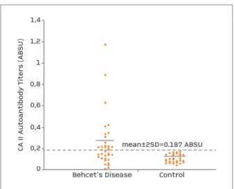
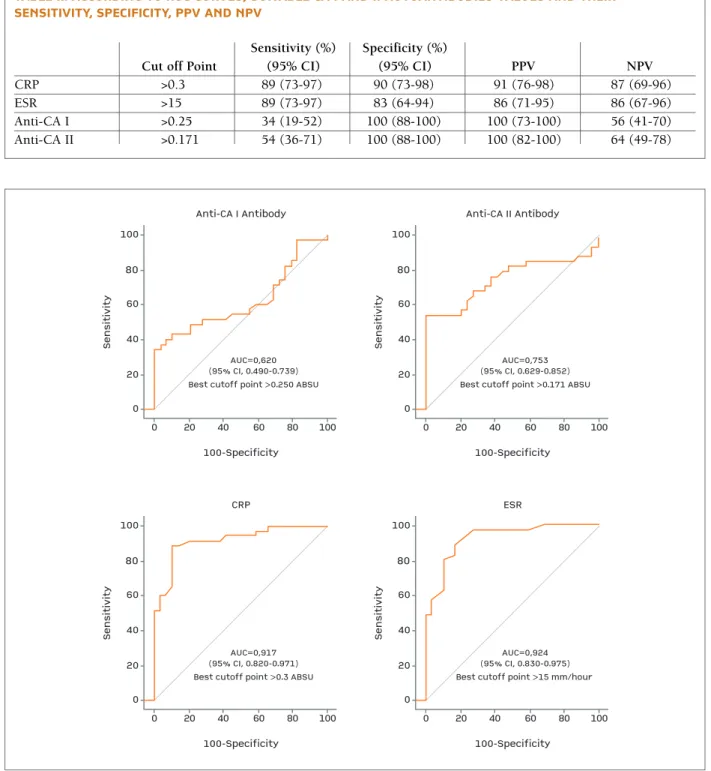
35 patients with BD and 29 healthy peers were in-cluded in this study. In the BD group, 25 patients were in active and 10 patients were in inactive state of the disease. Demographic characteristics of both groups are presented in Table I. The CA I and II autoantibody levels of both groups are shown in Table I. The mean absorbance value of CA I autoantibody for the control subjects was 0.137±0.056 and the absorbance values higher than 0.249, the mean absor bance + 2SD of con-trol subjects, were defined as positive. Positive results were obtained in 12 out of 35 patients with BD (Figu-re 1). The mean absorbance value of CA I autoantibody of BD group (0.237±0.217) was found to be si -gnificantly higher compared with that of the control group (p=0.013) (Table I). The mean absorbance va-lue of CA II autoantibody for the control subjects was 0.107±0.04 and the absorbance values higher than 0.187, the mean absorbance + 2SD of control subjects, were defined as positive. Positive results were obtained in 16 out of 35 patients with BD (Fi gure 2). The me-dian absorbance value of CA II autoantibody of BD group was found to be significantly higher compared with that of the control group (p=0.0001) (Table I). Also, ESR and CRP values of patients with BD was
DIscUssION
BD is a chronic inflammatory disorder and it is cha-racterized by ulcerations, ocular, arthritic, vascular and neurological involvement. There is no specific bioche-mical parameter for diagnosis of BD. How ever, CRP, ESR, and leukocyte count are used13,14. ESR and CRP
le-vels usually are found higher in patients with BD com-pared to healthy controls14,15. Similar to previous
re-ports, ESR and CRP values of patients with BD were found to be significantly higher than in the control group
(p=0.0001) (Table I). CA I and II autoanti bo dies, CRP and ESR le vels were also evaluated with ROC curve analysis. Cut off points, sensitivity %, specificity %, PPV and NPV for these parameters are demonstra ted in Table II and Figu re 3. Significant positive correlations both between CA I and II autoantibody titers in pa -tients with BD (r=0.972, p=0.0001) and between anti-CA I and ESR ti ters in patients with BD (r=0.117, p=0.037) were found.
Behcet’s Disease Control
CA I A u to an ti bo dy T it er s (A B SU ) 1,2 1 0,8 0,6 0,4 0,2 0 mean±2SD=0.249 ABSU
fIGUrE 1.The dotted line indicates the mean value plus 2 SD of healthy control sera (A480 = 0.249)
Behcet’s Disease Control
CA II A u to an ti bo dy T it er s (A B SU ) 1,4 1 1,2 0,8 0,6 0,4 0,2 0 mean±2SD=0.187 ABSU
fIGUrE 2.The dotted line indicates the mean value plus 2 SD of healthy control sera (A480 = 0.187)
tAblE I. DEMOGrAPHIc cHArActErIstIcs AND lAbOrAtOry fINDINGs Of GrOUPs
Clinical Characteristics BD Group (n=35) Control Group (n=29) p
Age (year) 36 (35-44) 37 (34-42) >0.05
Oral aphthous ulceration % 100 –
Genital aphthous ulceration % 80 –
Eye involvement % 51 – Arthritis % 54 – Vascular involvement % 37 – Erythema nodosum % 57 – Pathergy positivity % 51 – HLA-B5 positivity % 51 – CRP 0.54 (0.32-1.87) 0.16 (0.085-0.25) 0.0001* ESR 32.1±18.4 8.62±6.89 0.0001** Anti-CA I Ab 0.237±0.217 0.137±0.056 0.013** Anti-CA II Ab 0.184 (0.109-0.257) 0.094 (0.071-0.146) 0.0001*
The results were expressed as CRP: mg/dL, ESR: mm/1st hour, anti-CA I Ab: ABSU, anti-CA II Ab: ABSU Data were expressed as: mean ± SD, median (interquartile range for 25-75%)
**p shows differences between Control and BD according to student t test *p shows differences between Control and BD according to Mann Whitney U test
that BD may actually represent one type of autoin-flammatory disease, based largely on certain clinical features, including recurrent mucocutaneous lesions and non-deforming arthritis, along with elevated pro-found significantly higher (p=0.0001) than in the
con-trol group in our study. The etiology of BD is unclear, but viruses, autoimmunity, oxidative stress and toxic agent have been implicated13,14. Some experts main tain
tAblE II. AccOrDING tO rOc cUrvEs, sUItAblE cA I AND II AUtOANtIbODIEs vAlUEs AND tHEIr sENsItIvIty, sPEcIfIcIty, PPv AND NPv
Sensitivity (%) Specificity (%)
Cut off Point (95% CI) (95% CI) PPV NPV
CRP >0.3 89 (73-97) 90 (73-98) 91 (76-98) 87 (69-96) ESR >15 89 (73-97) 83 (64-94) 86 (71-95) 86 (67-96) Anti-CA I >0.25 34 (19-52) 100 (88-100) 100 (73-100) 56 (41-70) Anti-CA II >0.171 54 (36-71) 100 (88-100) 100 (82-100) 64 (49-78) 0 Se n si ti vi ty Anti-CA I Antibody 100-Specificity AUC=0,620 (95% CI, 0.490-0.739) Best cutoff point >0.250 ABSU 100 80 60 40 20 0 20 40 60 80 100 0 Se n si ti vi ty CRP 100-Specificity AUC=0,917 (95% CI, 0.820-0.971) Best cutoff point >0.3 ABSU 100 80 60 40 20 0 20 40 60 80 100 0 Se n si ti vi ty Anti-CA II Antibody 100-Specificity AUC=0,753 (95% CI, 0.629-0.852) Best cutoff point >0.171 ABSU 100 80 60 40 20 0 20 40 60 80 100 0 Se n si ti vi ty ESR 100-Specificity AUC=0,924 (95% CI, 0.830-0.975) Best cutoff point >15 mm/hour 100 80 60 40 20 0 20 40 60 80 100
tes27. ROS are physiological activators of many
trans-cription factors such as pro-inflammatory cytokines and adhesion molecules and can play a critical role in apoptosis and autoimmune reactions28. The increased
ROS and it is result of protein peroxidation have been extensively demonstrated in pathogenesis of many au-toimmune diseases28,29. We speculate that increased CA
antibody titers in BD patients may be a result of in-creased oxidative damage in these patients.
Autoimmunity against many antigens such as S-an-tigen, interphotoreceptor retinoid binding protein, heatshock protein 60, aenolase, kinectin, atropo -myosin, heparan sulfate, annexin-V, cardiolipin, sele-nium binding protein, PTEN-induced putative kinase 1 and switch associated protein 70 are reported in BD and it is believed that these autoantibodies may have a potential diagnostic marker and help in understanding the etiology of BD30,31. However, cause of increased
au-toimmunity against these antigens have not been elu-cidated clearly13.
In the present study, CA I and II autoantibody are de-tected in BD subjects, but the pathogenic role of these antibodies remains uncertain. It shows the need for further studies to evaluate the significance of CA autoan tibody production in BD subjects.
cOrrEsPONDENcE tO
Ahmet Mentese
Program of Medical Laboratory Techniques, Vocational School of Health Sciences, Karadeniz Technical University,
Trabzon, Turkey
E-mail: amentese028@gmail.com
rEfErENcEs
1. Dalvi SR, Yildirim R, Yazici Y. Behcet’s Syndrome. Drugs 2012; 72(17): 2223-2241.
2. Mendes D, Correia M, Barbedo M, et al. Behçet’s disease - a con-temporary review. J Autoimmun 2009; 32: 178-188. 3. Korkmaz S, Erturan I, Naziroglu M, et al. Colchicine
modu-lates oxidative stress in serum and neutrophil of patients with Behçet Disease through regulation of Ca2+release and antioxi
-dant system. J Membrane Biol 2011; 244: 113-120.
4. Saadoun D, Wechsler B. Behçet’s disease. Orphanet J Rare Dis 2012;7:20.
5. Direskeneli H. Behcet’s disease: Infectious aetiology, new au-toantigens, and HLA-B51. Ann Rheum Dis 2001; 60: 996-1002. 6. Supuran CT. Carbonic anhydrases: Novel therapeutic applica-tions for inhibitors and activators. Nat Rev Drug Discov 2008;7(2):168-181.
7. Mentese A, Guven S, Sumer A, et al. Serum anti-carbonic an-hydrase I and II antibodies and polycystic ovary syndrome. Turk J Biochem 2013; 38(1): 43-48.
8. Adamus G. Autoantibody targets and their cancer relationship in the pathogenicity of paraneoplastic retinopathy. Autoimmun Rev 2009; 8: 410-414.
inflammatory cytokines1. Inflammation is one of the
main characteristics of BD and it is believed that reac-tive oxygen species (ROS) are one of the major causes and/or results of this inflammation3. Disrupted
oxi-dant/antioxidant balance has been reported in BD patients and it is suggested that this failure may res -pon sible for tissue injury in BD11.
Carbonic anhydrase isozymes are nearly ubiqui tous in living systems and have various functions. CA I and II are the most widely distributed members of the CA family, being present in almost all tissues16,17. Recently,
autoantibodies against CA I and II were demonstrated in many autoimmune and idiopathic diseases7,9,12,18-23.
The present study is the first report which shows an in-creased autoimmune response to both CA I and II in BD patients. We found CA I and II autoantibody pre-valence to be 34% and 46%, respectively. As mentio-ned previously, CA II autoantibodies have been deter-mined in various autoimmune and idiopathic diseases, such as systemic lupus erythematosus (SLE) (28.5%), primary biliary cirrhosis (PBC) (25%), Sjögren’s syndrome (SS) (62%), rheumatoid arthritis (27%), en-dometriosis (70%), type 1 diabetes (65%), Graves’ di-sease (25%), metabolic syndrome (16%) and acute an-terior uveitis (15.6%)9,12,18-25. Also, CA I autoantibodies
have been observed in many diseases, such as Graves’ disease (5%), polycystic ovary syndrome (26%) and acute anterior uveitis (20%)7,12,25. Also, we determined
a significant correlation between CA I and II autoanti-body levels in BD patients. This situation may arise from cross-reactivity due to homology of the enzymes. Similar situation is reported in previous reports such as Graves’ disease, SLE, SS and PBC12,21,22. The high
pre-valence of CA I autoantibody may indicate an epiphe-nomenon secondary to the vascular structural chan-ges due to its settlement in vessel and it may have an important role in regulation of vessel tone24. CA II is
present in every tissue, and autoimmune sialoadenitis was induced in mice by immunization with CA II16,24.
Thus, it could be a target antigen in the autoimmune diseases related to some tissues or organs. However, increased autoimmunity against these isozymes have been reported in cancer and autoimmune diseases, but no mechanisms have been identified8. Iuchi et al
de-monstrated that superoxide dismutase (SOD)-knock out mouse developed CA II antibodies as a result of in-creased oxidative stress26. 4-Hydroxy-2-Nonenal
(HNE) modifies proteins and alters their anti genic pro-perties. Uchida and coworkers reported that CA II is a target for HNE in their study releated with
erythrocy-21. Kino-Ohsaki J, Nishimori I, Morita M, et al. Serum antibodies to carbonic anhydrase I and II in patients with idiopathic chro -nic pancreatitis and Sjögren’s syndrome. Gastroenterology 1996; 110: 1579-1586.
22. Invernizzi P, Battezzati PM, Crosignani A, et al. Antibody to carbonic anhydrase II is present in primary biliary cirrhosis irres -pective of antimitochondrial antibody status. Clin Exp Immunol 1998; 144: 448-454.
23. Taniguchi T, Okazaki K, Okamoto M, et al. High prevalence of autoantibodies against carbonic anhydrase II and lactoferrin in type 1 diabetes: Concept of autoimmune exocrinopathy and endocrinopathy of pancreas. Pancreas 2003; 27: 26-30. 24. Alver A, Mentese A, Erem C, et al. Serum carbonic anhydrase
autoantibodies in metabolic syndrome. Diabet Met Syndr Clin Res Rev 2009; 3: 211-213.
25. Turk A, Aykut M, Akyol N, et al. Serum anti-carbonic anhydrase antibodies and oxidant-antioxidant balance in patients with acute anterior uveitis. Ocul Immunol Inflamm 2014; 22(2): 127-132.
26. Iuchi Y, Okada F, Onuma K, et al. Elevated oxidative stress in erythrocytes due to SOD1 deficiency causes anemia and triggers autoantibody production. Biochem J 2007; 402: 219-227. 27. Uchida K, Hasui Y, Osawa T. Covalent attachment of
4-Hy-droxy-2-Nonenal to erythrocyte proteins. J Biochem 1997; 122: 1246-1251.
28. Mostafa GA, El-Hadidi ES, Hewedi DH, et al. Oxidative stress in Egyptian children with autism: Relation to autoimmunity. J Neuroimmunol 2010; 219(1-2): 114-118.
29. Kannan S. Molecular basis of the drug resistance induced au-toimmunity: A redox theory. Med Hypotheses 2005; 64: 882--883.
30. Kanakis MA, Vaiopoulos GA, Kaklamanis PG. Etiopathogene-sis of Behçet’s disease with emphaEtiopathogene-sis on the role of immuno-logical aberrations. Clin Rheumatol 2010; 29: 1211-1216. 31. Pineton de Chambrun M, Wechsler B, Geri G, et al. New
in-sights into the pathogenesis of Behçet’s disease. Autoimmun Rev 2012; 11: 687-698.
9. Alver A, Senturk A, Cakirbay H, et al. Carbonic anhydrase II au-toantibody and oxidative stress in rheumatoid arthritis. Clin Biochem 2011; 44(17-18): 1385-1389.
10. International Study Group for Behçet’s Disease. Criteria for diagno sis of Behçet’s disease. Lancet 1990; 335: 1078-1080. 11. Isik A, Koca SS, Ustundag B, et al. Decreased total antioxidant
response and increased oxidative stress in Behcet’s Disease. To-hoku J Exp Med 2007; 212: 133-141.
12. Alver A, Mentese A, Karahan SC, et al. Increased serum anti-car-bonic anhydrase II antibodies in patients with Graves’ disease. Exp Clin Endocrinol Diabetes 2007; 115(5): 287-291. 13. Okunuki Y, Usui Y, Takeuchi M, et al. Proteomic surveillance of
autoimmunity in Behcet’s disease with uveitis: Selenium bind-ing protein is a novel autoantigen in Behcet’s disease. Exp Eye Res 2007; 84: 823-831.
14. Akcay YD, Sagin FG, Aksu K, et al. A panel of oxidative stress assays does not provide supplementary diagnostic information in Behcet’s disease patients. J Inflamm 2012; 9: 13.
15. Akturk S, Akturk E, Kurtoglu E, et al. Association between red cell distribution width and disease activity in patients with Beh -cet’s Disease. J Clin Exp Cardiolog 2012; 3: 9.
16. Sly WS, Hu PY. Human carbonic anhydrases and carbonic an-hydrase deficiencies. Annu Rev Biochem 1995; 64: 375-401. 17. Boztas M, Cetinkaya Y, Topal M, et al. Synthesis and carbonic
anhydrase isoenzymes I, II, IX, and XII inhibitory effects of dimethoxybromophenol derivatives incorporating cyclo-propane moieties. J Med Chem 2015; 58(2): 640-650. 18. Inagaki Y, Jinno-Yoshida Y, Hamasaki Y, et al. A novel
autoan-tibody reactive with carbonic anhydrase in sera from patients with systemic lupus erythematosus and Sjögren’s syndrome. J Dermatol Sci 1991; 2: 147-154.
19. Itoh Y, Reichlin M. Antibodies to carbonic anhydrase in sys-temic lupus erythematosus and other rheumatic disease. Arthri-tis Rheum 1992; 35: 73-82.
20. Kiechle FL, Quattrociocchi-Longe TM, Brinton DA. Carbonic anhydrase antibody in sera from patients with endometriosis. Am J Clin Pathol 1994; 101: 611-615.