ABSTRACT
Purpose: To report the visual outcomes of non-arteritic central retinal artery occlusion (NA-CRAO) patients submitted to hyperbaric oxygen therapy (HBOT).
Methods: Retrospective analysis of patients with CRAO submitted to HBOT between 2010 and 2017. Patients with non-transient NA-CRAO, absence of a patent cilioretinal artery and less of 24 hours of evolution were included. All patients were submitted to 90-minute sessions with 100% oxygen at 2.4 atm. Primary outcome was visual acuity (VA) change (logMar) before and after treatment. Adverse effects from the treatment were reported.
Results: Fourteen patients met the inclusion criteria, 57% males (n=8) and 43% females (n=6), with a median age of 68 years (range from 41 to 92 years). VA at baseline was count-fingers (CF) or worse in 85.7% (n=12) of the patients. The median time between the symptoms onset and the beginning of the treatment was 7.0 hours (range from 2 to 24 hours).
It was observed a significant improvement in best-corrected visual acuity (BCVA) between baseline and post HBOT, which was 2.34±1.16 and 1.39±0.94 logMar respectively (p=0.007). VA gain was greater than 0.3 logMar in 71.4% of the patients.
No serious adverse effects from HBOT were reported. One patient experienced mild ear barotrauma.
Conclusions: Patients with NA-CRAO experienced VA improvement after HBOT, although with significant limitations. The implementation of strategies to reduce the time between the onset of symptoms and treatment may result in a higher functional gain.
RESUMO
Objetivos: Comparar a variação da acuidade visual nos doentes com oclusão da artéria central da retina (OACR) não arterítica após oxigenoterapia hiperbárica.
Métodos: Realizámos uma análise retrospectiva dos doentes com OACR referenciados ao serviço de Medicina hiperbárica do Hospital Pedro Hispano entre 2010 e 2017. Apenas foram incluídos doentes com oclusão arterial documentada por angiografia fluoresceínica, ausência de artéria ciliorretiniana patente e com tempo de isquemia inferior a 24h. Todos os doentes tratados foram submetidos a terapêutica com oxigénio a 100%, a 2,4 atm durante 90 minutos por sessão. Para a análise estatística comparou-se a MAVC em logMAR antes e após o tratamento.
Resultados: No total quatorze doentes foram incluídos no estudo, 57% do sexo masculino (n=8) e 43% do sexo feminino (n=6), com uma mediana de idades de 68 anos (min.41; máx 92). A MACV pré tratamento era conta-dedos ou inferior em 85,7% (n=12) dos doentes. A mediana do tempo entre o início dos sintomas e o tratamento foi de 7.0 horas (min.2; máx.24h) A MACV pré e pós tratamento foi 2,34±1,16 e 1,39±0,94 logMar respectivamente. A melhoria da AV foi estatisticamente significativa (p=0,007), e 71,4% dos doentes obtiveram um ganho superior a 0.3 logMar. Um doente desenvolveu barotrauma ligeiro com resolução espontânea. Conclusões: Verificou-se uma melhoria da MAVC nos doentes com OACR submetidos a oxigenoterapia hiperbárica, no entanto, permanecendo um impacto funcional importante. A implementação de estratégias com o objetivo de reduzir o tempo entre o início dos sintomas e o tratamento permitirá obter melhores resultados funcionais.
INTRODUCTION
Central retinal artery occlusion (CRAO) is described in the literature as an ophthalmological emergency, due to the immediate catastrophic functional effects that are often associated. However, it be considered as a paradox, since no effective treatment is currently available.
As previously demonstrated, time of ischemia is the critical factor for visual prognosis.1 Animal models of CRAO have demonstrated that an ischemic insult to the retinal tissue longer than 90 minutes will lead to some degree of inner retina damage and, if longer than 240 minutes, that damage will be irreversible.1 Additionally, visual outcome is influenced by several other factors, such as the type of occlusion (arteritic versus non-arteritic), the occurrence of a transient occlusion in patients with risk factors for low perfusion pressure in the central retinal artery, the level of obstruction and anatomical variations,
such as, the presence of a cilioretinal artery which provides a macular backup for blood supply.2 Recently, fundoscopic changes have been described as a reliable marker of irreversible damage, namely, the presence of cherry-red spot macula.3 Spontaneous visual recovery can be expected in about 39% of the patients following non-arteritic central retinal artery occlusion NA-CRAO,1 yet permanent visual loss is generally the rule.
Hyperbaric oxygen therapy (HBOT) enhances body tissue oxygenation in the presence of a correct vascular supply. It delivers 100% oxygen by a pressure greater than one atmosphere (atm), increasing plasma oxygen transportation and diffusion, with positive effects in vascular perfusion and neuroprotection.4-7 The theoretical scientific basis for its use in CRAO lies on the anatomical structure of the retina, a low thickness tissue, with double vascular supply, in which the choroidal capillary vessels that supply the outer retina will bypass the occlusion and
oxygenate inner ischemic retina until spontaneous reperfusion occurs. In animal models of CRAO, the apoptotic index of retinal cells was demonstrated to be lower in the group submitted to HBOT in the reperfusion phase than in control group.8
The largest published study so far evaluating HBOT for CRAO, included 128 patients with NA-CRAO, with a mean time delay from symptoms to treatment of 7.8±3.8 hours. Results demonstrated a significant mean improvement in best-corrected visual acuity (BCVA, logMar) of 0.526±0.688, from 2.14±0.50 to 1.61±0.78 (P<0.01), with 67% of the patients achieving a BCVA gain > 0.3 logMar.3
The purpose of this work was to describe the visual outcomes of NA-CRAO patients submitted to HBOT in our hospital.
METHODS
We retrospectively analyzed patient records with CRAO submitted to HBOT from 2010 to 2017 in Hospital Pedro Hispano Hyperbaric Medicine Unit, which is the referral center of approximately half of the Portuguese mainland. The protocol of this retrospective study was approved by the Ethical Committee of our Hospital and was conducted in compliance with the tenets of the Declaration of Helsinki.
POPULATION
The majority of CRAO patients treated in our HBOT department was referred from other hospitals. We included patients with NA-CRAO documented by fluorescein angiography (FA), and evidence of arterial perfusion delay or box scarring, normal choroidal and optic disc perfusion. Patients with fundoscopic signs of CRAO and normal perfusion times on FA, suggestive of transient retinal ischemia due to low perfusion pressure were excluded, as it has been proved that these patients tend to have higher rates of spontaneous visual recovery.2 Other exclusion criteria were branch retinal artery occlusion, clinical symptoms or inflammatory markers suggestive of arteritic
occlusion, no documented BCVA, time between symptoms onset and treatment longer than 24 hours, inhalation of high percentage of normobaric oxygen before HBOT, and the presence of a patent cilioretinal artery.
Information about demographics, personal history of dyslipidemia, arterial hypertension, diabetes, obesity, smoking habits or other relevant information for arterial occlusion was obtained.
Primary ophthalmological outcome was BCVA quantified in Snellen chart in decimal scale. In case of very low vision (<20/400 in Snellen chart), it was assessed by semiquantitative scale, the capability to count-fingers (CF), to see hand movement (HM) or light perception (LP) at a distance of 30 cm. Decimal value attributed to CF and HM was 0.014 and 0.005 respectively, as described elsewhere.9 For statistical analysis, BCVA was converted to logarithmic scale. A clinically significant visual improvement was defined as a decrease in logMAR of 0.3. Improvement from HM to CF has been described to be clinically relevant, and corresponds to an interval of 4 lines (in 0.1 log-unit steps).9
HBOT PROTOCOL
Patients were initially submitted to two daily sessions of 100% oxygen at 2.4 atm for 90 minutes during the first 72 hours. Additional treatments, that included 1 session per day, were performed if BCVA improvement occurred and until it stabilized. Treatment was interrupted if no subjective BCVA gain occurred after the first three consecutive sessions. All treatments were performed in the same multiplace chamber.
STATISTICS
Statistical analysis was performed using SPSS (version 21.0). Continuous variables were presented as median with minimum and maximum values. BCVA variation before and after treatment was assessed with Wilcoxon signed-rank test. A p value <0.05 was considered to be statistically significant.
RESULTS
A total of fourteen patients with NA-CRAO submitted to HBOT, 57% males (n=8) and 43% females (n=6), with a median age of 68 years (min.41; max.92), met the inclusion criteria. The most prevalent systemic diseases reported were arterial hypertension (85.6%), dyslipidemia (71.4%) and type 2 diabetes (50%). One patient had atrial fibrillation and another hyperhomocysteinemia.
BCVA at baseline was CF or worse in 85.7% (n=12) of the patients. Fundoscopic evaluation revealed arterial embolus in one patient (7.1%). Regarding conservative therapy prior to HBOT, 43% (n=6) of patients received ocular massage and topical ocular hypotensive medication at the discretion of the attending ophthalmologist. The median time between the symptoms onset and the beginning of the treatment was 8.0 hours (min.2;
max.24h), and the median number of hyperbaric treatments was 4.5 (min.3; max.16).
VISUAL ACUITY OUTCOMES
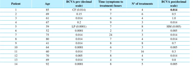
The baseline and after-HBOT BCVA was 2.34±1.16 and 1.39±0.94 logMar, respectively. This BCVA improvement following treatment was statistical significant (p=0.007). Overall, three patients did not experience any BCVA gain and one continued to worsen despite therapy. All the patients with BCVA gain (n=10; 71.4%) achieved a gain greater than 0.3 logMar, and 5 patients (35.7%) achieved a final BCVA better than 1.0 logMar (0.1 in decimal scale). Table 1 describes the characteristics and visual outcomes of patients with CRAO submitted to HBOT.
Table 1 - BCVA comparison between pre and post-HBOT.
Patient Age BCVA pre (decimal
scale)
Time (symptoms to
treatment) hours Nº of treatments
BCVA pos(decimal scale) 1 85 CF (0.014) 9 4 0.014 2 69 0.15 7 6 0.5 3 61 0.014 6 4 1.0 4 67 0.2 9 3 0.014 5 59 LP (0.0001) 7 8 HM (0.005) 6 52 0.0001 2 5 0.005 7 75 0.014 24 3 0.014 8 80 0.014 7 4 0.014 9 41 0.014 8 8 0.7 10 64 0.0001 6 3 0.005 11 60 0.014 7 16 0.3 12 70 0.005 8 4 0.014 13 69 0.014 4 9 0.8 14 92 0.0001 8 5 0.005
BCVA, best-corrected visual acuity; CF, counting-fingers; HM, hand-motion; LP, Light perception;
COMPLICATIONS FROM HBOT
Overall no serious complications related to HBOT were registered. Only one patient (7.1%) experienced mild ear barotrauma, with full spontaneous recovery in few days.
DISCUSSION
In this retrospective analysis, we aimed to report the clinical outcomes of patients with CRAO following HBOT. It is known that some spontaneous visual recovery occurs in the first 7 days after occlusion2 depending on the type of CRAO. In order to reduce the risk of bias associated with the natural history of CRAO, we only included patients with NA-CRAO, with delayed perfusion times on FA and without a patent cilioretinal artery. Our results demonstrated a significant positive impact of HBOT in visual function in patients with this condition,
where without treatment there is little chance of visual improvement. These results are in agreement with those published in the literature.3,10
In the non-transient NA-CRAO´s type Hayreh and Zimmerman have described some VA spontaneous recovery in about 22% of the patients.2 We observed a clinical significant VA improvement in about 71.4% of the patients following HBOT. Similar results were observed by Hadanny et al in which VA gain was achieved in 67.2% of the patients, with a time between symptoms and treatment of 7.8±3.8 hours.3
It is known that neuronal cells have little energy storage and high metabolic demands, which makes them very vulnerable to ischemic damage. Although Hayreh et al demonstrated massive irreversible damage occuring after 240 minutes (4 hours) of CRAO,1 the true retinal survival time limit doesn´t appear to be completely known. A delay of 7 hours until treatment may explain the limited VA gain experienced in our patients, where VA after treatment was 1.39±0.94 logMar (approximately 0.4/10 in decimal scale). Better results would probably be obtained if delay time was shorter than 4 hours.
When considering HBOT effects, distinction must be made between short and long-term therapy. Short term therapeutic effects in animal models simulating acute ischemic disease elicit a multiplicity of mechanisms with a positive impact, such as apoptotic and anti-inflammatory effects,4,11,12 probably secondary to amelioration of ischemic cells’ energy metabolismo.7 Other effect includes edema reduction by decreased expression of metalloproteinase (reducing vascular basement membrane degradation), preventing the decline of tissue perfusion after occlusion.5 These effects may help reduce the degeneration of neighboring cells secondary to the death of injured cells.11 However, extrapolations of metabolic results from animal models must be made with caution, as in these models HBOT is initiated briefly after ischemia, generally in the first 60 minutes, a virtually impossible scenario in clinical practice.
In spite of the limited experimental and clinical data available, we believe that HBOT may have a major beneficial effect on the visual acuity following CRAO. Strategies to reduce the delay in initiation of HBOT should be implemented, and widespread application of HBOT may eventually provide more robust evidence of its efficacy.
CONCLUSION
HBOT provided VA improvement in patients with NA-CRAO. In the absence of an effective treatment in CRAO, patients should be referred without delay to a hyperbaric medicine department. Strategies aiming to reduce the time between occlusion and treatment may potentially increase the visual outcome of this devastating condition.
REFERENCES
1. Hayreh SS, Zimmerman MB, Kimura A, Sanon A. Central retinal artery occlusion. Retinal survival time. Exp Eye Res. 2004;78(3):723–36.
2. Hayreh SS, Zimmerman MB. Central retinal artery occlusion: Visual outcome. Am J Ophthalmol. 2005;140(3).
3. Hadanny A, Maliar A, Fishlev G, Bechor Y, Bergan J, Friedman M, et al. Reversibility of retinal ischemia due to central retinal artery occlusion by hyperbaric oxygen. Clin Ophthalmol. 2017;11:115–25.
4. Wang R, Xu J, Xie J, Kang Z, Sun X, Chen N, et al. Hyperbaric oxygen preconditioning promotes survival of retinal ganglion cells in a rat model of optic nerve crush. J Neurotrauma. 2010;27(April):763–70.
5. Pushkov D, Nicholson JD, Michowiz S, Novitzky I, Weiss S, Ben Hemou M, et al. Relative neuroprotective effects hyperbaric oxygen treatment and TLR4 knockout in a mouse model of temporary middle cerebral artery occlusion. Int J Neurosci. 2016;126(2):174–81.
6. de Smet GHJ, Kroese LF, Menon AG, Jeekel J, van Pelt AWJ, Kleinrensink G-J, et al. Oxygen therapies and their effects on wound healing. Wound Repair Regen. 2017;
7. Hu Q, Manaenko A, Bian H, Guo Z, Huang JL, Guo ZN, et al. Hyperbaric Oxygen Reduces Infarction Volume and Hemorrhagic Transformation Through ATP/NAD+/Sirt1 Pathway in Hyperglycemic Middle Cerebral Artery Occlusion Rats. Stroke. 2017;48(6):1655–64.
8. Karaman S, Ozkan B, Yazir Y, Yardimoglu M. Comparison of hyperbaric oxygen versus iloprost treatment in an experimental rat central retinal artery occlusion model. 2016;
9. Schulze-Bonsel K, Feltgen N, Burau H, Hansen L, Bach M. Visual acuities “hand motion” and “counting fingers” can be quantified with the Freiburg Visual Acuity Test. Investig Ophthalmol Vis Sci. 2006;47(3):1236–40.
10. Cope A, Eggert J V, O’Brien E. Retinal artery occlusion: visual outcome after treatment with hyperbaric oxygen. Diving Hyperb Med. 2011;41(3):135–8.
11. Gaydar V, Ezrachi D, Dratviman-Storobinsky O, Hofstetter S, Avraham-Lubin BCR, Goldenberg-Cohen N. Reduction of apoptosis in ischemic retinas of two mouse models using hyperbaric oxygen treatment. Investig Ophthalmol Vis Sci. 2011;52(10):7514–22.
12. Migita H, Yoshitake S, Tange Y, Choijookhuu N, Hishikawa Y. Hyperbaric Oxygen Therapy Suppresses Apoptosis and Promotes Renal Tubular Regeneration After Renal Ischemia/Reperfusion Injury in Rats. Nephrourol Mon. 2016;8(1):e34421.
CONTACT
Pedro Coelho
Department of Ophthalmology Hospital Pedro Hispano Rua Dr. Eduardo Torres 4464-513 Senhora da Hora Portugal
E-mail: [email protected]
The authors declare having followed the protocols in use at their working center regarding patients’ data publication.
The authors have no conflicts of interest neither the work was published elsewhere. No subsidies or grants contributed to this work.